. 2025 Dec;16(6):1966-1974.
doi: 10.1007/s12975-025-01360-2. Epub 2025 Jun 6.

Clinical Significance of Somatic PIK3CA and MAP3K3 Mutations in Cerebral and Spinal Cavernous Malformations

Affiliations

Clinical Significance of Somatic PIK3CA and MAP3K3 Mutations in Cerebral and Spinal Cavernous Malformations

Hiroki Hongo et al. Transl Stroke Res. 2025 Dec.

Abstract

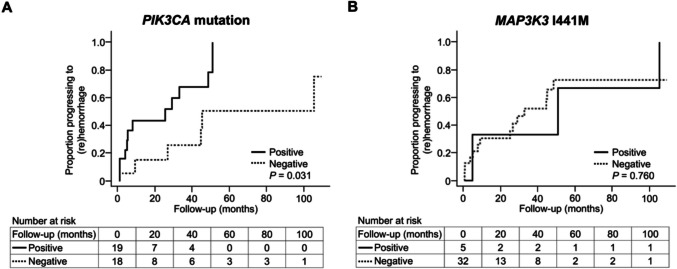
Somatic PIK3CA and MAP3K3 mutations in cerebral and spinal cavernous malformations (CMs) have been identified in recent studies. However, their significance in the clinical presentation and risk of hemorrhage in CMs remains poorly understood. We aimed to analyze the association between these mutations and the clinical characteristics of CMs. Among patients with CMs who underwent surgical resection of lesions between July 2002 and March 2022, those with complete clinical and radiological data at the time of initial surgery were included. Somatic PIK3CA and MAP3K3 mutations were detected using droplet digital polymerase chain reaction. Subsequently, the clinical and radiological characteristics correlated with these mutations were assessed. Furthermore, the effect of these mutations on the first symptomatic intraparenchymal hemorrhage during follow-up was evaluated. In total, 72 patients were included; among them, 50 had sufficient mutation data. PIK3CA E542K, E545K, and H1047R mutations were identified in 7 (14%), 7 (14%), and 15 (30%) patients, respectively. MAP3K3 I441M was identified in 10 (20%) patients (8 [16%] had both PIK3CA and MAP3K3 mutations). MAP3K3 I441M was more common in patients with Zabramski classification type II lesions than in those with CMs of other types (p = 0.024). Multivariate Cox regression analyses identified the presence of a PIK3CA mutation as a risk factor for early (re)hemorrhage. The results suggest that PIK3CA and MAP3K3 mutations are associated with clinical and radiological characteristics in patients with CMs and that the presence of a somatic PIK3CA mutation increases susceptibility to hemorrhage. These findings may help guide future therapeutic strategies.

Keywords: MAP3K3; PIK3CA; Cavernous malformations; Hemorrhage; Somatic mutations.


Conflict of interest statement

Declarations. Ethics Approval: This study was conducted in accordance with the Declaration of Helsinki and was approved by the Human Genome, Gene Analysis Research Ethics Committee of the Faculty of Medicine, The University of Tokyo (G10026, G10028, 2019211G), Ethics Committee of Tokyo Metropolitan Neurological Hospital (TS-R02-003), Ethics Committee of Fuji Brain Institute and Hospital (202103), and the Ethics Committee of Kanto Neurosurgical Hospital (2021–016). Consent to Participate: Written informed consent was obtained from each patient for inclusion in this study. Consent to Publish: Not applicable. Competing Interests: The authors declare no competing interests.

Figures

Fig. 1
Fig. 1
Typical appearance on MRI for each Zabramski classification type. A Type I lesion in the brainstem (Patient 16 at the time of surgery), defined as a unilocular lesion that appears hyperintense on T1-weighted imaging (T1WI) and shows a hyperintense or hypointense core on T2-weighted imaging (T2WI), consistent with acute or subacute hemorrhage. B Type II lesion in the right frontal lobe (Patient 32 at the time of surgery), defined as a lesion exhibiting a reticulated signal core on both T1WI and T2WI, representing hemorrhage and thrombosis of varying ages. C Type III lesion in the left temporal lobe (white arrows; Patient 26 at the time of surgery), defined as a lesion that appears isointense on T1WI and hypointense on T2WI, consistent with chronic hemorrhage. T1WI images are shown on the left and T2WI images on the right. Type IV CM lesions were not identified in this study
Fig. 2
Fig. 2
Kaplan–Meier plots of progression to intracerebral/spinal hemorrhage in the analysis of risk factors for hemorrhage during follow-up (n = 37). A Thirty-seven patients were divided into two groups based on the presence or absence of PIK3CA mutations. The"Positive"group consisted of patients with PIK3CA mutations, while the "Negative" group consisted of those without PIK3CA mutations. B Thirty-seven patients were divided into two groups based on the presence or absence of MAP3K3 I441M mutations. The "Positive" group consisted of patients with the MAP3K3 I441M mutation, and the "Negative" group consisted of those without it. Note: In both panels, the groups were defined solely based on the presence or absence of the specific mutation mentioned, regardless of the presence or absence of the other mutation. As a result, a patient could belong to the "Positive" or "Negative" group for each mutation independently. The horizontal axis represents follow-up time (months), and the vertical axis represents the proportion of patients progressing to hemorrhage after diagnosis. These plots illustrate the cumulative hemorrhage rate (1 − survival rate) over time. The “number at risk” at each time point indicates the number of individuals who have not yet experienced the event up to that point. In panel A, the difference first reached statistical significance at 33 months (P = 0.037). It temporarily lost significance at 45 months (P = 0.065) but became significant again at 51 months (P = 0.031) and remained so thereafter

References

    1. Hong T, Xiao X, Ren J, et al. Somatic MAP3K3 and PIK3CA mutations in sporadic cerebral and spinal cord cavernous malformations. Brain. 2021;144:2648–58. 10.1093/brain/awab117. - PubMed
    1. Zafar A, Quadri SA, Farooqui M, et al. Familial cerebral cavernous malformations. Stroke. 2019;50:1294–301. 10.1161/STROKEAHA.118.022314. - PMC - PubMed
    1. Akers AL, Johnson E, Steinberg GK, Zabramski JM, Marchuk DA. Biallelic somatic and germline mutations in cerebral cavernous malformations (CCMs): evidence for a two-hit mechanism of CCM pathogenesis. Hum Mol Genet. 2009;18:919–30. 10.1093/hmg/ddn430. - PMC - PubMed
    1. Zhou Z, Tang AT, Wong WY, et al. Cerebral cavernous malformations arise from endothelial gain of MEKK3-KLF2/4 signalling. Nature. 2016;532:122–6. 10.1038/nature17178. - PMC - PubMed
    1. Peyre M, Miyagishima D, Bielle F, et al. Somatic PIK3CA mutations in sporadic cerebral cavernous malformations. N Engl J Med. 2021;385:996–1004. 10.1056/NEJMoa2100440. - PMC - PubMed

Substances

LinkOut - more resources