. 2025 Jun 6;6(1):36.
doi: 10.1186/s43556-025-00276-5.

The EZH2 selective inhibitor ZLD1039 attenuates UUO-induced renal fibrosis by suppressing YAP activation

Affiliations

The EZH2 selective inhibitor ZLD1039 attenuates UUO-induced renal fibrosis by suppressing YAP activation

Qingling Xia et al. Mol Biomed. .

Abstract

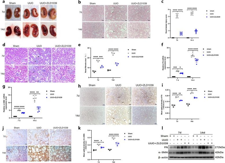
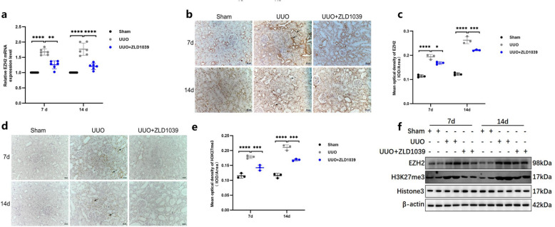
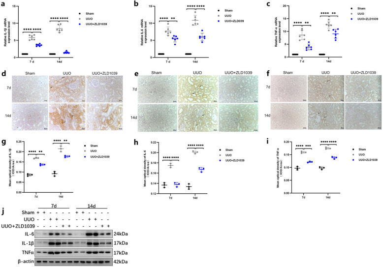
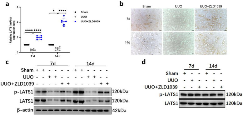
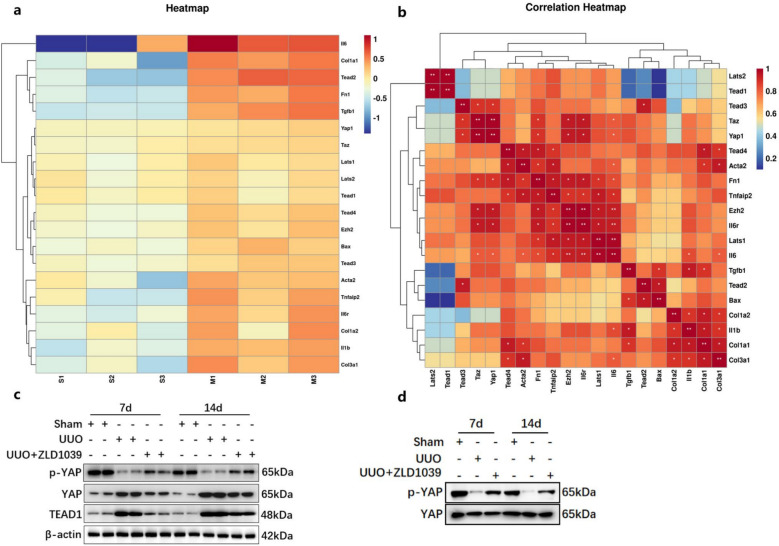
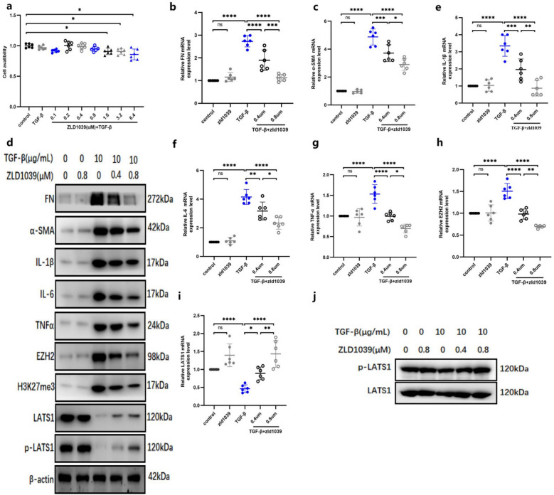
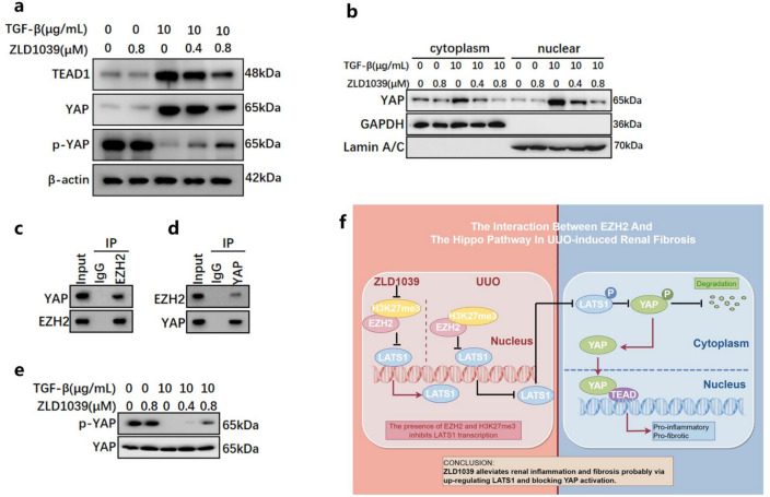
Renal fibrosis is a manifestation of the progression of chronic kidney disease (CKD) and chronic inflammation is a main driver in the development of renal fibrosis. Yes-associated protein (YAP), acting as a transcriptional co-activator within the Hippo signaling pathway, has been implicated in renal fibrosis. Enhancer of zeste homolog 2 (EZH2) exhibits high expression level in renal fibrosis induced by unilateral ureteral obstruction (UUO), yet the interplay between YAP and EZH2 in renal fibrosis remains to be elucidated. ZLD1039, a selective inhibitor of EZH2, has demonstrated protective effects against cancer and acute kidney injury (AKI). In this study, we conducted a systemic pharmacological investigation to determine if ZLD1039 treatment mitigates UUO-induced renal inflammation and fibrosis through modulation of the Hippo-YAP pathway. Our results revealed that UUO triggered renal inflammation and collagen deposition, with significant activation of YAP. Notably, ZLD1039 treatment effectively alleviated renal inflammation and fibrosis, while inhibiting the expression and nuclear translocation of YAP. Mechanically, we observed a notable down-regulation of large tumor suppressor homolog 1 (LATS1) in parallel with the up-regulation of EZH2. Furthermore, inhibition of EZH2 by ZLD1039 was linked to the up-regulation of LATS1 expression and YAP inactivation. Similarly, in vitro pharmacological inhibition of EZH2 by ZLD1039 resulted in elevated LATS1 expression and diminished YAP activation. Collectively, our findings suggest that ZLD1039, a selective inhibitor of EZH2, likely attenuates renal inflammation and fibrosis probably by up-regulating LATS1 and inhibiting YAP activation. This mechanistic link between EZH2 and YAP provides a fresh perspective on treating renal fibrosis.

Keywords: Enhancer of zeste homolog 2; Inflammation; Large tumor suppressor 1; Renal fibrosis; Yes-associated protein 1.


Conflict of interest statement

Declarations. Ethics approval and consent to participate: The experimental scheme was approved by the Ethics Committee of Southwest Medical University (NO.20240715-008) and followed the Guide for the Care and Use of Laboratory Animals. Consent for publication: Not applicable. Competing interests: The authors declare no conflict of interest.

Figures

Fig. 1
Fig. 1
ZLD1039 inhibits UUO-induced renal fibrosis. a The appearance of kidney tissues following unilateral ureteral ligation; b, c Representative sections of H&E staining of kidney tissues (original magnification × 200) and renal tubular injury scores were calculated according to the criterion in materials and methods; Data are represented as means ± SDs (n = 6); d, e Representative sections of Masson’s trichrome staining of kidney tissues (original magnification × 200) and renal fibrosis scores were calculated. Data are represented as means ± SDs (n = 3); f, g The mRNA levels of FN and α-SMA; Data are represented as means ± SDs (n = 6); h, i Representative sections of IHC staining of FN (original magnification × 200) and mean optical density of FN; j, k Representative sections of IHC staining of α-SMA (original magnification × 200) and mean optical density of α-SMA; Data are represented as means ± SDs (n = 3); l The protein expression levels of FN and α-SMA were quantified by normalized with β-actin. * p < 0.05; ** p < 0.01; *** p < 0.001; **** p < 0.0001. All western blotting analyses were performed in two randomized mice from each group, and the experiments were repeated in triplicate
Fig.2
Fig.2
ZLD1039 indeed suppresses EZH2 and H3K27me3 expression in UUO rats. a The mRNA level of EZH2; b-e Photomicrographs (original magnification × 200) illustrates EZH2 and H3K27me3 IHC staining of kidney tissues from sham or UUO mice with/without ZLD1039 administration and the mean optical density of EZH2 and H3K27me3; Data are represented as means ± SDs (n = 3); f The protein expression levels of EZH2 and H3K27me3 were quantified by normalized with β-actin and Histone H3; * p < 0.05; ** p < 0.01; *** p < 0.001; **** p < 0.0001. All western blotting analyses were performed in two randomized mice from each group, and the experiments were repeated in triplicate
Fig.3
Fig.3
EZH2 inhibition by ZLD1039 attenuates renal inflammation in UUO rats. a-c The mRNA levels of IL-1β, IL-6 and TNFα; Data are represented as means ± SDs (n = 6); d, g Representative sections of IHC staining of IL-1β (original magnification × 200) and the mean optical density of IL-1β; e, h Representative sections of IHC staining of IL-6 (original magnification × 200) and the mean optical density of IL-6; f, i Representative sections of IHC staining of TNFα (original magnification × 200) and the mean optical density of TNFα; Data are represented as means ± SDs (n = 3); j The protein expression levels of IL-1β, IL-6 and TNFα were quantified by normalized with β-actin; ** p < 0.01; *** p < 0.001; **** p < 0.0001. All western blotting analyses were performed in two randomized mice from each group, and the experiments were repeated in triplicate
Fig.4
Fig.4
EZH2 inhibition by ZLD1039 increases LATS1 expression in UUO rats. a The mRNA level of LATS1; Data are represented as means ± SDs (n = 6); b Representative sections of IHC staining of p-LATS1 (original magnification × 200). c The protein expression level of LATS1 and p-LATS1 were quantified by normalized with β-actin; d The protein expression level of p-LATS1 were quantified by normalized with LATS1; * p < 0.05; **** p < 0.0001. All western blotting analyses were performed in two randomized mice from each group, and the experiments were repeated in triplicate
Fig.5
Fig.5
EZH2 inhibition by ZLD1039 blocks YAP activation in UUO rats. a, b The cluster heatmap and correlation heatmap were conducted to present differentially expressed genes in the GSE181380 dataset (n = 3). c The protein expression levels of YAP, p-YAP and TEAD1 were quantified by normalized with β-actin; d The protein expression level of p-YAP were quantified by normalized with YAP; All western blotting analyses were performed in two randomized mice from each group, and the experiments were repeated in triplicate
Fig.6
Fig.6
ZLD1039 alleviates TGFβ-induced renal TECs inflammation and fibrosis by inhibiting EZH2 and elevating the expression of LATS1. a The cell viability of TECs under the stimulation of TGFβ and ZLD1039. Data are represented as means ± SDs (n = 6); b-e, f-i The mRNA levels of FN, α-SMA, IL-1β, IL-6, TNFα, EZH2 and LATS1; Data are represented as means ± SDs (n = 6); d The protein expression levels of FN, α-SMA, IL-1β, IL-6, TNFα, EZH2, H3K27me3, LATS1 and p-LATS1 were quantified by normalized with β-actin; j The protein expression level of p-LATS1 were quantified by normalized with LATS1 in TGFβ-induced TECs; * p < 0.05; ** p < 0.01; *** p < 0.001; **** p < 0.0001; nsp > 0.05. All Western Blotting analyses were repeated in triplicate
Fig.7
Fig.7
ZLD1039 treatment represses YAP activation in TGFβ-induced TECs. a The protein expression levels of YAP, p-YAP and TEAD1 were quantified by densitometry and normalized with β-actin; b The expression levels of YAP in cytoplasm and nucleus; c,d The interactions between EZH2 and YAP were detected by Co-Immunoprecipitation (Co-IP) in NRK-52E cells. e The protein expression level of p-YAP was quantified by normalized with YAP in TGFβ-induced TECs; f The interaction between EZH2 and the Hippo pathway in UUO-induced renal fibrosis (This picture is drawn using Figdraw). All Western Blotting analyses were repeated in triplicate

Similar articles

References

    1. Kaur R, Singh R. Mechanistic insights into CKD-MBD-related vascular calcification and its clinical implications. Life Sci. 2022;311(Pt B): 121148. 10.1016/j.lfs.2022.121148. - PubMed
    1. Lohia S, Vlahou A, Zoidakis J. Microbiome in Chronic Kidney Disease (CKD): an omics perspective. Toxins (Basel). 2022;14(3): 176. 10.3390/toxins14030176. - PMC - PubMed
    1. Nastase MV, Zeng-Brouwers J, Wygrecka M, Schaefer L. Targeting renal fibrosis: mechanisms and drug delivery systems. Adv Drug Deliv Rev. 2018;129:295–307. 10.1016/j.addr.2017.12.019. - PubMed
    1. Liu BC, Tang TT, Lv LL, Lan HY. Renal tubule injury: a driving force toward chronic kidney disease. Kidney Int. 2018;93(3):568–79. 10.1016/j.kint.2017.09.033. - PubMed
    1. McLeod DJ, Sebastião YV, Ching CB, Greenberg JH, Furth SL, Becknell B. Longitudinal kidney injury biomarker trajectories in children with obstructive uropathy. Pediatr Nephrol. 2020;35(10):1907–14. 10.1007/s00467-020-04602-7. - PMC - PubMed

MeSH terms

Substances

LinkOut - more resources