. 2025 Jul;14(18):e2500971.
doi: 10.1002/adhm.202500971. Epub 2025 Jun 6.

Design and Validation of Hybrid Polymer-Lipid Nanoparticles as Novel Transfection Vectors for MicroRNA Delivery to Human Cardiac Fibroblasts

Affiliations

Design and Validation of Hybrid Polymer-Lipid Nanoparticles as Novel Transfection Vectors for MicroRNA Delivery to Human Cardiac Fibroblasts

Letizia Nicoletti et al. Adv Healthc Mater. 2025 Jul.

Abstract

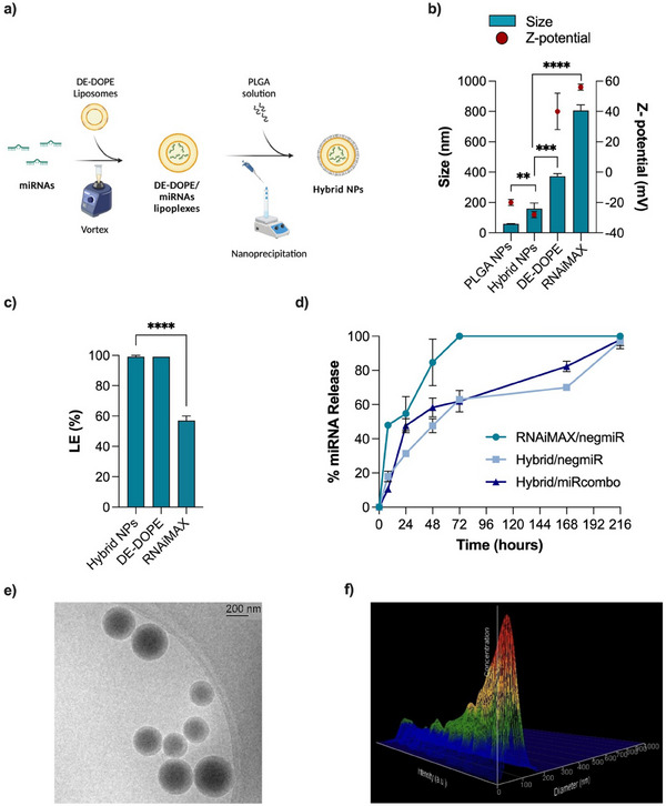
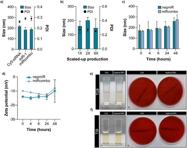
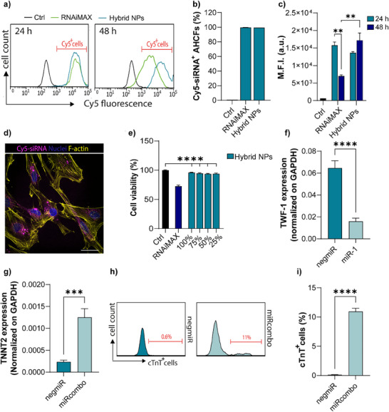
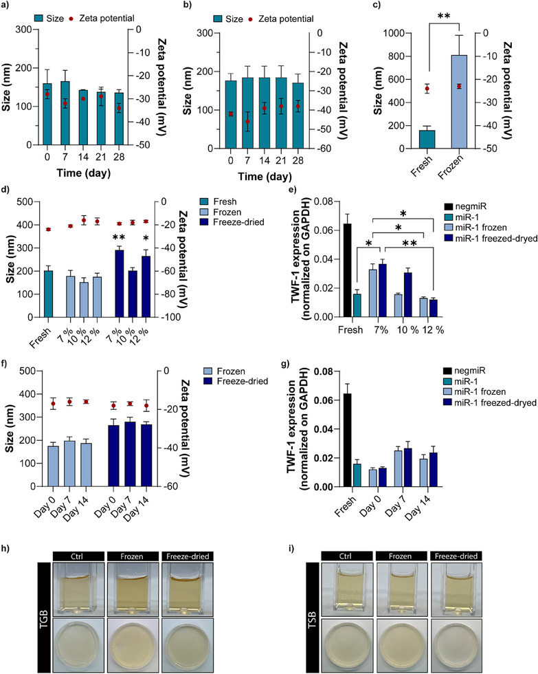
Hybrid polymer-lipid nanoparticles (hybrid NPs) are developed as novel in vitro transfection vectors for microRNAs (miRNAs) delivery to overcome the poor stability, incomplete loading efficiency and fast release kinetics of commercial transfection agents. Hybrid NPs with nanometric size are prepared by a scalable high-yield nanoprecipitation method. They consisted of a lipoplex core, composed of the cationic lipid [2-(2,3-didodecyloxypropyl)-hydroxyethyl] ammonium bromide (DE) and helper lipid dioleoyl phosphatidylethanolamine (DOPE), providing 99% miRNA loading, and a poly(lactic acid-co-glycolic acid) (PLGA) shell, ensuring NPs colloidal stability and controlled miRNA release kinetics. Adult human cardiac fibroblasts (AHCFs), transiently transfected with miR-1 loaded hybrid NPs versus RNAiMAX showed superior viability and higher miRNA content over 48 h. Hybrid NPs could be stored up to 14 days at -20 °C, upon freeze-drying with trehalose cryoprotectant (12% w/v), regaining their physicochemical and biological properties when redispersed. Hybrid NPs are assessed in a miRcombo model of fibroblast-to-cardiomyocyte reprogramming. At 15 days post-transfection with reprogramming miRNAs (miRcombo: miRs-1, 133, 208 and 499), cardiac troponin T marker expression is significantly increased at gene and protein level. These results pave the way to hybrid NP use as transfection vectors for the in vitro testing of miRNAs targeting AHCFs.

Keywords: human cardiac fibroblasts; hybrid nanoparticles; microRNA; nanoparticles storage; transfection vectors.


Conflict of interest statement

The authors declare no conflict of interest.

Figures

Figure 1
Figure 1
Physicochemical properties of hybrid NPs: a) Schematic illustration of the nanoprecipitation protocol for the preparation of hybrid NPs. Created with Biorender. b) Size and Z‐potential of control PLGA NPs (PLGA NPs), hybrid NPs, and RNAiMAX, measured by DLS. NegmiR was loaded as model miRNA. c) NegmiR loading efficiency (LE) measured by Qubit fluorometric quantification assay for hybrid NPs compared to RNAiMAX. d) Release profile of negmiR and miRcombo from hybrid NPs and of negmiR from RNAiMAX NPs as a function of their incubation time (24, 48, 72, and 168 h) in PBS at 37 °C under dynamic conditions. Data are expressed as mean ± SD. NegmiR was loaded as model miRNA. Statistical analysis was performed by 1‐way ANOVA. e) Representative Cryo‐TEM images of hybrid NPs. f) 3D topographic image of hybrid NPs by NTA analysis. Data are expressed as mean ± SD. Statistical analysis was performed by a two‐sided t‐test.
Figure 2
Figure 2
Physicochemical properties of hybrid NPs incubated in different media, scalability of the production process, and sterility of hybrid NPs. a) Hydrodynamic diameter and PDI of hybrid NPs loaded with different oligonucleotides (Cy5‐siRNA, miR‐1, and miRcombo) measured by DLS. b) Hydrodynamic diameter and PDI of hybrid NPs prepared using 1, 2, and 8X volumes and measured by DLS. c,d) Stability study at physiological temperature in DMEM+FBS by measuring: hydrodynamic size (c) and Z‐potential (d) of hybrid NPs as a function of their incubation time (0, 4, 6, 24, and 48 h). e,f) Analysis of microbial contamination of freshly prepared negmiR‐loaded hybrid NPs kept in TBG at 37 °C (e) and TSB at 25 °C (f) for 14 days. In both figures f,g) the images on the left show turbidity assessment by visual inspection (i.e., cuvettes with clear broth indicating the absence of microbial contamination), while the images on the right show the absence of CFU on agar solid plates. Control (Ctrl) samples are TGB and TSB with no hybrid NPs, respectively. Data are expressed as mean ± SD. Statistical analysis was performed by a two‐sided t‐test.
Figure 3
Figure 3
Hybrid NPs showed efficient uptake, miRNA release, and gene expression regulation in AHCFs. a) Representative flow cytometry analysis showing cellular uptake of hybrid NPs and RNAiMAX loaded with Cy5‐siRNA in AHCFs after 24 and 48 h of treatment. b) Bar graphs showing the percentage of Cy5‐siRNA positive cells (Cy5‐siRNA+ cells) after 24 h treatment with hybrid NPs and RNAiMAX. c) M.F.I. after 24 h and 48 h of treatment with hybrid NPs and RNAiMAX. Untreated cells were used as control for all experiments. d) Representative fluorescence microscopy images showing Cy5‐siRNA (magenta) uptake by AHCFs, mediated by hybrid NPs after 24 h treatment. Nuclei were counterstained with DAPI (blue) and F‐actin with Phalloidin (yellow). Scale bar = 50 µm. e) Viability of AHCFs treated with negmiR‐loaded hybrid NPs and RNAiMAX NPs at 24 h. Hybrid NPs were tested at four concentrations: 100, 75, 50, and 25 (%) (Table 1). Cell viability was analyzed by resazurin assay. The viability of transfected AHCFs was normalized to that of non‐transfected AHCFs. f) Gene expression of TWF‐1 mRNA target in AHCFs treated with negmiR or miR‐1‐loaded hybrid NPs at 48 h, analyzed by ddPCR. g) Gene expression of TNNT2 mRNA in AHCFs treated with negmiR or miRcombo‐loaded hybrid NPs after 15 days of culture post‐treatment, analyzed by ddPCR. h,i) Representative flow plots (h) and percentage (i) of cTnT+ cells for AHCFs treated with negmiR or miRcombo using hybrid NPs after 15 days of culture. Reported data are average values ± SEM of three independent experiments. Statistical differences between the groups were determined by two‐sided t‐tests.
Figure 4
Figure 4
Stability and microbial contamination analysis of hybrid NPs under storage conditions. a,b) Hydrodynamic diameter and Z‐potential of hybrid NPs stored in RNase‐free water suspension at 4 °C encapsulating (a) negmiR and (b) miRcombo evaluated over time (0, 7, 14, 21, and 28 days). c) Hydrodynamic diameter and Z‐potential of frozen hybrid NPs loaded with negmiR compared to fresh hybrid NPs. Statistical analysis was performed by two‐sided t‐test. d) Hydrodynamic diameter and Z‐potential of frozen and freeze‐dried hybrid NPs with different concentrations of trehalose (7, 10, and 12% w/v) compared to fresh hybrid NPs. e) Gene expression of TWF‐1 mRNA target (normalized on GAPDH) in AHCFs transfected with negmiR or miR‐1 using fresh, frozen, and freeze‐dried hybrid NPs with different concentrations of trehalose (7, 10, and 12% w/v). Gene expression was analyzed at 48 h post‐transfection by ddPCR. f) Hydrodynamic diameter and Z‐potential of frozen and freeze‐dried hybrid NPs with 12% w/v of trehalose evaluated over time (0, 7, and 14 days). g) Gene expression of TWF‐1 mRNA target in AHCFs transfected with negmiR or miR‐1 using frozen and freeze‐dried hybrid NPs with 12% w/v of trehalose after 0, 7, and 14 days of storage compared to fresh hybrid NPs. Gene expression was analyzed at 48 h post‐transfection by ddPCR. h,i) Analysis of microbial contamination of frozen and freeze‐dried negmiR hybrid NPs stored for 14 days cultured in TBG at 37 °C (h) and TSB at 25 °C (i) for 14 days. In both figures, the images at the top represent the turbidity assessment (i.e., cuvettes with clear broth indicating the absence of microbial contamination), while the images at the bottom show the absence of CFU on agar solid plates. Ctrl samples are TGB and TSB with no hybrid NPs, respectively. For physicochemical hybrid NPs characterizations, data are expressed as mean ± SD of three independent experiments. Statistical analysis was performed by 1‐way ANOVA, unless otherwise specified in the text. For in vitro cells study, data are expressed as mean ± SEM of three independent experiments. Statistical analysis was performed by two‐sided t‐test.

Similar articles

References

    1. Ha M., Kim V. N., Nat. Rev. Mol. Cell Biol. 2014, 15, 509. - PubMed
    1. Jayawardena T. M., Egemnazarov B., Finch E. A., Zhang L., Alan Payne J., Pandya K., Zhang Z., Rosenberg P., Mirotsou M., Dzau V. J., Circ. Res. 2012, 110, 1465. - PMC - PubMed
    1. Jayawardena T. M., Finch E. A., Zhang L., Zhang H., Hodgkinson C. P., Pratt R. E., Rosenberg P. B., Mirotsou M., Dzau V. J., Circ. Res. 2014, 116, 418. - PMC - PubMed
    1. Zhou S. S., Jin J. P., Wang J. Q., Zhang Z. G., Freedman J. H., Zheng Y., Cai L., Acta Pharmacol. Sin. 2018, 39, 1073. - PMC - PubMed
    1. Nicoletti L., Paoletti C., Tarricone G., Andreana I., Stella B., Arpicco S., Divieto C., Mattu C., Chiono V., Nanomedicine 2022, 45, 102589. - PubMed

LinkOut - more resources