. 2025 Jun 6;20(6):e0324238.
doi: 10.1371/journal.pone.0324238. eCollection 2025.

Human umbilical cord mesenchymal stem cell-derived exosomes mitigate acute radiation-induced intestinal oxidative damage via the Nrf2/HO-1/NQO1 signaling pathway

Affiliations

Human umbilical cord mesenchymal stem cell-derived exosomes mitigate acute radiation-induced intestinal oxidative damage via the Nrf2/HO-1/NQO1 signaling pathway

Hongyu Wang et al. PLoS One. .

Abstract

Acute radiation-induced intestinal injury (ARII), a prevalent complication of abdominal radiotherapy, remains clinically challenging due to limited therapeutic options. This study demonstrates the therapeutic efficacy of human umbilical cord mesenchymal stem cell-derived exosomes (hucMSC-Exos) in mitigating ARII through Nrf2/HO-1/NQO1 pathway activation. In a rat model receiving 12 Gy abdominal irradiation, systemic hucMSC-Exos administration significantly restored intestinal mucosal integrity and reduced oxidative damage markers. Mechanistically, hucMSC-Exos potentiated the antioxidant axis by upregulating Nrf2 signaling, as evidenced by histopathological, biochemical, and molecular analyses. Complementary in vitro experiments revealed hucMSC-Exos protected irradiated IEC-6 cells from oxidative dysfunction while enhancing proliferation, effects substantially attenuated upon Nrf2 silencing via siRNA. These findings establish that hucMSC-Exos orchestrate redox equilibrium through targeted Nrf2 pathway modulation, effectively counteracting radiation-induced enterocyte apoptosis. The elucidated mechanism expands the therapeutic paradigm of MSC-derived exosomes in radioprotection and provides a clinically translatable strategy for managing ARII in oncological radiotherapy.


Conflict of interest statement

The authors have declared that no competing interests exist.

Figures

Fig 1
Fig 1. Identification of hucMSCs and exosomes.
(A) hucMSCs exhibited a spindle shape microscopically. Scale bar: 50 μm. (B-F) Flow cytometric analysis of stem cell markers CD73, CD90, and CD105 was positive, while hematopoietic stem cell markers CD34 and CD45 were negative. (G-H) Transmission electron microscopy(TEM) was used to examine the morphology of hucMSC-Exos. (I-J) Nanoparticle tracking analysis (NTA) was performed to determine the diameter, concentration, and particle size distribution of hucMSC-Exos. (K) Western blot analysis was used to detect exosome-related markers in hucMSC-Exos and hucMSCs.
Fig 2
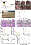
Fig 2. HucMSC-Exos remodel the intestinal mucosal barrier and improves survival.
(A) Rats were administered exosomes both before and after irradiation, and intestinal segments were collected on day 5 after irradiation, as shown in the figure (n = 6 animals/group). (B) Representative images of intestinal conditions in each group. (C-D) Colonic lengths in each group. (E) Hematoxylin and eosin (H&E) staining showing representative images of the cross-sectional structure of the small intestine. Magnification: × 200 (top), × 400 (bottom). (F) Representative image of periodic acid-Schiff (PAS) staining. Magnification: × 200 (top), × 400 (bottom). (G-I) Length of small intestinal villi, depth of crypts, and number of cup cells based on H&E and PAS staining analysis. (J) Changes in body weight in rats of different groups (n = 6/group). (K) Kaplan-Meier survival analysis of rats following 12 Gy irradiation (n = 10/group).The data are representative of three independent experiments; one-way ANOVA (Tukey’s multiple comparisons test) ; *p < 0.05; **p < 0.01, ***p < 0.001, ****p < 0.0001.
Fig 3
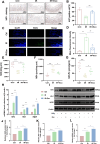
Fig 3. HucMSC-Exos promote proliferation and reduces oxidative stress injury in the rat intestinal epithelium.
(A-B) Immunohistochemistry (IHC) staining showing representative images of Ki67 + expression in a cross-section of the small intestine on day 5 after irradiation, along with analysis of the positive rate. Magnification: × 200 (top), × 400 (bottom). (C-D) Expression of reactive oxygen species (ROS) in the epithelial tissue of the rat small intestine and analysis of fluorescence intensity. Scale bar: 100 μm. (E-G) Biochemical assay kits was performed to detect the expression of SOD, GSH-Px, and MDA redox markers in the tissues. (H) qRT-PCR was performed to detect the expression levels of Nrf2, HO-1, and NQO1 in small intestinal tissues. (I) Western blot analysis of Nrf2, HO-1, and NQO1 expression in small intestinal tissues. (J-L) Relative quantitative analysis of Nrf2, HO-1, and NQO1 protein levels in small intestinal tissues. The data are representative of three independent experiments; one-way ANOVA (Tukey’s multiple comparisons test) ; *p < 0.05; **p < 0.01, ***p < 0.001, ****p < 0.0001.
Fig 4
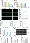
Fig 4. HucMSC-Exos alleviate oxidative stress injury in IEC-6 cells.
(A) The effects of different radiation doses on IEC-6 cell activity were assessed using the CCK-8 assay to determine the optimal radiation dose. (B) The CCK-8 assay was used to determine the optimal exosome dose under a 10 Gy irradiation dose. (C-D) Representative fluorescence images and fluorescence intensity analysis of ROS in different groups of IEC-6 cells. (E-G) Biochemical assay kits for the expression of SOD, GSH-Px, and MDA redox markers in IEC-6 cells. (H) qRT-PCR to detect the expression levels of Nrf2, HO-1, and NQO1 in IEC-6 cells. (I) Western blot analysis of Nrf2, HO-1, and NQO1 expression in IEC-6 cells. (J-L) Relative quantitative analysis of Nrf2, HO-1, and NQO1 protein levels in IEC-6 cells.The data are representative of three independent experiments; one-way ANOVA (Tukey’s multiple comparisons test) ; *p < 0.05; **p < 0.01. ***p < 0.001, ****p < 0.0001.
Fig 5
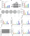
Fig 5. HucMSC-Exos mitigate IR-induced oxidative damage and enhance cellular proliferative capacity by activating the Nrf2/HO-1/NQO1 antioxidant signaling pathway.
We silenced the Nrf2 transcription factor using siRNA and verified and analyzed the silencing efficiency through qRT-PCR (A), and Western blot analysis (B and C) to identify the optimal knockdown strand. Colony formation assay (D-E) and CCK-8 assay (F) were performed to assess the impact of hucMSC-Exos treatment on the proliferation of transfected cells. (G-H) qRT-PCR analysis to compare the expression levels of HO-1 and NQO1 before and after silencing. (I) Western blot analysis to compare the expression levels of HO-1 and NQO1. (J-K) Relative quantitative analysis of HO-1 and NQO1 protein levels before and after silencing.The data are representative of three independent experiments; one-way ANOVA (Tukey’s multiple comparisons test) ; *p < 0.05; **p < 0.01. ***p < 0.001, ****p < 0.0001.
Fig 6
Fig 6. The antioxidant mechanism of hUCMSC-Exos in ARII.
HucMSC-Exos alleviate radiation-induced acute oxidative damage by inhibiting ROS accumulation, reducing lipid peroxidation, and further amplifying the Nrf2/HO-1/NQO1 signaling pathway.

Similar articles

References

    1. Wang K, Tepper JE. Radiation therapy-associated toxicity: Etiology, management, and prevention. CA Cancer J Clin. 2021;71(5):437–54. doi: 10.3322/caac.21689 - DOI - PubMed
    1. Hauer-Jensen M, Denham JW, Andreyev HJN. Radiation enteropathy--pathogenesis, treatment and prevention. Nat Rev Gastroenterol Hepatol. 2014;11(8):470–9. doi: 10.1038/nrgastro.2014.46 - DOI - PMC - PubMed
    1. Zhou Y, Bao L, Gong S, Dou G, Li Z, Wang Z, et al.. T Cell-Derived Apoptotic Extracellular Vesicles Hydrolyze cGAMP to Alleviate Radiation Enteritis via Surface Enzyme ENPP1. Adv Sci (Weinh). 2024;11(31):e2401634. doi: 10.1002/advs.202401634 - DOI - PMC - PubMed
    1. Larrey EK, Pathak R. Radiation-Induced Intestinal Normal Tissue Toxicity: Implications for Altered Proteome Profile. Genes (Basel). 2022;13(11):2006. doi: 10.3390/genes13112006 - DOI - PMC - PubMed
    1. Chaves-Pérez A, Yilmaz M, Perna C, de la Rosa S, Djouder N. URI is required to maintain intestinal architecture during ionizing radiation. Science. 2019;364(6443):eaaq1165. doi: 10.1126/science.aaq1165 - DOI - PubMed

MeSH terms

LinkOut - more resources