. 2025 Jun 6;53(11):gkaf459.
doi: 10.1093/nar/gkaf459.

Bacterial RecD2 is a processive single-stranded DNA translocase with strand-switching capacity at DNA forks

Affiliations

Bacterial RecD2 is a processive single-stranded DNA translocase with strand-switching capacity at DNA forks

Silvia Hormeño et al. Nucleic Acids Res. .

Abstract

RecD2 is a superfamily 1B helicase involved in DNA replication and repair, modulating replication restart, fork progression, and RecA recombinase activity. In this work, we have characterized the functions of Bacillus subtilis RecD2 using biochemical and single-molecule approaches. ATPγS binding and low MgCl2 concentrations enhance DNA association, with a preference for forked structures and unstructured DNA longer than 30 nucleotides. RecD2 binds to end-less single-stranded DNA stretched at 8-20 pN and translocates through ATP hydrolysis over long distances (>20 kb) with 5'-3' polarity at high rates. RecD2 shows limited unwinding activity on fork structures, strongly dependent on protein concentration and duplex length, reflecting low processivity. However, processivity improves significantly when force is applied to the translocating strand or unwound DNA ends, enabling the unwinding of thousands of base pairs at rates up to 160 bp/s. Single-molecule assays reveal frequent strand switching on fork substrates, resulting in a non-productive cycle of unwinding and rewinding, likely mediated by the N-terminal domain. This behavior explains the low helicase activity observed in bulk assays. We propose that regulation of strand-switching activity may be relevant for RecD2's in vivo function.


Conflict of interest statement

None declared.

Figures

Graphical Abstract
Graphical Abstract
Figure 1.
Figure 1.
ssDNA-dependent ATPase activity of RecD2. (A) ATPase activity in the presence of different ssDNAs. Reactions containing 5 nM RecD2 and the indicated ssDNA cofactor (3 μM in nucleotides) were incubated at 37°C in the presence of 10 mM MgCl2. The level of ATP hydrolysis was measured by the spectrophotometric coupled ATP/NADH assay. Plots are the mean of three independent experiments. (B) Average ATPase rate determined from the linear slope of the curves with different ssDNA as effectors. (C) Effect of MgCl2 concentrations on the ATPase activity of RecD2 in the presence of dT80 or circular ssDNA [sspGEM3Zf (+)]. (D) Michaelis–Menten plot of ATP hydrolysis for RecD2 in the presence of circular ssDNA or dT80 as cofactors and 2 or 10 mM MgCl2. The kinetic parameters obtained are listed in the text. The data shown are the averages of three assays, and the errors represent the SD.
Figure 2.
Figure 2.
DNA binding of RecD2. Increasing amounts of RecD2 were incubated for 15 min at 37°C with radiolabeled ssDNA (0.25 nM in molecules) in a binding buffer containing 2 mM MgCl2 and 1 mM ATPγS. Then, glutaraldehyde (0.05% v/v) was added, samples were incubated for another 15 min, and then separated on 8% PAGE run in 1× TBE. DNA substrates: (A) dT20 (lanes 1–6) and dT30 (lanes 7–12); (B) dT60 (lanes 1–6) and dT80 (lanes 7–12). (C) Summary table with apparent binding constants Kapp (nM), which represent the concentration of RecD2 that binds 50% of DNA, shown as mean ± SD for at least three independent experiments for all substrates assayed (data from this figure and Supplementary Figs S5 and S6): (D) 60-nt ssDNA, (E) 80-nt ssDNA, and (F) fork 30–30. FD: free DNA; C: protein–DNA complexes.
Figure 3.
Figure 3.
Analysis of RecD2 DNA unwinding activity from bulk assays. In all assays, 0.25 nM DNA molecules of radiolabeled structures were incubated with increasing concentrations of RecD2 (0.75–100 nM) for 10 min at 37°C. Then, reactions were stopped and separated on 10% PAGE in 0.5× TBE buffer. The percentage of DNA unwound (%) was quantified from the gels by the ImageLab software. (A) RecD2 unwinds 5′-tailed and forked DNA. The substrates used are fork 30–30 (blue), 5′-tailed (red), 3′-tailed (green), and dsDNA (pink). (B) The effect of the tail length and structure in the unwinding of forked structures. The substrates used are fork 30–8 (pink), fork 30–16 (red), fork 30–30 (blue), and fork 30-dT30 (green). (C) Unwinding activity of RecD2 with longer dsDNA regions. Helicase assays were performed with the radiolabeled fork structures: 30–30 (blue), 40–30 (red), 50–30 (green), and 50-dT37 (pink). (D) Long tails do not further increase the unwinding of forked structures. Substrates used: 50-dT80 (blue), 50-dT37 (pink), 70-dT80 (green), and 70-dT37 (red). The graphs represent the mean ± SD of at least three independent experiments for each substrate, and representative gels are shown in Supplementary Fig. S8.
Figure 4.
Figure 4.
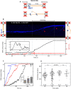
RecD2 is a fast ssDNA translocase with high processivity. (A) Illustration of the experimental setup to measure the translocation activity of RecD2 in the C-Trap. Individual 22.3-knt ssDNA tethers were attached between two streptavidin-coated beads trapped by two optical traps. A confocal laser scanned the DNA tethers to detect RecD2 conjugated to one quantum dot. (B) Representative kymograph of RecD2 movement (blue) in the presence of 1 mM ATP under 15 pN of tension. No changes in DNA extension are detected (lower panel). (C) Interval scatter plots of RecD2 translocation rates on ssDNA as a function of force in the presence of 1 mM ATP. The central bar represents the mean of the data, and the error bars represent the SD. The number of individual trajectories measured at each force is indicated below the data. Statistical analysis revealed no statistical significance of the mean rates at forces ranging from 8 to 25 pN (P >0.05). (D) Interval scatter plots for RecD2 at different protein concentrations in the presence of 1 mM ATP. The tension on the DNA was kept constant at 15 pN. The central bar represents the mean of the data, and the error bars represent the SD.
Figure 5.
Figure 5.
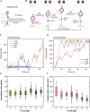
RecD2 unwinds duplex DNA only if assisted by force. (A) Illustration of the experimental setup to measure the unwinding activity of RecD2 in the C-Trap. Individual 5′-tailed long DNA tethers (2.3-knt ssDNA and 15-kb dsDNA) were attached between two streptavidin-coated optically trapped beads. A confocal laser scanned the DNA tethers to detect QD-labeled RecD2. (B) Representative kymograph of RecD2 activity (blue) in the presence of 1 mM ATP on a hybrid DNA, with force increased stepwise from 20 to 21 and 22 pN. At 22 pN, the unwinding of the double helix by RecD2 is detected as an increment in DNA extension (lower panel). Inset: Extension fluctuations characterized by unwinding and rewinding of short DNA segments. Red arrows indicate translocating RecD2. (C) Representative unwinding traces at different forces. The force assists in the unwinding by reducing the number of pauses and increasing the overall unwinding rate (inset, N = 17). Note that in all the cases, RecD2 unwinds the complete 15-kb dsDNA fragment of these hybrid substrates. (D) RecD2 unwinding rates as a function of force. Each data point corresponds to the slope measured by linear regression of discrete segments along single RecD2 unwinding traces. The central bar represents the mean unwinding rate, and the error bars represent the SD. The total number of data points considered is included in the graph. Statistical analysis revealed no statistical significance of the unwinding rates at 20–25 and 30–40 pN (P >0.05).
Figure 6.
Figure 6.
RecD2 exhibits strand switch at ss–dsDNA junctions. (A) Schematic representation of the DNA hairpin substrate used in the magnetic tweezers experiment. (B) Illustration showing the interpretation of the time courses obtained in MT experiments. RecD2 is loaded in the ssDNA region adjacent to the hairpin and unwinds the dsDNA until it reaches the hairpin loop. Then, it translocates on ssDNA for a short time before switching strands returning to the unwinding mode. (C) Representative time courses of the activity of RecD2 in the DNA hairpin at low forces. At forces below 12 pN, RecD2 often fails to fully unwind the hairpin. (D) Representative time courses of the activity of RecD2 in the DNA hairpin at higher forces, where RecD2 typically succeeded in fully unwinding the hairpin, but strand-switching events occur. (E) Unwinding rate of RecD2 as a function of the applied unzipping force. (F) Translocation rate of RecD2 as a function of the applied unzipping force.

Similar articles

References

    1. Gilhooly NS, Gwynn EJ, Dillingham MS Superfamily 1 helicases. Front Biosci. 2013; 5:206–16.10.2741/S367. - DOI - PubMed
    1. Singleton MR, Dillingham MS, Wigley DB Structure and mechanism of helicases and nucleic acid translocases. Annu Rev Biochem. 2007; 76:23–50.10.1146/annurev.biochem.76.052305.115300. - DOI - PubMed
    1. Walsh BW, Bolz SA, Wessel SR et al. . RecD2 helicase limits replication fork stress in Bacillus subtilis. J Bacteriol. 2014; 196:1359–68.10.1128/JB.01475-13. - DOI - PMC - PubMed
    1. Wang J, Julin DA DNA helicase activity of the RecD protein from Deinococcus radiodurans. J Biol Chem. 2004; 279:52024–32.10.1074/jbc.M408645200. - DOI - PubMed
    1. Montague M, Barnes C, Smith HO et al. . The evolution of RecD outside of the RecBCD complex. J Mol Evol. 2009; 69:360–71.10.1007/s00239-009-9290-x. - DOI - PubMed

MeSH terms

LinkOut - more resources