. 2025 Jun 6;53(11):gkaf462.
doi: 10.1093/nar/gkaf462.

Unlocking the serine mischarging paradox and inhibiting lactyltransferase activity of AlaRS by a single-point mutation

Affiliations

Unlocking the serine mischarging paradox and inhibiting lactyltransferase activity of AlaRS by a single-point mutation

Wooyoung Park et al. Nucleic Acids Res. .

Abstract

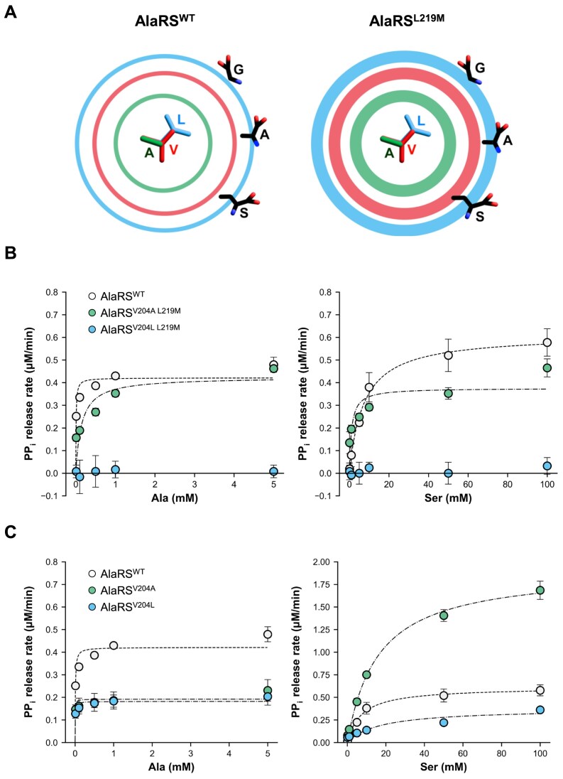
Aminoacyl-tRNA synthetases are critical for accurate genetic translation, attaching amino acids to their corresponding transfer RNA molecules. Alanyl-tRNA synthetase (AlaRS) often misactivates Ser or Gly instead of Ala, which is detrimental unless corrected by its editing functions. The paradox of misactivating larger Ser by AlaRS was considered inevitable due to its inherent design, sharing an essential acidic residue to accommodate the activated adenylated intermediates from both cognate and non-cognate amino acids. Here we show a groundbreaking discovery where a single-point mutation, L219M, in AlaRS from Methylomonas sp. DH-1, effectively eliminates Ser misactivation. Structural analysis of the pre-activation state unveiled that the flexibility of Val204 is the key to preventing Ser binding in AlaRSL219M. This research elucidates the amino acid discrimination mechanism in AlaRS, independent of editing domain. Remarkably, the AlaRSL219M mutation was initially identified as a causal mutation enhancing lactate tolerance in a strain developed through adaptive laboratory evolution. We showed that AlaRSL219M also eliminates the enzyme's inherent lactyltransferase activity, suggesting that the lactate tolerance observed might result from preventing excessive protein lactylation under lactate stress. This opens possibilities for developing high-fidelity and lactylation-deficient AlaRS mutants across various organisms, facilitating studies on their potential benefits in different physiological scenarios.


Conflict of interest statement

None declared.

Figures

Graphical Abstract
Graphical Abstract
Figure 1.
Figure 1.
AlaRSL219M increases lactate tolerance in Methylomonas sp. JHM80 and DH-1. Growth curves of the indicated strains cultured in NMS media containing lactate (A, B, D) and in control NMS media (C, E). Twenty percent (v/v) methane was used as the carbon source in all conditions. Error bars represent standard deviation (SD) from three biological replicates. (F) Schematic illustration of AlaRS protein domains highlighting the position of Leu219.
Figure 2.
Figure 2.
AlaRSL219M mutation enhances both the activity and fidelity of the enzyme. (A) Schematic representation of enzymatic reactions catalyzed by AlaRS using either the cognate substrate Ala or the non-cognate substrate Ser. (B) PPi release assay evaluating the enzyme activity of wild-type AlaRS (AlaRSWT) and the AlaRSL219M mutant in the presence of tRNAAla and various amino acids. (C) PPi release assay of AlaRS and AlaRSL219M in the absence of tRNA using Ala or Ser as substrates. (D) Spotting assay comparing the growth of Methylomonas sp. DH-1 strains expressing AlaRSWT or AlaRSL219M on NMS minimal medium and NMS supplemented with 50 mM Ser. (E) PPi release assay of AlaRSL219F and AlaRSL219A in the presence of tRNAAla with Ala or Ser. The AlaRSWT data shown for comparison correspond to panel (B). Error bars represent SD (n = 3).
Figure 3.
Figure 3.
Overall structure of Methylomonas sp. AlaRS429L219M and structural comparison among Methylomonas sp. AlaRS429, its L219M variant (AlaRS429L219M), and E. coli AlaRS. (A) Ribbon diagram of AlaRS429L219M showing bound ATP and Ala as stick models. The catalytic domain is colored yellow and the tRNA recognition domain green. The Cα atoms of Met219 and Val204 are highlighted with red and green spheres, respectively. Motifs 1, 2, and 3 are colored in gray, orange, and blue, respectively. (B) Structural superposition of AlaRS429 (purple) and AlaRS429L219M (yellow). (C) Superposition of AlaRS429L219M with E. coli AlaRS in both the closed (cyan, PDB: 3HXU) and open (magenta, PDB code: 3HY1) conformations. The arrow indicates the ∼11 Å conformational shift between the open and closed states of E. coli AlaRS.
Figure 4.
Figure 4.
The binding modes of ATP and Ala and local structure around Leu219/Met219. (A) Surface representation of AlaRS429L219M showing ATP, Ala, and Leu219/Met219 as sticks. A magenta dotted line indicates the distance between the phosphorus atom of the α-phosphate and the Cα atoms of Leu219/Met219. (B) Close-up view of the boxed region showing interactions between active site residues (yellow) and ATP/Ala ligands (white). Final 2Fo–Fc electron density maps (blue) are contoured at 1σ for ATP and Ala. Distances (Å) are labeled; green and red dotted lines represent polar and hydrophobic interactions, respectively. (C) Close-up view of the dotted boxed region showing the local structure around Leu219/Met219. Residues from AlaRS429 and AlaRS429L219M are shown as purple and yellow sticks, respectively. (D) Multiple sequence alignment of the β7 and β8 strands of AlaRS. Residue numbers correspond to Methylomonas sp. DH-1 AlaRS. GenBank accession numbers: WP_197495997.1 (Methylomonas sp. DH-1), WP_105477077.1WP_197495997.1 (E. coli), GHM91772.1 (Saccharomyces cerevisiae), WP_223968742.1 (Thermus thermophilus), WP_262119243.1 (Bacillus subtilis), KAI2579449.1 (Homo sapiens), and WP_009990629.1 (Sulfolobus solfataricus).
Figure 5.
Figure 5.
Influence of Val204 flexibility on substrate binding. (A) Schematic illustration of the van der Waals excluded volumes for Ala204, Val204, and Leu204. Ring diameters represent the van der Waals sizes of Ala (green), Val (red), and Leu (blue), while ring thickness reflects side chain flexibility, inferred from the B-factors of AlaRSWT and AlaRSL219M. (B) PPi release assay for AlaRSV204A L219M and AlaRSV204L L219M double mutants in the presence of tRNAAla, using Ala and Ser as substrates. (C) PPi release assay of AlaRSV204A and AlaRSV204L single mutants under identical conditions. AlaRSWT data are shown for comparison and correspond to results in Fig. 2B. Error bars represent SD (n = 3).
Figure 6.
Figure 6.
AlaRSL219M mutation eliminates lactyltransferase activity of AlaRS. (A) Schematic representation comparing the canonical alanyl-tRNA synthetase activity of AlaRS with its proposed lactyltransferase activity (B) Quantification of lactate activation by AlaRSWT and the AlaRSL219M. Enzymatic reactions were performed with 10 mM lactate for 15 min, and the released PPi was measured as an indicator of activity. Error bars represent SD (n = 3). (C) Predicted structural model of lactate binding within the AlaRS active site compared to Ala. Salt bridge and hydrogen bonding interactions are shown as dotted lines.

Similar articles

References

    1. Ibba M, Söll D Aminoacyl-tRNA synthesis. Annu Rev Biochem. 2000; 69:617–50.10.1146/annurev.biochem.69.1.617. - DOI - PubMed
    1. Ling J, Reynolds N, Ibba M Aminoacyl-tRNA synthesis and translational quality control. Annu Rev Microbiol. 2009; 63:61–78.10.1146/annurev.micro.091208.073210. - DOI - PubMed
    1. Baldwin AN, Berg P Transfer ribonucleic acid-induced hydrolysis of valyladenylate bound to isoleucyl ribonucleic acid synthetase. J Biol Chem. 1966; 241:839–45.10.1016/S0021-9258(18)96841-9. - DOI - PubMed
    1. Eldred EW, Schimmel PR Rapid deacylation by isoleucyl transfer ribonucleic acid synthetase of isoleucine-specific transfer ribonucleic acid aminoacylated with valine. J Biol Chem. 1972; 247:2961–3.10.1016/S0021-9258(19)45304-0. - DOI - PubMed
    1. Yarus M Phenylalanyl-tRNA synthetase and isoleucyl-tRNAPhe: a possible verification mechanism for aminoacyl-tRNA. Proc Natl Acad Sci USA. 1972; 69:1915–9.10.1073/pnas.69.7.1915. - DOI - PMC - PubMed

LinkOut - more resources