. 2025 Jun 6;53(11):gkaf481.
doi: 10.1093/nar/gkaf481.

Selective engineering of condensation properties of single-stranded DNA binding (SSB) protein via its intrinsically disordered linker region

Affiliations

Selective engineering of condensation properties of single-stranded DNA binding (SSB) protein via its intrinsically disordered linker region

Péter Ecsédi et al. Nucleic Acids Res. .

Abstract

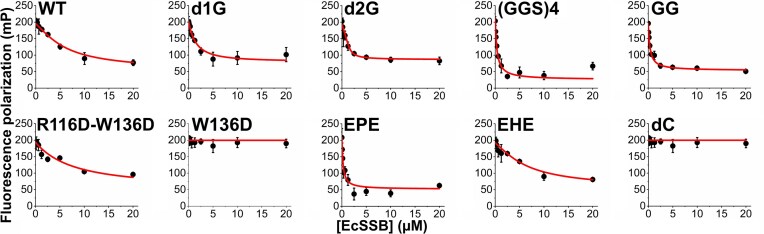
Single-stranded DNA binding (SSB) proteins are essential components of genome metabolism in both bacteria and eukaryotes. Recently demonstrated condensation propensities have placed SSB functions in a new context regarding the organization of nucleic acid-modifying complexes. In this work, we provide functional dissection of the condensation and partner binding properties of Escherichia coli (Ec) SSB via engineered modifications of its intrinsically disordered linker (IDL) region. We identify specific alterations in two glycine-rich regions as well as aromatic and/or positively charged residues of the IDL by which a broad-range, selective modification of condensation propensity and condensate thermal and chemical stability can be achieved, while leaving the single-stranded DNA and partner protein binding functions of SSB unchanged. AlphaFold 3-predicted structures of tetrameric wild-type and engineered EcSSB constructs identify multiple possible binding sites for the conserved C-terminal tip on the tetramer core of the IDL, establishing a link between condensation propensity and restrictions in IDL conformational dynamics. Besides defining the contributions of IDL-driven interactions to driving protein condensation, these results pave the way for the definition of in vivo roles of EcSSB condensation via genetic engineering and delineate ways for further development of liquid-liquid phase separation prediction algorithms.


Conflict of interest statement

None declared.

Figures

Graphical Abstract
Graphical Abstract
Figure 1.
Figure 1.
IDL sequences, structure schematics, and SDS–PAGE image of purified EcSSB constructs. (A) IDL sequences (starting at aa 114) of EcSSB constructs used in this study. [“OB” represents the OB domain (aa 1–113). The EcSSB CTP sequence is MDFDDDIPF.] (B) Structural schematics of EcSSB constructs. Gray sphere and gray line represent the OB domain and the IDL, respectively. CTP is shown as a red segment at the C-terminus of the IDL. Green and yellow sections represent the two G-rich regions of the IDL (green: GGRQGGG; yellow: GGNIGGG). IDLs from P. falciparum SSB and human SSB1 are shown in purple and teal, respectively. The (GGS)4 linker is presented as a dashed line. Red letters “D” indicate the introduced aspartates (positions 116 and 136, underlined in the WT sequence). (C) SDS–PAGE shows that preparations of EcSSB variants are free of WT EcSSB expressed by host bacteria. The lower electrophoretic mobility of the W136D and R116D–W136D constructs is presumably due to more extended IDL conformation resulting from changes in charge patterning.
Figure 2.
Figure 2.
ssDNA and RecQ helicase binding by IDL-engineered EcSSB constructs. FP titrations of 10 nM labeled ssDNA oligonucleotides (A, ssDNA36; B, dT79) with EcSSB constructs (tetramer concentrations stated throughout the article) showed retained ssDNA binding capability for all constructs. Lines show best fits based on a quadratic binding equation (see the “Materials and methods” section and Supplementary Table S1). (C) Twenty-five nanomolar labeled SSB C-terminal peptide (flCTP) was complexed with RecQ helicase (650 nM) and titrated with EcSSB constructs in competitive FP assays. Lines show best fits based on a competitive binding scheme ([26], see also the “Materials and methods” section and Supplementary Table S1). Data points show the mean ± standard deviation (SD) of three independent experiments in each case.
Figure 3.
Figure 3.
IDL engineering markedly alters the condensation propensity of EcSSB constructs. Turbidity (OD600) measurements were carried out in the (A) absence or (C) presence of molecular crowder [3% (m/v) PEG 20K]. Each data point represents the mean ± SD of three independent experiments. Error bars are within symbols. To determine whether the observed turbidity increase is caused by LLPS-based condensation or amorphous aggregation, epifluorescence microscopic images were recorded in the (B) absence or (D) presence of 3% PEG 20K. Twenty micromolar of each EcSSB construct was mixed with 0.3 μM fluorescently labeled WT EcSSB before imaging. Spherical droplets indicate LLPS (cf. those in WT samples), whereas amorphous aggregates were seen in GG samples. Scale bars, 20 μm.
Figure 4.
Figure 4.
Co-condensation ability of EcSSB constructs with the WT protein and the dC variant in the absence of molecular crowder. (A) Samples contained 5 or 10 μM of each EcSSB construct as indicated for the first two columns of each panel. “+ WT” and “+ dC” columns show turbidity values of 10 μM target construct mixed with 10 μM WT or dC, respectively, at 1:1 volume ratio (thus resulting in 10 μM total protein concentration). Gray and red dashed lines show the turbidity of 5 μM WT and dC constructs, respectively, providing references for co-condensation effects. “+ WT” values above the gray line, or “+ dC” values above the red line, are indicative of effective co-condensation. Each column represents the mean ± SD of three independent experiments. (B) Co-condensates imaged upon mixing 10 μM EcSSB variant (as indicated in each panel) with 10 μM dC at 1:1 volume ratio (thus resulting in 10 μM total protein concentration). Condensates were visualized using 0.3 μM fluorescently labeled WT EcSSB.
Figure 5.
Figure 5.
Intermolecular OB domain binding by CTP segments of EcSSB constructs, monitored in competitive titrations. Fluorescein-labeled SSB C-terminal peptide (flCTP, 25 nM) was complexed with dC (2.5 μM) (Kd = 2.1 ± 1.0 μM, cf. Supplementary Fig. S1), and this complex was titrated with the indicated EcSSB constructs in competitive FP assays. Decrease in FP values reveals the ability of the CTP segment of each EcSSB construct to compete with flCTP for binding to the OB fold of dC tetramers. Data points represent the mean ± SD of three independent experiments. Lines show best fits based on a competitive binding scheme ([26], see also the “Materials and methods” section and Supplementary Table S1).
Figure 6.
Figure 6.
Inhibition of condensation of EcSSB variants by ssDNA and ionic conditions. Condensates of indicated EcSSB constructs (20 μM) were titrated with ssDNA (dT79), NaCl, K-Glu, and l-Arg to determine their effects on EcSSB condensation. Asterisks (*) show the presence of 3% (m/v) PEG 20K molecular crowder in the experiments. Note that all variants used here, except for WT, required the presence of crowder for condensation (cf. Fig. 3). Data points represent the mean ± SD of three independent experiments. Error bars are within symbols. Lines show best fits based on the quadratic binding equation [8] in case of dT79 and Hill equation in case of others (see also Supplementary Table S2).
Figure 7.
Figure 7.
Temperature profiles of condensate formation by EcSSB constructs reflect modulation of condensation propensity. (A) Temperature profiles of turbidity (OD600) values of samples of indicated EcSSB constructs (20 μM) are shown. Samples were heated from 4°C to 60°C at a rate of 0.2°C/min. Data points represent the mean ± SD of three independent experiments. The horizontal dashed line shows the reference (buffer) condensation-free absorbance value, while vertical lines mark temperatures at which each EcSSB variant reached the condensation-free value (Tc). Profiles marked with * were recorded in the presence of 3% (m/v) PEG 20K molecular crowder. (B) Tc values determined from experiments in panel (A). Columns show the mean ± SD, with individual experimental values shown as dots.
Figure 8.
Figure 8.
EcSSB constructs show diverse profiles for enrichment of binding partners inside condensates. (A) Epifluorescence images indicating the extent of enrichment of different fluorescently labeled partner/control molecules in condensates of indicated EcSSB constructs (20 μM) 15 min after mixing. In these experiments, 10% (m/v) PEG 20K was applied to minimize the amount of dissolved (non-condensed) EcSSB molecules, thereby reducing background fluorescence. Scale bars, 20 μm. (B) Enrichment (mean droplet intensity/mean background intensity) values of fluorescent molecules in EcSSB condensates (flCTP, blue; fldT79, sand; eGFP as negative control, green; flRecQ, purple). Light and dark colors indicate results obtained after incubating EcSSB condensates with the fluorescent partner/control molecules for 1 and 15 min, respectively. Values >1 (dashed lines) indicate enrichment of the labeled molecules in EcSSB condensates. Mean ± standard error of the mean values for n = 15 are shown.
Figure 9.
Figure 9.
Intra- and intertetramer OB–CTP interactions in EcSSB identified by AF3. (A) Tetrameric model structure of WT #0 (cf. Supplementary Table S3) in cartoon representation, showing the CTP (encircled in red) of chain A (blue) binding to site S1 (highlighted in orange in panel (C) and in Supplementary Table S3) on the tetramer surface of the opposite dimer (chains CD) in “trans” configuration (cf. Supplementary Table S3). (B) Tetrameric model structure of WT #2, showing the CTP binding to site S2 (highlighted in yellow in panel (C) and in Supplementary Table S3) in “trans” configuration. (C) Key residues within sites S1, S2, and S3 (orange, yellow, and blue, respectively; cf. Supplementary Table S3) mapped onto the EcSSB OB tetramer surface (PDB: 1EYG) with the bound ssDNA segment hidden (left) or shown (green, right). It is apparent from the models that ssDNA occupies all predicted CTP binding sites. (D) Model WT #0 of the AF3 prediction encompassing 12 EcSSB WT chains depicting three tetramers, with the CTP of chain I of tetramer 3 (blue) forming an intertetramer OB–CTP interaction with tetramer 2.

Similar articles

References

    1. Nowicka B, Kruk J Powered by light: phototrophy and photosynthesis in prokaryotes and its evolution. Microbiol Res. 2016; 186–187:99–118.10.1016/j.micres.2016.04.001. - DOI - PubMed
    1. Shin Y, Brangwynne CP Liquid phase condensation in cell physiology and disease. Science. 2017; 357:eaaf4382.10.1126/science.aaf4382. - DOI - PubMed
    1. Banani SF, Lee HO, Hyman AA et al. . Biomolecular condensates: organizers of cellular biochemistry. Nat Rev Mol Cell Biol. 2017; 18:285–98.10.1038/nrm.2017.7. - DOI - PMC - PubMed
    1. Hirose T, Ninomiya K, Nakagawa S et al. . A guide to membraneless organelles and their various roles in gene regulation. Nat Rev Mol Cell Biol. 2023; 24:288–304.10.1038/s41580-022-00558-8. - DOI - PubMed
    1. Spannl S, Tereshchenko M, Mastromarco GJ et al. . Biomolecular condensates in neurodegeneration and cancer. Traffic. 2019; 20:890–911.10.1111/tra.12704. - DOI - PubMed

MeSH terms

LinkOut - more resources