. 2025 Jun 6;16(1):5254.
doi: 10.1038/s41467-025-60536-4.

Cryo-EM uncovers a sequential mechanism for RNA polymerase I pausing and stalling at abasic DNA lesions

Affiliations

Cryo-EM uncovers a sequential mechanism for RNA polymerase I pausing and stalling at abasic DNA lesions

Alicia Santos-Aledo et al. Nat Commun. .

Abstract

During synthesis of the ribosomal RNA precursor, RNA polymerase I (Pol I) monitors DNA integrity but its response to DNA damage remains poorly studied. Abasic sites are among the most prevalent DNA lesions in eukaryotic cells, and their detection is critical for cell survival. We report cryo-EM structures of Pol I in different stages of stalling at abasic sites, supported by in vitro transcription studies. Slow nucleotide addition opposite abasic sites occurs through base sandwiching between the RNA 3'-end and the Pol I bridge helix. Templating abasic sites can also cause Pol I cleft opening, which enables the A12 subunit to access the active center. Nucleotide addition opposite the lesion induces a translocation intermediate where DNA bases tilt to form hydrogen bonds with the new RNA base. These findings reveal unique mechanisms of Pol I stalling at abasic sites, differing from arrest by bulky lesions or abasic site handling by RNA polymerase II.


Conflict of interest statement

Competing interests: The authors declare no competing interest.

Figures
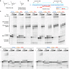
Fig. 1
Fig. 1. Ap site has distinct effects on Pol I and Pol II elongation.
a The natural Ap site exists in equilibrium between the closed sugar hemiacetal and the open aldehyde. The synthetic analog tetrahydrofuran (hereafter termed Ap for simplicity) was used in this study as a mimic of the natural Ap site. b Full mismatched bubble scaffold used for in vitro transcription assays. c In vitro RNA extension assay on four different mismatched bubbles. Each lane corresponds to incubation times of 0, 1, 15, and 60 min. d RNA extension assay with the same scaffold as in (a) but using individual NTPs. ‘T’ and ‘S’ state for ‘Time’ and ‘Start (12mer)’. These experiments were repeated twice. Source data are provided as a Source Data file.
Fig. 2
Fig. 2. Structure of Pol I paused by an Ap site at i + 1.
a Diagram of the nucleic acid scaffold with an Ap site at i + 1. Filled squares denote nucleotides visible in the cryo-EM map that were modelled. b Consensus Ap-PEC structure with subunits colored as in (c) and close-up view of the active center. c Two views of the Closed Ap-PEC structure indicating the different subunits and structural elements in the enzyme. d Superposition between Closed Ap-PEC (colors) and Pol I with undamaged DNA in a post-translocated state (PDB code 5M3F, grey). e Superposition of Closed Ap-PEC (colors) with Pol I stalled at a CPD lesion (PDB code 6H67, grey and CPD lesion in yellow). f Comparison between Closed (grey) and Open (blue) Ap-PEC structures in two different views.
Fig. 3
Fig. 3. Paused Pol I by Ap site at i + 1 with A12-Ct in the funnel.
a Bar diagram of subunit A12 and Ap-RCC structure showing the location of A12 structural elements. b Cryo-EM map of the Ap-RCC structure showing density for A12-linker and A12-Ct. The template and non-template strands are in blue and cyan, respectively, while RNA is in red. c Superposition of Ap-RCC (colors) with Closed Ap-PEC (grey). The A12-Ct catalytic acidic loop (residues 103–108) in Ap-RCC was modelled in poor density. The map around Y717 in subunit A135 is shown as surface in both cases. d Superposition of Ap-RCC (colors) with a Pol I pre-initiation complex where A12-Ct occupies the funnel (PDB code 6RUO, dark green) and free monomeric Pol I with A12-Ct in the funnel (PDB code 5M3M, pink). e Superposition of Ap-RCC (yellow) with Closed (purple) and Open (blue) Ap-PEC in the two different views.
Fig. 4
Fig. 4. Structures of Pol I paused by an Ap site at i + 1 lacking lobe-associated subunits.
a Overall structures of Ap-PEC* and Ap-PEC** with a close-up view around the active center of their superposition with Closed Ap-PEC (grey). b Structural differences around the lobe of Closed Ap-PEC, Ap-PEC*, and Ap-PEC**. Cryo-EM maps around the lobe and associated subunits are shown as transparent surfaces. c Superposition between Ap-PEC* (yellow) and Pol I* with undamaged DNA and GMPCPP (PDB code 6HLQ, dark blue). d Superposition between Ap-PEC* (salmon with A12 in purple) and Ap-RCC (grey with A12 in yellow).
Fig. 5
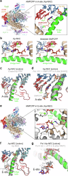
Fig. 5. Structures of Pol I paused by an Ap site at i + 1 in the presence of AMPCPP.
a Overall Ap-NAC structure and close-up view around AMPCPP in the A-site. H-bonds are shown as green dotted lines. b Comparison between Ap-NAC and the same structure where GMPCPP has been modelled. H-bonds and hydrophobic contacts are shown as green and orange dotted lines, while an orange star denotes a clash. c Superposition of Ap-NAC (colors) with Closed Ap-PEC (grey). d Superposition between Ap-NAC (colors) and Pol I with undamaged DNA in a pre-insertion state in the presence of GMPCPP in the A-site (PDB code 6HKO, grey). e Overall Ap-NEC structure and close-up view around AMPCPP in the E-site. The close-up view is rotated ~120° along the bridge helix with respect to that in (a). f Superposition of Ap-NEC (colors) with Ap-NAC (grey). g Superposition of Ap-NEC (colors) with Pol II with ATP in the E-site (PDB code 1R9T, grey). View is as in (e).
Fig. 6
Fig. 6. Structure of Pol I stalled by an Ap site at i–1.
a Schematic diagram of the nucleic acid scaffold with an Ap site at i–1. Filled squares denote nucleotides visible in the cryo-EM map that were modelled. b Overall view of Pol I stalled at an Ap site (Ap-SEC) indicating the different subunits and close-up view of the active center. Canonical Watson-Crick H-bonds and other H-bonds between bases are shown as black and green dotted lines, respectively. cf Close-up views comparing the position of nucleotides in the DNA/RNA hybrid between Ap-SEC (colors) and Closed Ap-PEC (grey, (c)), Ap-NAC (grey, (d)), Pol I EC with undamaged DNA (PDB code 5M3F, grey, (e)), paused bacterial RNA polymerase (PDB code 6ASX, pink; (f)), or paused human Pol II (PDB code 8UHA, grey; (f)).
Fig. 7
Fig. 7. Mechanistic model of Pol I pausing and stalling at Ap sites.
The DNA template and non-template strands are in blue and cyan, RNA is in red, the Ap site is in orange, the incoming nucleotide is in purple, A12 is in yellow, the bridge helix is in dark green and magnesium ions are in light green. Pol I is initially paused as the Ap site occupies the templating position (i + 1), which leads to cleft swiveling. This may allow access of A12-Ct into the funnel for subsequent RNA cleavage or lead to Pol I dissociation from DNA in the open cleft state. Pol I stalled at Ap site allows NTP entry into the E-site (entry), which enables access into the A-site (addition). In the A-site, purines are stabilized by sandwiching between the RNA 3′-end and the bridge helix, with preference for ATP. Phosphodiester bond formation likely leads to an altered hybrid configuration that induces RNA cleavage to minimize lesion bypass. Alternatively A49/A34.5 are lost, which hampers RNA cleavage but induces an intermediate of translocation that compromises nucleotide addition, thus stalling Pol I.

Similar articles

References

    1. Lindahl, T. Instability and decay of the primary structure of DNA. Nature362, 709–715 (1993). - PubMed
    1. Loeb, L. A. & Preston, B. D. Mutagenesis by apurinic/apyrimidinic sites. Annu. Rev. Genet.20, 201–230 (1986). - PubMed
    1. Strauss, B. S. The ‘A rule’ of mutagen specificity: a consequence of DNA polymerase bypass of non-instructional lesions?. BioEssays13, 79–84 (1991). - PubMed
    1. Zahn, K. E., Belrhali, H., Wallace, S. S. & Doublié, S. Caught bending the A-rule: crystal structures of translesion DNA synthesis with a non-natural nucleotide. Biochemistry46, 10551–10561 (2007). - PubMed
    1. Obeid, S. et al. Replication through an abasic DNA lesion: structural basis for adenine selectivity. EMBO J.29, 1738–1747 (2010). - PMC - PubMed

LinkOut - more resources