. 2025 Jun 6;16(1):5261.
doi: 10.1038/s41467-025-60563-1.

TANGO2 binds crystallin alpha B and its loss causes desminopathy

Affiliations

TANGO2 binds crystallin alpha B and its loss causes desminopathy

Maike Stentenbach et al. Nat Commun. .

Abstract

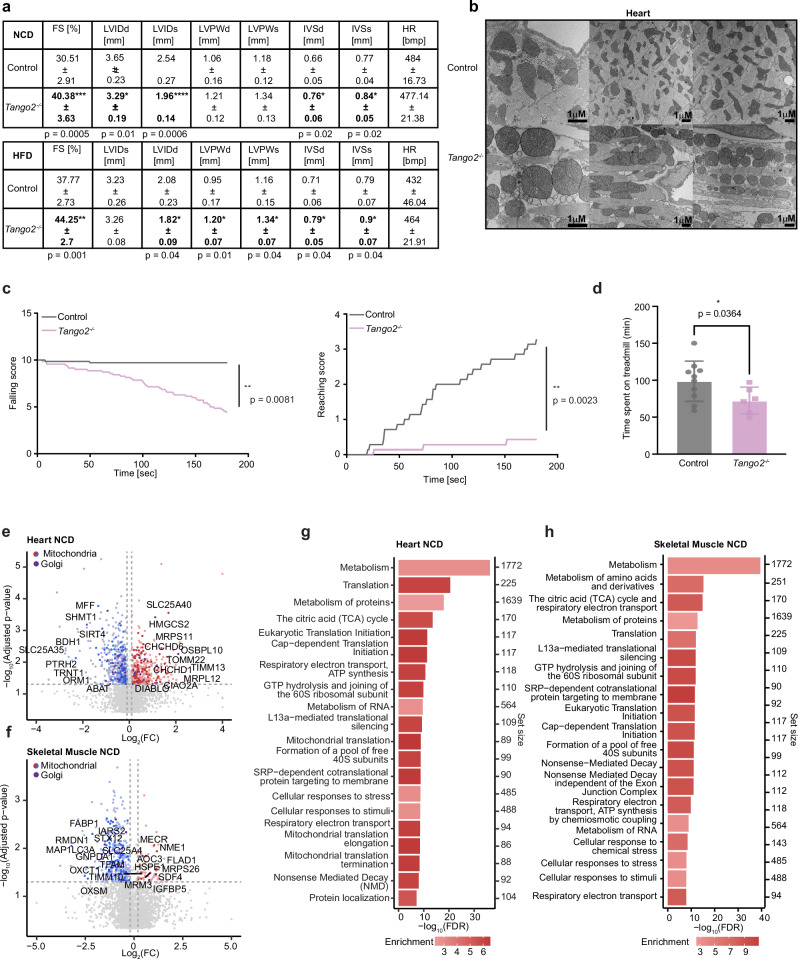
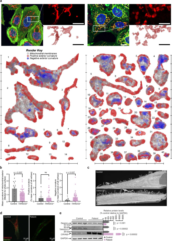
Mutations in the TANGO2 gene cause an autosomal recessive disorder characterised by developmental delay, stress-induced episodic rhabdomyolysis, and cardiac arrhythmias along with severe metabolic crises. Although TANGO2 mutations result in a well characterised disease pathology, the function of TANGO2 is still unknown. To investigate the function of TANGO2, we knocked out the TANGO2 gene in human cells and mice. We identify that loss of TANGO2 impairs intermediate filament structure, resulting in fragmented mitochondrial networks and formation of cup-like mitochondria. In male mice, loss of TANGO2 caused heart defects, reduced muscle function and glucose intolerance by remodelling of intermediate filaments, which altered the mitochondrial and cytoplasmic proteomes, N-glycosylation and nucleocytoplasmic O-GlcNAcylation. We identify that TANGO2 binds the small heat shock protein crystallin alpha B (CRYAB) to prevent the aggregation of the intermediate filament desmin and in the absence of TANGO2, mice develop desminopathy, which is consistent with features found in patients carrying mutations in either desmin or CRYAB.


Conflict of interest statement

Competing interests: The authors declare no competing interests.

Figures

Fig. 1
Fig. 1. TANGO2 knockout decreases mitochondrial respiration and affects mitochondrial structure and morphology in vitro.
a Immunoblots of control and TANGO2–/– cells probed with total OXPHOS or COXII antibodies and normalised to TOMM20 (n = 6, biological replicates). b Immunoblots of proteins involved in mitochondrial translation (MRPS16, MRPL44, LRPPRC, TFAM, MRPP2) (n = 6, biological replicates). SDHA was used as a loading control. c De novo mitochondrial protein synthesis was measured by 35S methionine/cystine incorporation in control and TANGO2–/– cells. Coomassie stained gels are shown as loading controls (n = 3). d Measurement of de novo biogenesis of OXPHOS complexes by incorporation of 35S-labelled cysteine and methionine. The results in c and d are representative of three independent experiments. e Oxygen consumption through the N-linked, S-linked or CIV-linked pathway using either pyruvate/glutamate/malate, succinate or TMPD/ascorbate as substrates was measured for leak (L) and OXPHOS capacity (P) in control and TANGO2–/– cells grown in galactose media. ET capacity (ET) was measured using carbonyl cyanide p-trifluoromethoxyphenylhydrazone (FCCP) (n = 4, biological replicates). f Membrane potential measurements in control and TANGO2–/– cells in the presence and absence of FCCP (n = 4, biological replicates). g Mitochondrial DNA copy number in control and TANGO2–/– cells grown in glucose was quantitated by qPCR of MT-CYB and HBB (n = 4, biological replicates). Values for (e, f and g) are means ± SD. *p < 0.05, **p < 0.01 ***p < 0.001, Student’s two-tailed t test. h Steady state levels of proteins involved in mitochondrial dynamics (LONP1, AFG3L2, CLPX, OPA1, YME1L1 and CHCHD3) (n = 6, biological replicates). TOMM20 was used as a loading control. Relative abundance of proteins shown in panels (a, b and h) was analysed relative to the loading control (n = 6). Values are means ± SD. *p < 0.05, **p < 0.01 ***p < 0.001, Student’s two-tailed t test. i Mitotracker staining of control and TANGO2–/ cells grown in glucose and galactose media. Cells were stained with Mitotracker Orange prior to fixation and scored for fragmentation (n = 100 per cell line and treatment). Source data are provided as a Source Data file.
Fig. 2
Fig. 2. Loss of TANGO2 affects cytoskeletal and membrane structure, protein transport and glycosylation.
a Transcriptome-wide changes in TANGO2–/– cells compared with controls (n = 3). Increased genes are shown in red and decreased genes in blue. b GO analysis based on transcriptomic changes showing significantly changing pathways involved in biological processes, molecular function and cellular component in TANGO2–/ cells compared with control cells. c Protein changes in TANGO2–/– cells compared with control cells identified by mass spectrometry (n = 5 per genotype), the inset shows the number of significantly increased (red) or decreased (blue) proteins and adjusted p values are shown in Supplementary Data 2. Significantly increased and decreased proteins are shown in light red and light blue, respectively, mitochondrial proteins are shown in green, Golgi proteins are shown in purple and intermediate filament proteins are shown in yellow. d GO analyses based on proteomic changes show significantly changing reactome pathways in TANGO2–/– cells compared with control cells. The results show the top 20 pathways with the highest –log10(FDR) and the colour scale represents fold change (FC) for each pathway and set size is the number of genes within each pathway. e N-glycomic profile of control and TANGO2–/– cells. f Changes in glycosylating enzymes identified by RNA-seq. The colour scale represents fold change (FC). All values shown in panels (e and f) are means ± SD *p < 0.05, **p < 0.01 ***p < 0.001, ****p < 0.0001, Student’s two-tailed t test (n = 5 biological replicates).
Fig. 3
Fig. 3. Tango2 deletion leads to cardiomyopathy and muscle weakness in vivo.
a Echocardiographic parameters for 20-week-old control and Tango2–/– mice either fed NCD or HFD. LVIDd left ventricular internal diameter during diastole, LVIDs left ventricular internal diameter during systole, FS fractional shortening, LVPWd left ventricular posterior wall in diastole, LVPWs left ventricular posterior wall in systole, IVSd intraventricular septum in diastole, IVSs intraventricular septum in systole, HR heart rate (n = 5). All values are means ± SD, Student’s two-tailed t test. b Electron microscopy images of heart sections from 20-week-old control andTango2–/– mice fed a NCD. The results are representative of at least three independent mice per genotype. c Hanging wire test in 20-week-old control and Tango2–/– mice. Falling and reaching score was determined after 3 min (n = 7). d Involuntary treadmill exercise of 20-week-old control and Tango2–/– mice (n = 7 per genotype). All values are means ± SD *p < 0.05, **p < 0.01 ***p < 0.001, ****p < 0.0001 Student’s two-tailed t test or Welch’s t test for (d). Proteomic changes in (e) heart and (f) skeletal muscle from 20-week-old Tango2–/– mice fed a NCD compared to control mice (n = 5), and adjusted p values are shown in Supplementary Data 4. Significantly increased and decreased proteins were shown in red and blue, respectively, mitochondrial proteins are shown in green and Golgi proteins in purple. Gene ontology analyses show significantly changing reactome pathways in (g) heart and (h) skeletal muscle from 20-week-old Tango2–/– mice fed a NCD compared to control mice (n = 5). The results show the top 20 pathways with the highest –log10(FDR) and the colour scale represents fold change (FC) for each pathway and set size is the number of genes within each pathway.
Fig. 4
Fig. 4. TANGO2 associates with CRYAB and regulates the protein-folding of desmin.
a Protein fragments of CRYAB that interact with TANGO2 identified from yeast two-hybrid screens of a human heart cDNA library. The common region found in different clones encoding CRYAB includes, at a minimum, amino acids 28-168. b Interaction of wild-type or mutant CRYAB and TANGO2 proteins in yeast two-hybrid assays. Interactions were assessed by survival on media lacking histidine and methionine (SC-L-T-M-H). The SMAD-SMURF interaction was used as a positive control. c AlphaFold model of the TANGO2 (green)—CRYAB (purple) interaction. d Confirmation of a direct interaction between recombinant proteins. SH3GL2 was used as a negative control. The results are representative of three independent experiments. e Protein binding affinity between TANGO2 and hemin (grey) or TANGO2 and CRYAB (pink) (n = 3 biological replicates). Values are means ± SD. f Immunoblots of CAL51 cells, heart and skeletal muscle lysates probed for CRYAB, desmin or vimentin. GAPDH was used as a loading control (n = 3 biological replicates). All values are means ± SD *p < 0.05, **p < 0.01 ***p < 0.001, Student’s two-tailed t test. g In vitro co-sedimentation assay of desmin, CRYAB and TANGO2. Filament assembly was initiated at 22 °C, 37 °C and 44 °C, and pellet (P) or aggregated fractions and supernatant (S) or soluble fractions analysed by SDS-PAGE and Coomassie Blue staining. Results are representative of three independent experiments. h Split-GFP protein-protein interaction assay for desmin and CRYAB variants (S59E, R120G and T170A) binding in control and TANGO2–/– cells (n = 3 biological replicates). All values are means ± SD ***p < 0.001, ****p < 0.0001, Student’s two-tailed t test. (i) BioID identification of CRYAB client proteins in control and TANGO2–/– cells (n = 3 biological replicates). Values are fold changes in abundance relative to adjusted p values, listed in Supplementary Data 2; significantly increased or decreased binding proteins are shown in red and blue, respectively; unique client proteins in each of the cell lines are shown to the left and right of the volcano plot; mitochondrial and cytoskeletal organisation proteins are highlighted in green and yellow, respectively. Source data are provided as a Source Data file.
Fig. 5
Fig. 5. TANGO2 is required for intermediate filament structure that is used by the mitochondrial network.
a Three dimensional renders of mitochondria detected in the control and TANGO2–/– cells. For each cell, a full fluorescence micrograph, inset of the region isolated for FIB-SEM imaging, and the maximum value projection of raw AIVE data are shown; the reconstructed mitochondria and their distribution from the FIB-SEM dataset are shown. Scale bars, 2 μm (fluorescence micrographs) and 1 μm (fluorescence micrograph insets, electron micrographs and raw AIVE data); each notch around the reconstructed datasets represents 1 μm. The positive and negative curvature of mitochondria are shown in red and blue, respectively. b Quantitation of the mean curvature of the mitochondrial exterior. Values are means ± SD, *p < 0.05, **p < 0.01, Student’s two-tailed t test (n = 40). c Sum projected images of 3D voxel values showing surface filopodia detected in the control and TANGO2–/– cells. d Immunostaining of control and TANGO2 patient fibroblasts with desmin (green) and TANGO2 (red) antibody. The results are representative of three independent biological samples. e Immunoblots of control and TANGO2 patient fibroblasts probed with desmin, vimentin, TANGO2 and CRYAB. GAPDH was used as loading control (n = 3 biological replicates). All values are means ± SD *p < 0.05, **p < 0.01, ***p < 0.001, Student’s two-tailed t test. Source data are provided as a Source Data file.

References

    1. Dines, J. N. et al. TANGO2: Expanding the clinical phenotype and spectrum of pathogenic variants. Genet Med.21, 601–607 (2019). - PMC - PubMed
    1. Jennions, E. et al. TANGO2 deficiency as a cause of neurodevelopmental delay with indirect effects on mitochondrial energy metabolism. J. Inherit. Metab. Dis.42, 898–908 (2019). - PubMed
    1. Milev, M. P. et al. The phenotype associated with variants in TANGO2 may be explained by a dual role of the protein in ER-to-Golgi transport and at the mitochondria. J. Inherit. Metab. Dis.44, 426–437 (2021). - PubMed
    1. Mingirulli, N. et al. Clinical presentation and proteomic signature of patients with TANGO2 mutations. J. Inherit. Metab. Dis.43, 297–308 (2020). - PMC - PubMed
    1. Kremer, L. S. et al. Bi-allelic truncating mutations in TANGO2 cause infancy-onset recurrent metabolic crises with encephalocardiomyopathy. Am. J. Hum. Genet.98, 358–362 (2016). - PMC - PubMed

Supplementary concepts

LinkOut - more resources