. 2025 Jun 6;11(1):268.
doi: 10.1038/s41420-025-02507-8.

GAS6/AXL signaling promotes M2 microglia efferocytosis to alleviate neuroinflammation in sepsis-associated encephalopathy

Affiliations

GAS6/AXL signaling promotes M2 microglia efferocytosis to alleviate neuroinflammation in sepsis-associated encephalopathy

Yuedong Tang et al. Cell Death Discov. .

Abstract

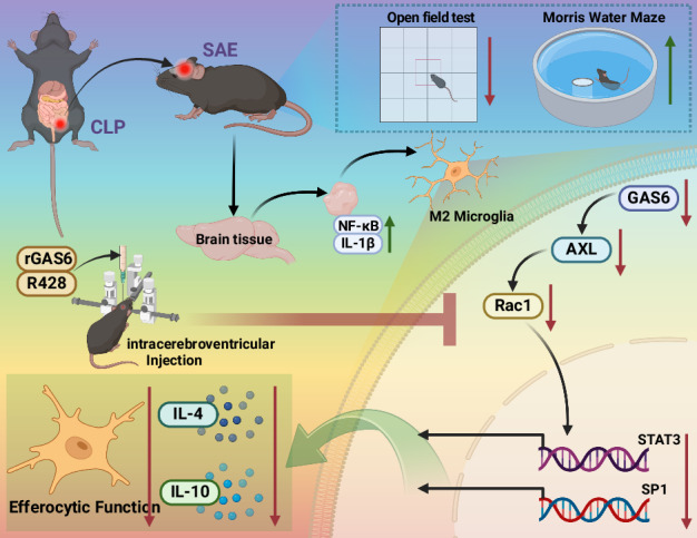
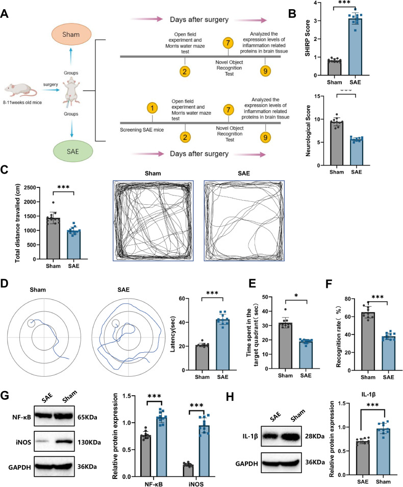
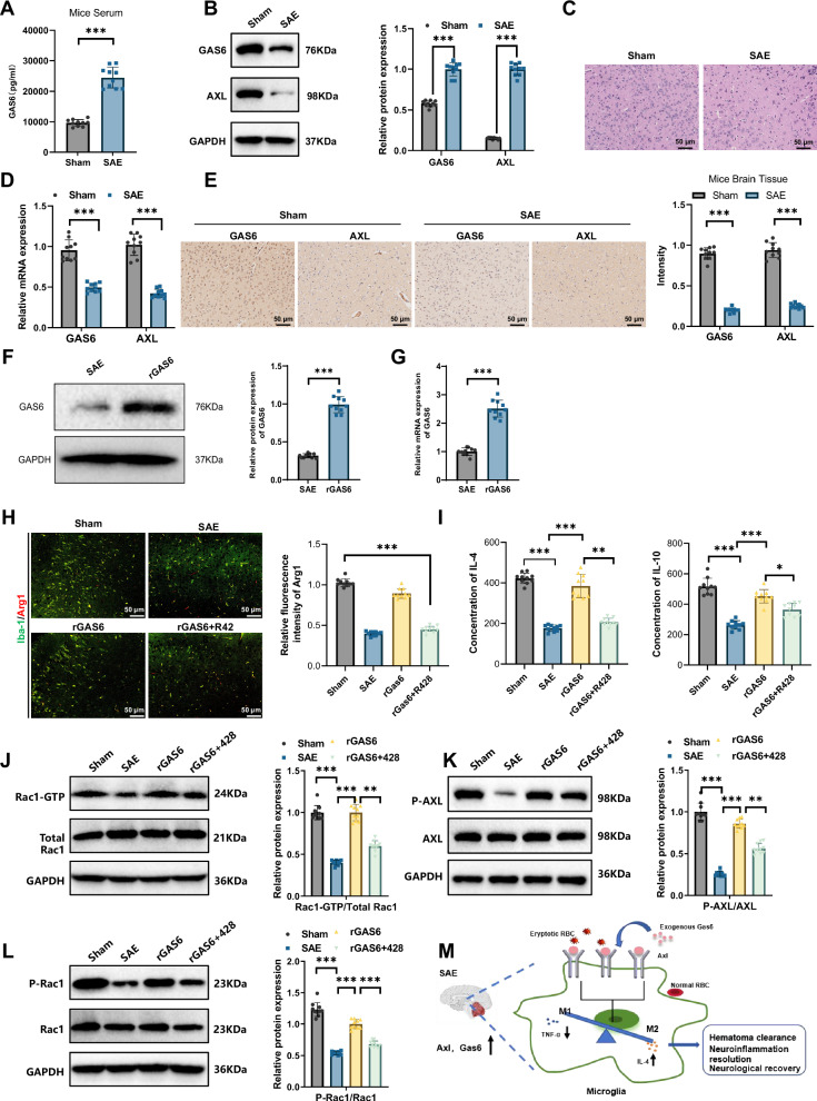
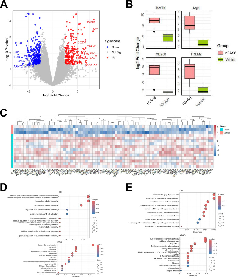
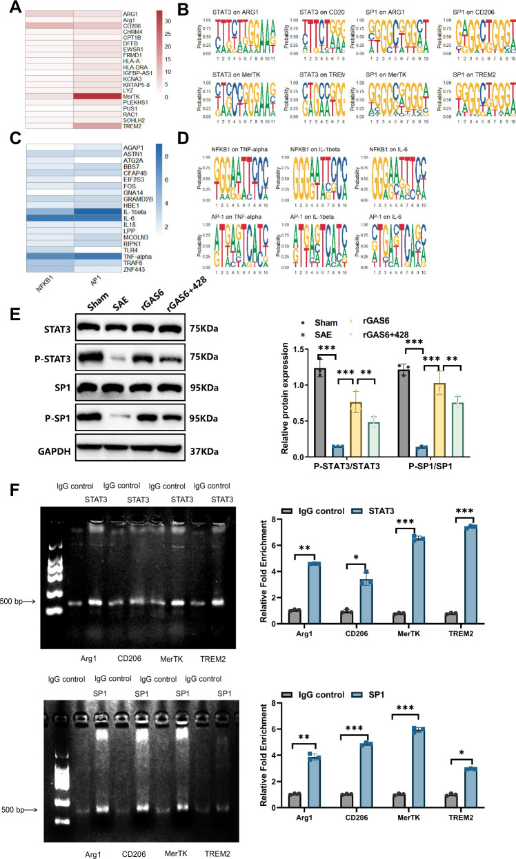
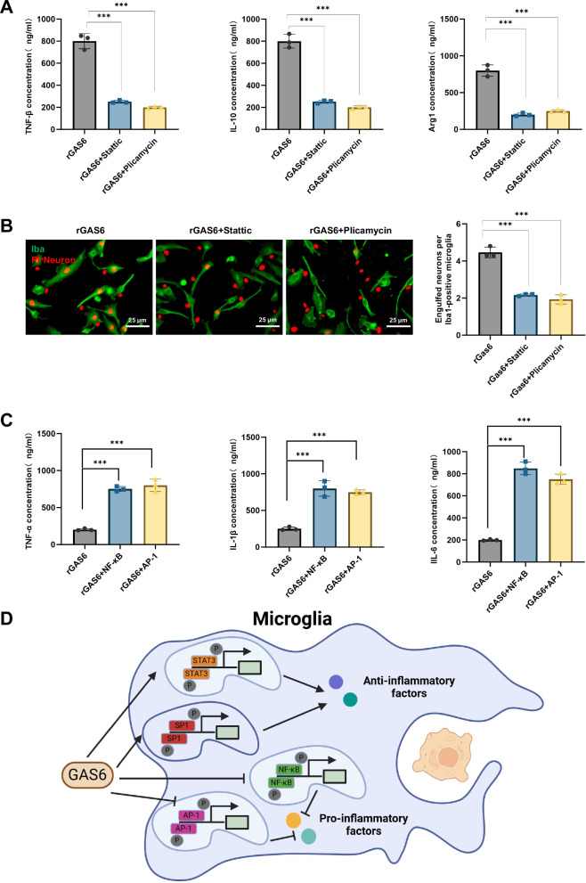
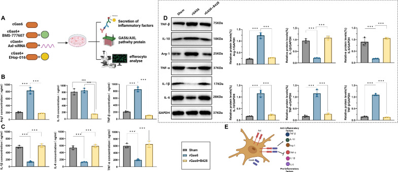
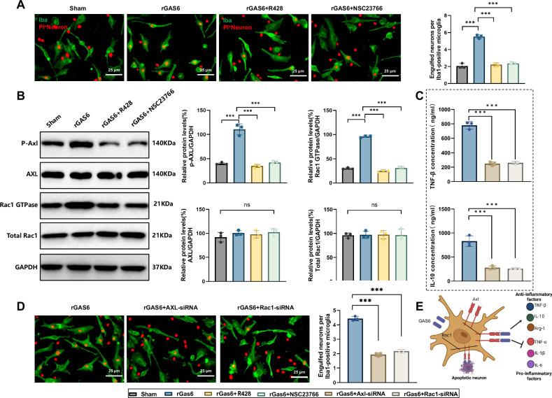
Sepsis-associated encephalopathy (SAE) is a severe complication marked by acute central nervous system (CNS) injury and neuroinflammation. M2 microglia efferocytosis is essential for resolving neuroinflammation, but its regulatory mechanisms remain unclear. This study explored the GAS6/AXL signaling pathway in SAE, hypothesizing its role in enhancing anti-inflammatory responses and efferocytosis. A mouse model of SAE was established via cecal ligation and puncture (CLP), and cognitive impairments were assessed through behavioral tests. Brain tissues and microglia were isolated for RNA sequencing (RNA-Seq) to identify genes associated with the GAS6/AXL pathway. Recombinant GAS6 (rGAS6) protein and an AXL inhibitor were used to examine the pathway's effects on microglial Rac1 activity and functionality. Results demonstrated that GAS6/AXL activation significantly upregulated anti-inflammatory cytokines, enhanced efferocytosis, and suppressed pro-inflammatory responses, improving cognitive outcomes. These findings highlight GAS6/AXL as a critical modulator of microglial functions, providing a promising molecular target for treating SAE. GAS6/AXL Pathway Reduces Neuroinflammation in SAE via Regulation of Anti-Inflammatory and Efferocytic Function in M2 Microglia.


Conflict of interest statement

Competing interests: The authors declare no competing interests. Ethics approval: All animal experiments were approved by the Animal Ethics Committee of Shanghai Public Health Clinical Center Laboratory Animal Welfare & Ethics Committee (No. 2024-A035-01). All methods were performed in accordance with the relevant guidelines and regulations.

Figures

None
GAS6/AXL Pathway Reduces Neuroinflammation in SAE via Regulation of Anti-Inflammatory and Efferocytic Function in M2 Microglia.
Fig. 1
Fig. 1. Establishment and behavioral assessment of the SAE mouse model.
Note: (A) Schematic of the animal experiment process; (B) Comparison of SHIRPA and neurological scores between SAE and Sham groups 24 h post-CLP surgery; (C) comparison of spontaneous activity distance in the Open Field Test between SAE and Sham groups; (D) comparison of escape latency during training sessions in the Morris Water Maze between SAE and Sham groups; (E) comparison of time spent in the target quadrant in the Morris Water Maze probe test between SAE and Sham groups; (F) comparison of novel object recognition rate between SAE and Sham groups; (G) western blot analysis of NF-κB and iNOS expression levels in brain tissue of SAE and Sham groups; (H) western blot analysis of IL-1β expression levels in brain tissue of SAE and Sham groups. N = 10. Results are presented as mean ± SEM, with statistical significance determined by one-way ANOVA: *p < 0.05, **p < 0.01, ***p < 0.001.
Fig. 2
Fig. 2. The GAS6/AXL signaling pathway activation mitigates neuroinflammation in SAE by regulating Rac1 activity.
Note: (A) ELISA analysis of serum GAS6 levels in Sham and SAE groups; (B) western blot analysis of serum GAS6 levels in Sham and SAE groups; (C) H&E staining of brain tissue to observe histological changes in each group; (D) RT-qPCR analysis of relative mRNA expression levels of GAS6 and AXL in each group; (E) immunohistochemical analysis of GAS6 and AXL in brain tissue (scale bar: 50 μm); (F) western blot analysis of GAS6 in the SAE and rGAS groups; (G) RT-qPCR analysis of the relative mRNA expression of GAS6 in each group of mice; (H) immunofluorescence staining revealed the proportion of Arg1-positive cells in the brain tissues of mice from different groups; (I) ELISA analysis of anti-inflammatory cytokines IL-4 and IL-10 concentrations in brain tissue; (J) Rac1 activity assay indicating changes in Rac1-GTP binding in brain tissue across groups; (K) western blot analysis of phosphorylated AXL levels in brain tissue of each group; (L) western blot analysis of phosphorylated Rac1 levels in brain tissue of each group; (M) schematic representation of the GAS6/AXL signaling pathway’s anti-inflammatory and protective roles in microglia via Rac1 activity regulation. N = 10. Data are presented as mean ± SEM, with statistical significance determined by one-way ANOVA: *p < 0.05, **p < 0.01, ***p < 0.001.
Fig. 3
Fig. 3. Differential gene expression and functional enrichment analysis of microglia in the rGAS6 and vehicle groups.
Note: (A) differential gene expression in microglia between the rGAS6-treated and Vehicle groups. rGAS6 treatment significantly altered gene expression in microglia, upregulating anti-inflammatory and efferocytosis-related genes (e.g., Arg1, CD206, MerTK, TREM2) and downregulating pro-inflammatory genes (e.g., TNF-α, IL-1β, IL-6); (B) rGAS6 treatment enhanced the expression of genes associated with anti-inflammatory and efferocytic function in microglia, promoting immune regulation and apoptotic cell clearance through the upregulation of key genes (e.g., Arg1, MerTK); (C) cluster analysis illustrating distinct gene expression profiles in microglia between the rGAS6-treated and Vehicle groups; (D) GO and KEGG enrichment analyses showing significant enrichment of upregulated genes in pathways related to immune response, efferocytosis, and anti-inflammatory activity; (E) GO and KEGG enrichment analyses showing significant enrichment of downregulated genes in pathways associated with NF-κB signaling and pro-inflammatory cytokine signaling. n = 3.
Fig. 4
Fig. 4. Molecular mechanism analysis of GAS6/AXL pathway-mediated activation of anti-inflammatory and efferocytic function in microglia through STAT3 and SP1.
Note: (A) JASPAR database predictions indicate significant enrichment of STAT3 and SP1 binding sites in upregulated genes (e.g., Arg1, CD206, MerTK, and TREM2) in the rGAS6-treated microglia; (B) sequence logo for STAT3 and SP1 binding motifs within significantly upregulated genes; (C) JASPAR database predictions reveal significant enrichment of NF-κB and AP-1 binding sites in downregulated genes (e.g., TNF-α, IL-1β, and IL-6); (D) sequence logo for NF-κB and AP-1 binding motifs within significantly downregulated genes; (E) western blot analysis of STAT3 and SP1 phosphorylation levels across different treatment groups; (F) ChIP analysis of STAT3 and SP1 binding to promoter regions of target genes (Arg1, CD206, MerTK, and TREM2) in rGAS6-treated microglia. All cell experiments were repeated three times, n = 3. Results are presented as mean ± SEM, with statistical significance determined by one-way ANOVA: *p < 0.05, **p < 0.01, ***p < 0.001.
Fig. 5
Fig. 5. Molecular mechanism analysis of GAS6/AXL pathway activation of anti-inflammatory and efferocytic functions in microglia via STAT3 and SP1.
Note: (A) ELISA results showing secretion levels of anti-inflammatory factors TNF-β, IL-10, and Arg1 in rGAS6-induced microglia following treatment with specific STAT3 and SP1 inhibitors, and expression levels of pro-inflammatory factors TNF-α, IL-1β, and IL-6 in rGAS6-induced microglia following NF-κB and AP-1 inhibition; (B) fluorescence microscopy analysis demonstrating efferocytic capacity of rGAS6-induced microglia for PI-labeled neurons following treatment with STAT3 and SP1 inhibitors; (C) ELISA analysis of pro-inflammatory cytokine secretion, specifically TNF-α, IL-1β, and IL-6; (D) schematic diagram illustrating GAS6/AXL pathway activation via STAT3 and SP1, enhancing anti-inflammatory and efferocytic responses in M2-polarized microglia, alongside NF-κB and AP-1 inhibition reducing pro-inflammatory gene expression. All cell experiments were performed in triplicate, n = 3. Results are presented as mean ± SEM, with statistical significance determined by one-way ANOVA: ***p < 0.001.
Fig. 6
Fig. 6. Mechanism analysis of GAS6/AXL pathway regulation of inflammatory response in microglia.
Note: (A) flowchart of the cell experiment; (B) ELISA analysis of anti-inflammatory factor secretion levels (TNF-β, IL-10, and Arg1) across experimental groups (Sham, rGAS6, and rGAS6 + R428 groups); (C) ELISA analysis of pro-inflammatory cytokine secretion levels (TNF-α, IL-1β, and IL-6) across experimental groups; (D) western blot analysis of protein expression levels for TNF-β, IL-10, Arg1, TNF-α, IL-1β, and IL-6 across groups; (E) schematic illustrating the mechanism by which the GAS6/AXL pathway modulates the inflammatory state in microglia. All experiments were performed in triplicate (n = 3). Data are presented as mean ± SEM, with statistical significance determined by one-way ANOVA: ***p < 0.001.
Fig. 7
Fig. 7. GAS6/AXL enhances anti-inflammatory and efferocytic function in microglia via the Rac1 signaling pathway.
Note: (A) fluorescence microscopy analysis showing phagocytosis of PI-labeled neurons by microglia across experimental groups (Sham, rGAS6, rGAS6 + R428, and rGAS6 + NSC23766); (B) western blot analysis of AXL and Rac1 phosphorylation levels and Rac1 GTP-binding activity across groups; (C) secretion levels of anti-inflammatory factors TNF-β and IL-10 in microglia after AXL and Rac1 knockdown via siRNA; (D) efferocytic capacity of microglia after AXL and Rac1 knockdown via siRNA; (E) schematic overview of the GAS6/AXL pathway’s regulation of anti-inflammatory and efferocytic function in microglia via the Rac1 pathway. All experiments were conducted in triplicate (n = 3). Results are presented as mean ± SEM, with statistical significance determined by one-way ANOVA: ***p < 0.001.

Similar articles

References

    1. Gao Q, Hernandes MS. Sepsis-associated encephalopathy and blood-brain barrier dysfunction. Inflammation. 2021;44:2143–50. - PMC - PubMed
    1. Hong Y, Chen P, Gao J, Lin Y, Chen L, Shang X. Sepsis-associated encephalopathy: From pathophysiology to clinical management. Int Immunopharmacol. 2023;124:110800. - PubMed
    1. Fülesdi B, Molnar L, Németh N, Molnár C. Sepsis-associated encephalopathy: a review of literature. Neurol India. 2018;66:352. - PubMed
    1. Yan X, Yang K, Xiao Q, Hou R, Pan X, Zhu X Central role of microglia in sepsis-associated encephalopathy: From mechanism to therapy. Front Immunol. 2022; 13. 10.3389/fimmu.2022.929316. - PMC - PubMed
    1. Zhao L, Gao Y, Guo S, Lu X, Yu S, Ge ZZ, et al. Sepsis-associated encephalopathy: insight into injury and pathogenesis. CNSNDDT. 2021;20:112–24. - PubMed

LinkOut - more resources