. 2025 Jun 6;16(1):5274.
doi: 10.1038/s41467-025-60617-4.

Developmental features and unique characteristics of peptide-specific PLZF+ innate-like T cells in mice

Affiliations

Developmental features and unique characteristics of peptide-specific PLZF+ innate-like T cells in mice

Ahmed Hassan et al. Nat Commun. .

Abstract

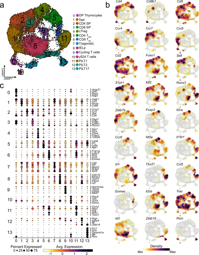
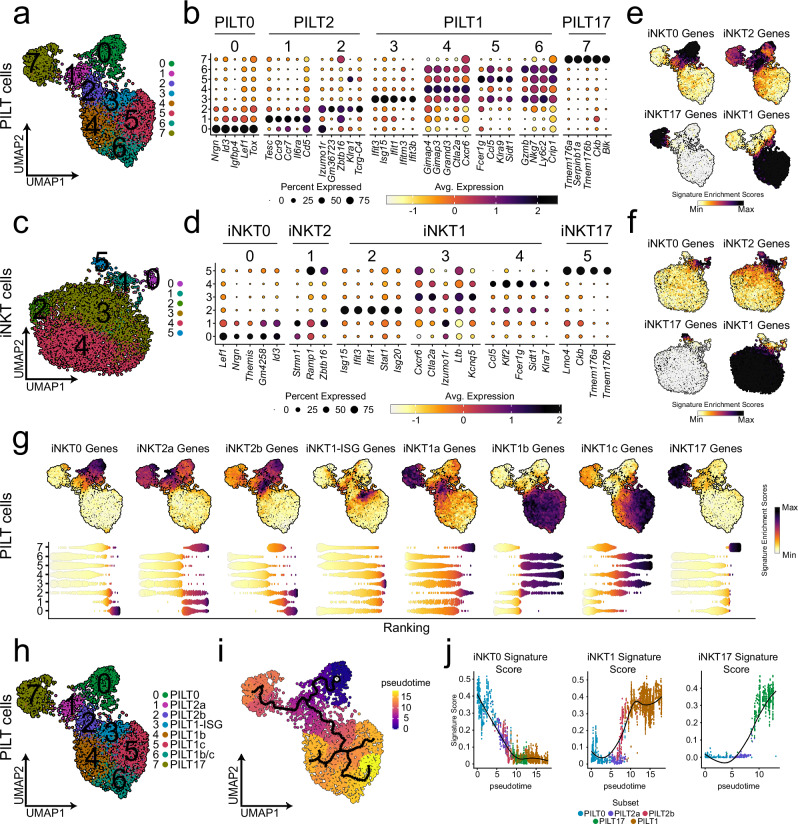
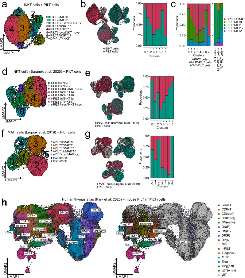
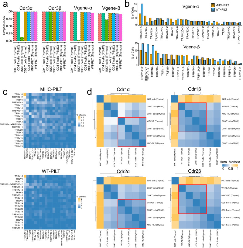
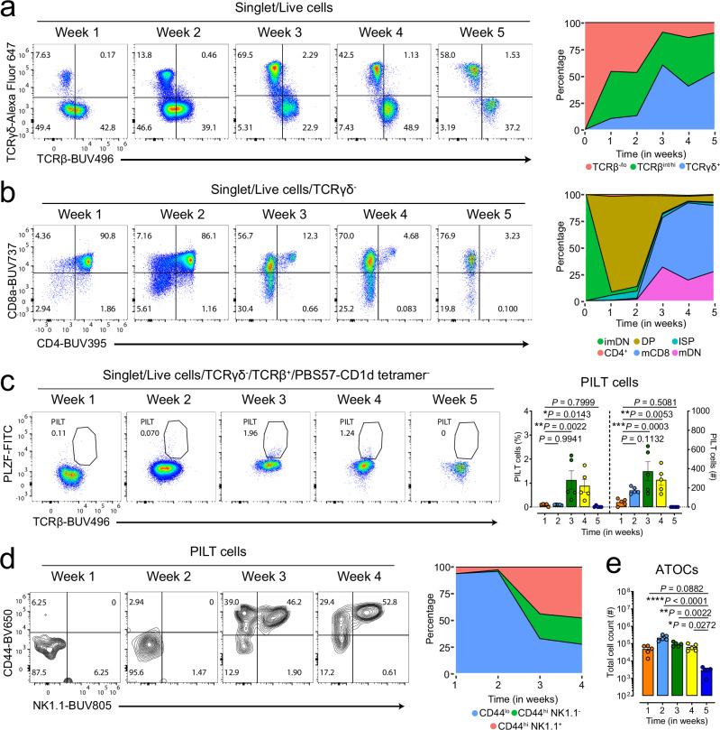
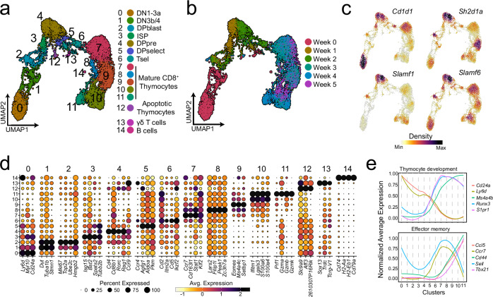
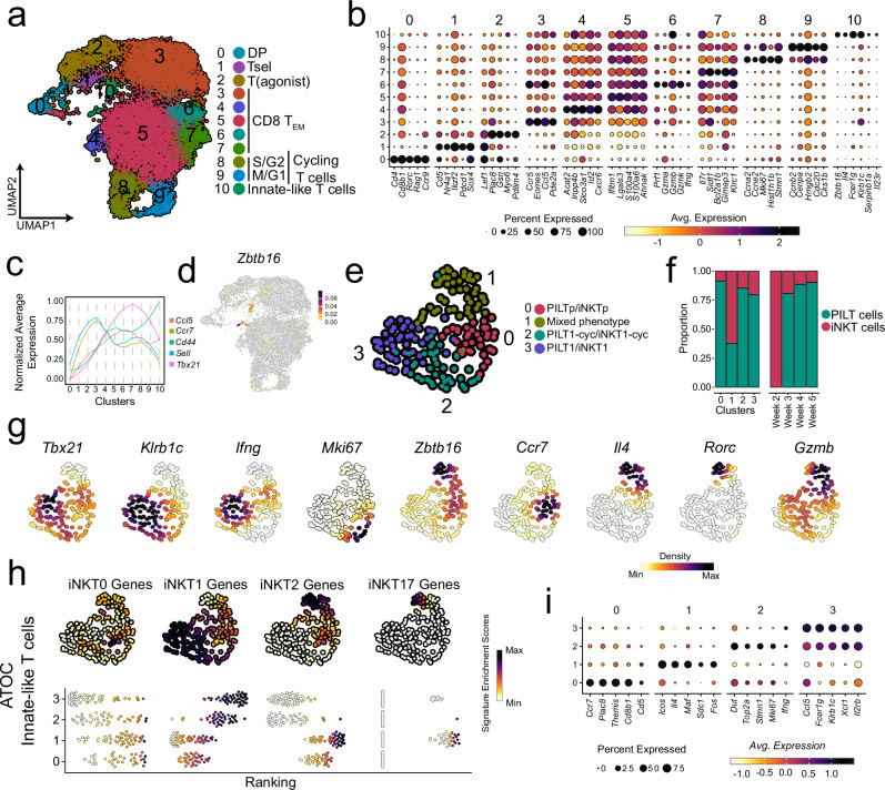
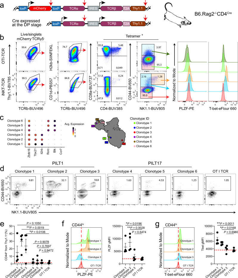
Peptide-specific PLZF+ innate-like T (PILT) cells are a member of the innate-like T cell family utilizing a diverse set of T cell receptor (TCR) Vβ chains. Yet there are no present studies providing clues into the developmental features of PILT cells at a transcriptome level. Here, we performed single-cell transcriptomic analyses of PILT cells and compared them to other members of the innate-like T cell family. We show that PILT cells share similar transcriptional profiles and overlapping developmental trajectories with invariant Natural Killer T (iNKT) cells. However, in contrast to iNKT cells, PILT cells display a polyclonal TCR repertoire closely resembling the one of conventional CD8 T cells, inferring MHC I restriction and a broader range of antigen specificity. We further show that artificial thymic organoid cultures (ATOC) support selection and development of PILT cells in vitro exhibiting similar transcriptional profiles to their counterparts maturing in the thymus. Moreover, using an "on-time" TCR retrogenic ATOC system, we provide evidence for an instructive role of TCR specificity in PILT cell lineage commitment and functional differentiation. Altogether, our findings provide further insights into the PILT cells unique characteristics and molecular mechanisms governing their development.


Conflict of interest statement

Competing interests: The authors declare no competing interests.

Figures

Fig. 1
Fig. 1. Transcriptional landscape of sorted thymic PILT cell-enriched population.
a UMAP of sorted thymic PILT cell-enriched population from T-MHC and Cd1d–/– mice after QC. Displayed are 26148 cells derived from three biologically independent experiments as depicted in Supplementary Fig. 1b. DP is an abbreviation for double positive thymocytes; Tsel is for thymocytes undergoing positive selection; CD4 SP for CD4 single positive thymocytes; CD8 SP for CD8 single positive thymocytes; cTreg for circulating regulatory T cells; CD4 TEM for CD4 effector memory T cells; CD8 TML for CD8+ Memory-like T cells; T(agonist) for agonist-signaled thymocytes; IELp for intraepithelial lymphocyte progenitors; γδ24 T cells for CD24+ gamma-delta T cells; and PILT for peptide-specific PLZF+ innate-like T cells. b Dot plot showing the scaled log normalised average expression of selected differentially expressed genes (DEGs) and publicly available curated markers for each of the clusters depicted in (a). c Density plots of selected genes.
Fig. 2
Fig. 2. Transcriptional landscape of PILT and iNKT cells.
a UMAP of subsetted PILT cells (cells from clusters 11, 12 and 13 from Fig. 1a) n = 3995 cells. b Dot plot showing the scaled log normalised average expression of the top 5 differentially expressed gene markers for each PILT cell cluster. c UMAP of sorted iNKT cells n = 5403 cells derived from two biologically independent experiments as depicted in Supplementary Fig. 1b. d Dot plot showing the scaled log normalised average expression of the top 5 differentially expressed gene markers for each iNKT cell cluster. Feature plot showing the k-nearest neighbors (KNN) smoothed UCell signature enrichment score for iNKT subsets signature gene markers (Supplementary Table 1) for PILT cells in (e) and iNKT cells in (f). g Feature (top) and bee swarm (bottom) plots for KNN smoothed UCell signature enrichment score of the top 50 iNKT subsets gene markers reported by Baranek et al. for PILT cells. h UMAP of subsetted PILT cells showing the annoatiation of each cluster. i UMAP showing PILT cell pseudo-time trajectory built using Monocle3. The cells are ranked in pseudo-time based on Egr1, Egr2, Cd24a, Rag1 and Rag2 expression. j Gene signature score dynamics in pseudotime. PILT is an abbreviation for peptide-specific PLZF+ innate-like T cells; iNKT for invariant natural killer T cells and ISG for interferon-stimulated genes.
Fig. 3
Fig. 3. PILT cells share a similar transcriptional profile with other innate-like T cells.
a UMAP of integrated PILT (from Fig. 2a) and iNKT cells (from Fig. 2b) with n = 9398 cells. b Distribution of iNKT and PILT cells shown as UMAP overlay (left panel). Right panel, filled bar plot displaying iNKT and PILT-cell distribution across the different clusters as annotated in (a). c Filled bar plots showing the distribution of iNKT, MHC-PILT and WT-PILT cells in each cluster (left panel). A filled bar plot showing the subset composition of iNKT, MHC-PILT and WT-PILT cells (right panel). WT-PILT is an abbreviation for derived from CD1d–/– mice and MHC-PILT for derived from T-MHC I mice peptide-specific PLZF+ innate-like T. d UMAP of integrated PILT cells (from Fig. 2a) and iNKT cells from Baranek et al. n = 7226 cells. e Distribution of iNKT (Baranek et al.) and PILT cells shown as UMAP overlay (left panel). Right panel, filled bar plot displaying iNKT and PILT-cell distribution across the different clusters as annotated in (d). f UMAP of integrated PILT cells (from Fig. 2a) and MAIT cells from Legoux et al.. n = 7339 cells. g Distribution of MAIT (Legoux et al.) and PILT cells shown as UMAP overlay (left panel). Right panel, filled bar plot displaying iNKT and PILT-cell distribution across the different clusters as annotated in (f). h UMAP of integrated PILT cells (from Fig. 2a) and an atlas of human thymic development dataset with thymocyte-cell populations as annotated by Park et al. n = 80989 (left UMAP). Unconventional thymocyte-cell populations are highlighted in the second UMAP. CD8αα is an abbreviation for CD8 alpha-alpha; DN(early) for double negative early thymic progenitor; DN(P) for double negative proliferating; DN(Q) for double negative quiescent; DP(P) for double positive proliferating; DP(Q) for double positive quiescent; NKT for natural killer T-like cells; mPILT for mouse peptide-specific PLZF+ innate-like T; T(agonist) for agonist-signaled; Th17 for T helper 17-like cells; Treg for regulatory T cells; Treg(diff) for differentiating regulatory T cells; αβT(entry) for alpha-beta T cells entering single positive stage and γδT for gamma-delta T cells. Source Data are provided as a Source Data file.
Fig. 4
Fig. 4. Characteristics of PILT cell TCR repertoire.
a A bar plot showing the Simpson diversity index score of Cdr3α, Cdr3β, Vgene-α and, Vgene-β usage for down sampled PILT cells and other selected populations from the thymus (this experiment) and PBMCs (10x genomics published datasets). b A bar plot showing the Vgene-α and Vgene-β usage for MHC-PILT and WT-PILT cells. c A Heatmap shows the Vgene-α and Vgene-β pairing of MHC-PILT and WT-PILT cells. d A heatmap showing the Horn-Morisita index score of Cdr1α, Cdr1β, Cdr2α and Cdr2β for PILT cells and other selected populations from the thymus (this experiment) and PBMCs (10x genomics published datasets). WT-PILT is an abbreviation for derived from CD1d–/– mice; MHC-PILT for derived from T-MHC I mice peptide-specific PLZF+ innate-like T; iNKT for invariant natural killer T cells and PBMC for peripheral blood mononuclear cells. Source Data are provided as a Source Data file.
Fig. 5
Fig. 5. Kinetics of DN thymocyte development in ATOC system over five weeks.
a Representative flow cytometry plots (left) and a stacked area plot (right) showing the dynamics of TCRγδ+ and TCRαβ+ cells development in the ATOC system. b Representative flow cytometry plots (left) and a stacked area plot (right) showing the dynamics of CD8+ and CD4+ T cells development in the ATOC system over time. DP is an abbreviation for double positive; imDN is for immature double negative; ISP for immature single positive; CD4+ for CD4 single positive; mCD8 for mature CD8 single positive and mDN for mature double negative T cells. c Representative flow cytometry plots (left) and a bar plot (right) showing the dynamics of PILT cells development in the ATOC system. d Representative flow cytometry plots (left) and a bar plot (right) showing the dynamics of CD44 and NK1.1 expression on developing PILT cells in the ATOC system. Cells were gated as shown in Supplementary Fig. 7c. e Total cell counts of thymocytes developing in the ATOC system over time. Each point represents one ATOC in (c, e): n = 4 ATOCs per time point (a–e). Data are representative of two biologically independent experiments (a–e). Statistical significance was calculated using one-way ANOVA followed by Fisher’s LSD multiple comparisons test; not significant (P ≥ 0.05), *P < 0.05, **P < 0.01, ***P < 0.001 and ****P < 0.0001. Data are presented as mean values ± SEM. Source data are provided as a Source Data file.
Fig. 6
Fig. 6. Transcriptional landscape of ATOC-generated thymocytes.
UMAP of sorted DN thymocytes as shown in Supplementary Fig. 7b (week 0) and ATOC-generated αβ thymocytes from weeks 1:5 (identified as total seq γδ TCR negative cells) after QC n= 6884cells grouped by clusters (a) and time in weeks (b). DN1-3a is an abbreviation for double negative 1-3a stage thymocytes; DN3b-4 for double negative 3b-4 stage thymocytes; DPblast for double positive blast; ISP for immature single positive; DPpre for double positive pre-selection; DPselect for double positive entering positive selection and Tsel for thymocytes undergoing positive selection. c Density plots for selected gene markers associated with innate-like T cell selection and development for cells in Fig. 6a. d Dot plot showing the scaled log normalised average expression of the top 5 differentially expressed gene markers for each cluster in Fig. 6a. e Changes in normalised average expression of selected genes in clusters from Fig. 6a.
Fig. 7
Fig. 7. Transcriptional landscape of ATOC generated innate-like thymocytes.
a UMAP of sorted PILT cell-enriched population (as shown in Supplementary Fig. 1a) from ATOCs at weeks 1:5 n = 14,019 cells. DP is an abbreviation for double positive thymocytes; T(agonist) for agonist-signaled thymocytes; Tsel is for thymocytes undergoing positive selection; CD8 TEM for CD8 effector memory T cells; S/G2 for cycling T cells in S/G2-phase and M/G1 for cycling T cells in M/G1-phase. b Dot plot showing the scaled log normalised average expression of selected genes for cells in Fig. 7a. c Changes in normalised average expression of selected genes in clusters from Fig. 7a. d Density plot of Zbtb16 in Fig. 7a. e UMAP of ATOC-generated innate-like thymocytes (cells from cluster 10 and cells with Zbtb16 expression >0) n = 173 cells. PILTp is an abbreviation for PILT-cell progenitors; iNKTp for invariant natural killer T-cell progenitors; PILT1-cyc for cycling PILT1 cells and iNKT1-cyc for cycling iNKT1 cells. f A filled bar plot showing PILT and iNKT cells distribution in clusters (left) and time by weeks (right) from Fig. 7e. g Density plots of selected genes in Fig. 7e. h Feature (top) and bee swarm (bottom) plots for KNN smoothed signature enrichment score for iNKT subsets gene markers in Supplementary Table 1 for PILT cells. i Dot plot showing the scaled log normalised average expression of selected genes for cells in Fig. 7e. Source data are provided as a Source Data file.
Fig. 8
Fig. 8. T-cell receptor retrogenic T cells develop in the ATOC system.
a Schematic drawing depicting the retroviral expression vector allowing “on-time” expression of TCRα in B6.Rag2–/–CD4Cre settings. Black arrows indicate translation initiation sites and red arrows indicate stop codons. b Flow cytometry evaluation of T cell development in the T-cell receptor retrogenic ATOC system at week 4. Shown are representative plots of ATOCs seeded with the OTI TCR clonotype (Top) and a selected iNKT1 TCR clonotype (bottom). ATOCs were generated as depicted in Supplementary Fig. 6b. c Dot plot showing the log normalised average expression of selected genes by each of the PILT selected clonotypes (right) and its UMAP coordinates in Fig. 2a (left). d Representative flow cytometry plots showing CD44 and NK1.1 expression on T cells generated in the T-cell receptor retrogenic ATOC system. ATOCs were generated as depicted in Supplementary Fig. 6c. Cells are gated on live/singlets/Thy1.1+. CD44hi cell frequency are shown in (e). Flow cytometry evaluation of PLZF expression in (f) and T-bet expression in (g) on CD44hi T-cell receptor retrogenic PILT1 cells. Each point represents one ATOC in (e–g): n = 5 ATOCs per group in (b) and n = 4 ATOCs per group in (d–g). Data are representative of three in (b) and two in (d–g) biologically independent experiments. Statistical significance was calculated using one-way ANOVA followed by Fisher’s LSD multiple comparisons test in (e–g); not significant (P ≥ 0.05), *P < 0.05 and **P < 0.01.Data are presented as mean values ± SEM. Source data are provided as a Source Data file.

Similar articles

References

    1. Bharadwaj, N. S. & Gumperz, J. E. Harnessing invariant natural killer T cells to control pathological inflammation. Front. Immunol. 13, 998378 (2022). - PMC - PubMed
    1. Hackstein, C.-P. & Klenerman, P. MAITs and their mates: “Innate-like” behaviors in conventional and unconventional T cells. Clin. Exp. Immunol.213, 1–9 (2023). - PMC - PubMed
    1. Trivedi, S. et al. Mucosal-associated invariant T (MAIT) cells mediate protective host responses in sepsis. eLife9, e55615 (2020). - PMC - PubMed
    1. Sag, D., Özkan, M., Kronenberg, M. & Wingender, G. Improved detection of cytokines produced by invariant NKT cells. Sci. Rep.7, 16607 (2017). - PMC - PubMed
    1. Wingender, G., Krebs, P., Beutler, B. & Kronenberg, M. Antigen-specific cytotoxicity by invariant NKT cells in vivo is CD95/CD178-dependent and is correlated with antigenic potency. J. Immunol.185, 2721–2729 (2010). - PMC - PubMed

MeSH terms

Substances

LinkOut - more resources