. 2025 Jun 6;23(1):423.
doi: 10.1186/s12951-025-03422-x.

Automatic target-seeking nanoparticle inhibiting orthotopic drug-resistant colon cancer and liver metastases via regulating cancer cell adhesion and proliferation

Affiliations

Automatic target-seeking nanoparticle inhibiting orthotopic drug-resistant colon cancer and liver metastases via regulating cancer cell adhesion and proliferation

Shaobo Bai et al. J Nanobiotechnology. .

Abstract

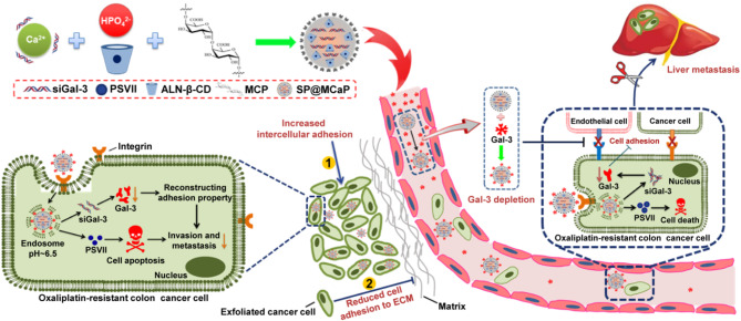
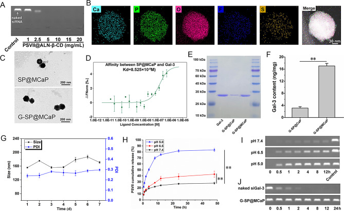
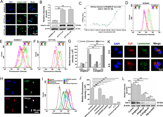
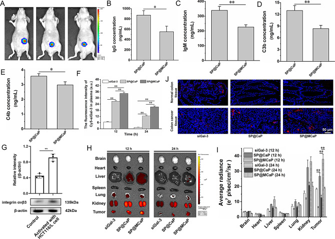
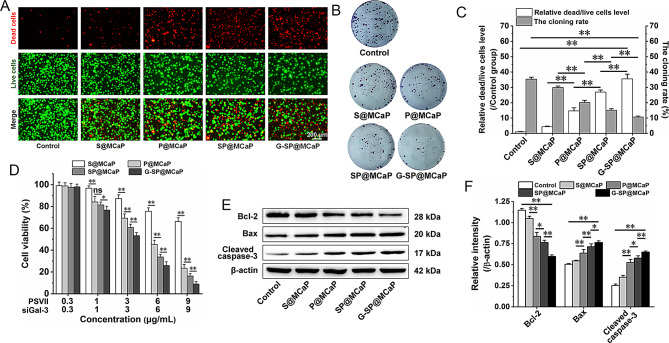
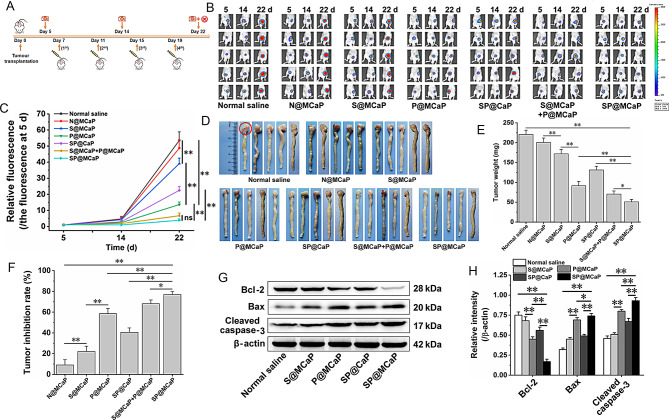
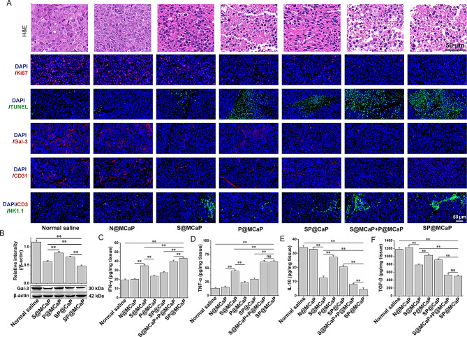
Galectin-3 (Gal-3) plays an important role in adhesion and proliferation of cancer cells. The level of Gal-3 in blood and the expression of Gal-3 in colon cancer tissue are significantly increased in patient with colon cancer. The elevated Gal-3 promotes the migration and drug resistance of colon cancer. Therefore, Gal-3 is a promising target to inhibit the growth and metastases of cancer cells. Besides, integrin αvβ3, a receptor of Gal-3, is highly expressed in colon cancer cell and blood vessel in colon cancer tissue. In this paper, an automatic target-seeking nanoparticle (SP@MCaP) contained siGal-3 and paris saponin VII (PSVII) was prepared. In vivo, by automatically capturing Gal-3 in the blood circulation, SP@MCaP actively recognized cancer tissue vessel and drug-resistant colon cancer cells with elevated integrin αvβ3 expression, resulting in specifical accumulation in orthotopic drug-resistant colon cancer tissue. SP@MCaP diminished Gal-3 level in serum and orthotopic drug-resistant colon cancer tissue, and then suppressed the proliferation of drug-resistant colon cancer cells. Importantly, SP@MCaP reconstructed the adhesion of drug-resistant colon cancer cells and reversed the immunosuppressive microenvironment in orthotopic drug-resistant colon cancer tissue and liver tissue. Finally, under the synergistic effect of siGal-3 and PSVII, SP@MCaP successfully inhibited the growth of orthotopic drug-resistant colon cancer and its liver metastases. In a word, this paper explored a novel concept of the active co-delivery of siGal-3 and PSVII by modification of nanoparticle, which holds promise for targeted therapy in orthotopic drug-resistant colon cancer and its liver metastases.

Keywords: Cell adhesion; Drug-resistant colon cancer; Galectin-3; Integrin αvβ3; Paris saponin VII.


Conflict of interest statement

Declarations. Ethics approval and consent to participate: All animal experiments were approved by the Air Force Medical University Institutional Animal Care and Utilization Committee (No: IACUC-20220812). Consent for publication: Not applicable. Competing interests: The authors declare no competing interests.

Figures

None
First, SP@MCaP automatically captured Gal-3 in the blood, actively recognized cancer tissue vessel and drug-resistant colon cancer cells with elevated integrin αvβ3 expression and specifically accumulated in orthotopic drug-resistant colon cancer tissue. Then, SP@MCaP successfully inhibited the growth of orthotopic drug-resistant colon cancer and its liver metastases by diminishing Gal-3 level in serum and orthotopic drug-resistant colon cancer tissue, suppressing the proliferation of drug-resistant colon cancer cells, reconstructing the adhesion of drug-resistant colon cancer cells and ameliorating the immunosuppressive microenvironment in orthotopic drug-resistant colon cancer tissue and liver tissue
Scheme 1
Scheme 1
The mechanism of an automatic target-seeking nanoparticle contained siGal-3 and PSVII
Fig. 1
Fig. 1
Characteristics of G-SP@MCaP. (A) The effect of PSVII@ALN-β-CD on siGal-3 loaded by calcium phosphate nanoparticle. (B) The TEM mapping element analysis of SP@MCaP. (C) TEM images of SP@MCaP and G-SP@MCaP. (D) The affinity between SP@MCaP and Gal-3 detected by microscale thermophoresis. (E) The capture of Gal-3 by SP@MCaP in PBS. (F) The capture of Gal-3 by SP@MCaP in serum of nude mice with orthotopic drug-resistant colon cancer. (G) Changes in particle size of G-SP@MCaP in normal saline solution. (H) Release of PSVII from G-SP@MCaP under different pH medium. (I) Release of siGal-3 from G-SP@MCaP under different pH medium. (J) The stability of siGal-3 in G-SP@MCaP. n = 3, mean ± SD, **P < 0.01
Fig. 2
Fig. 2
Uptake of G-SP@MCaP by HCT116/L cells and its mechanism. (A-B) Expression of integrin αvβ3 in normal colon epithelial cells (NCM460 cells) and colon cancer cells (HCT116 cells and HCT116/L cells). (C) The affinity between G-SP@MCaP and integrin αvβ3 detected by microscale thermophoresis. (D-G) Uptake of G-SP@MCaP by co-cultured NCM460 cells, RAW264.7 cells and HCT116/L cells. (H) Typical LSCM picture of colocalization of G-SP@MCaP and integrin αvβ3 in HCT116/L cells. (I-J) Mechanism of the uptake of G-SP@MCaP by HCT116/L cells. (K) Typical LSCM picture of siGal-3 escape from the lysosomes of HCT116/L cells. (L) The silencing effect of G-SP@MCaP on the expression of Gal-3 in HCT116/L cells. n = 3, mean ± SD, *P < 0.05, **P < 0.01, ns: no significant difference
Fig. 3
Fig. 3
SP@MCaP distribution in nude mice with orthotopic drug-resistant colon cancer. (A) Fluorescent images of orthotopic drug-resistant colon cancer in nude mice. (B-E) The adsorption of immune-related proteins and complement activated fragments by SP@MCaP. (F) Dynamic change of fluorescence intensity in serum of nude mice with orthotopic drug-resistant colon cancer. (G) Expression of integrin αvβ3 in normal HUVEC cells and HCT116/L activated HUVEC cells. (H-I) Distribution of SP@MCaP in organs and cancer tissues of nude mice with orthotopic drug-resistant colon cancer. (J) Typical LSCM picture of distribution of SP@MCaP in orthotopic drug-resistant colon cancer tissues and normal colon tissues from nude mice with orthotopic drug-resistant colon cancer. n = 3, mean ± SD, *P < 0.05, **P < 0.01
Fig. 4
Fig. 4
Effect of G-SP@MCaP on the proliferation of HCT116/L cells. (A-C) The dead/living cell staining and cloning formation of HCT116/L cells. (D) MTT results of G-SP@MCaP on HCT116/L cells. (E-F) The expression of apoptosis-related protein. n = 3, mean ± SD, *P < 0.05, **P < 0.01, ns: no significant difference
Fig. 5
Fig. 5
Effects of G-SP@MCaP on adhesion of HCT116/L cells. (A-B) The migration and invasion of HCT116/L cells. (C-D) Intercellular adhesion between HCT116/L cells. (E) The expression of invasion-related protein and motion-related protein in HCT116/L cells. (F) Effect of G-SP@MCaP on adhesion between HCT116/L cells and matrix. (G) Typical picture of homotypic aggregation of HCT116/L cells. (H) Typical picture of morphology and pseudopodia formation of HCT116/L cells. (I) Typical picture of adhesion between HCT116/L cells and HUVEC cells. (J) Statistical results of adhesion between HCT116/L cells and HUVEC cells. (K) ICAM-1 expression in HUVEC cells. n = 3, mean ± SD, *P < 0.05, **P < 0.01
Fig. 6
Fig. 6
Inhibitory effect of SP@MCaP on orthotopic drug-resistant colon cancer in nude mice. (A) Schematic diagram of treatment regimen for nude mice with orthotopic drug-resistant colon cancer. (B-C) The growth of orthotopic drug-resistant colon cancer tissues in nude mice during treatment, n = 5. (D) The typical pictures of colon cancer tissue (red circle indicates the location of drug-resistant colon cancer tissue), n = 5. (E) The weight of colon cancer tissue, n = 3. (F) Inhibition rate for orthotopic drug-resistant colon cancer, n = 3. (G-H) The expression of apoptosis-related protein in drug-resistant colon cancer tissue, n = 3. Mean ± SD, *P < 0.05, **P < 0.01
Fig. 7
Fig. 7
Effects of SP@MCaP on apoptosis and immune microenvironment of orthotopic drug-resistant colon cancer tissues. (A) Typical picture of H&E and immunofluorescence staining of Ki67, Gal-3, CD31, CD3/NK1.1 and TUNEL+ cells in orthotopic drug-resistant colon cancer tissues. (B) The expression of Gal-3 in orthotopic drug-resistant colon cancer tissues. (C-F) The contents of IFN-γ, TNF-α, IL-10 and TGF-β in orthotopic drug-resistant colon cancer tissues. n = 3, mean ± SD, **P < 0.01, ns: no significant difference
Fig. 8
Fig. 8
Effect of SP@MCaP on liver metastases of drug-resistant colon cancer cells. (A) Typical in vivo image of nude mice with liver metastasis of drug-resistant colon cancer (yellow circle indicates splenic tumors, and pink circle indicates hepatic tumors). (B) Typical pictures of cancer nodule in liver of nude mice (The red arrow represents hepatic metastasis). (C) Typical H&E staining pictures of liver tissue of nude mice (The red arrow represents the lesion of hepatic metastases). (D) Statistical results of the proportion of liver metastases. (E) Typical picture of immunofluorescence staining of Gal-3, CD31/ICAM-1 and NK cells (CD3/NK1.1+) in liver tissue of nude mice. (F) The content of Gal-3 in serum of cancer-bearing nude mice. n = 3, mean ± SD, *P < 0.05, **P < 0.01

References

    1. Yin R, Zhao H, Li L, Yang Q, Zeng M, Yang C, et al. Gra-CRC-miRTar: the pre-trained nucleotide-to-graph neural networks to identify potential MiRNA targets in colorectal cancer. Comput Struct Biotechnol J. 2024;23:3020–9. - PMC - PubMed
    1. Zhou J, Xu B, Shen Q, Zhang Z, Hu Y, Wang M, et al. Identification and biological evaluation of fused tetrahydroisoquinoline derivatives as Wnt/β-catenin signaling inhibitors to suppress colorectal cancer. Eur J Med Chem. 2024;276:116664. - PubMed
    1. Han BF, Zheng RS, Zeng HM, Wang SM, Sun KX, Chen R, et al. Cancer incidence and mortality in China, 2022. J Natl Cancer Cent. 2024;4(1):47–53. - PMC - PubMed
    1. Hu G, Che P, Deng L, Liu L, Liao J, Liu Q. MiR-378a-5p exerts a radiosensitizing effect on CRC through LRP8/β-catenin axis. Cancer Biol Ther. 2024;25(1):2308165. - PMC - PubMed
    1. Yadav R, Bhawale R, Srivastava V, Pardhi E, Bhalerao HA, Sonti R, et al. Innovative nanoparticulate strategies in colon cancer treatment: a paradigm shift. AAPS PharmSciTech. 2024;25(3):52. - PubMed

MeSH terms

LinkOut - more resources