. 2025 Jun 6;23(1):632.
doi: 10.1186/s12967-025-06619-8.

GPR56 function as a key repressor in hepatocyte pyroptosis and the pathogenesis of liver fibrosis

Affiliations

GPR56 function as a key repressor in hepatocyte pyroptosis and the pathogenesis of liver fibrosis

Zhemin Shi et al. J Transl Med. .

Abstract

Background: Given the rising prevalence of liver fibrosis, there is an urgent need to improve the effective diagnostic methods and treatment of liver fibrosis. Although GPCRs are involved in various physiological and pathological processes, however, the hepatic functions of GPR56 have rarely been explored. This study aims to investigate the role and underlying mechanisms of GPR56 in liver fibrosis.

Methods: The expression of GPR56 in carbon tetrachloride (CCl4) and bile duct ligation (BDL) induced mouse liver fibrosis, as well as human fibrotic liver tissues, was assessed by western blot, qRT-PCR and immunohistochemistry. Then, WGCNA combined with GO enrichment analysis were employed to predict the functions of GPR56. Additionally, Gain- and loss-of-function models (in vitro and in vivo) were established to explore GPR56's function and the signaling pathways involved in liver fibrosis and hepatocyte pyroptosis.

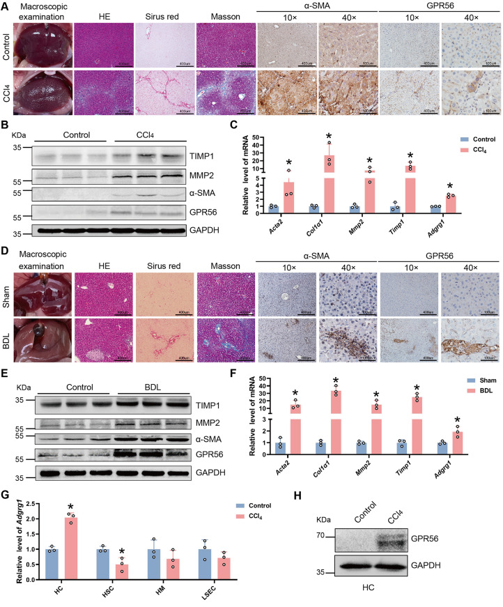
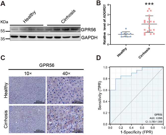
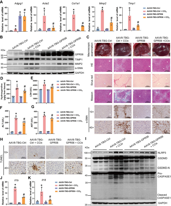
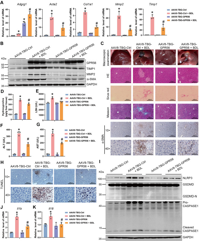
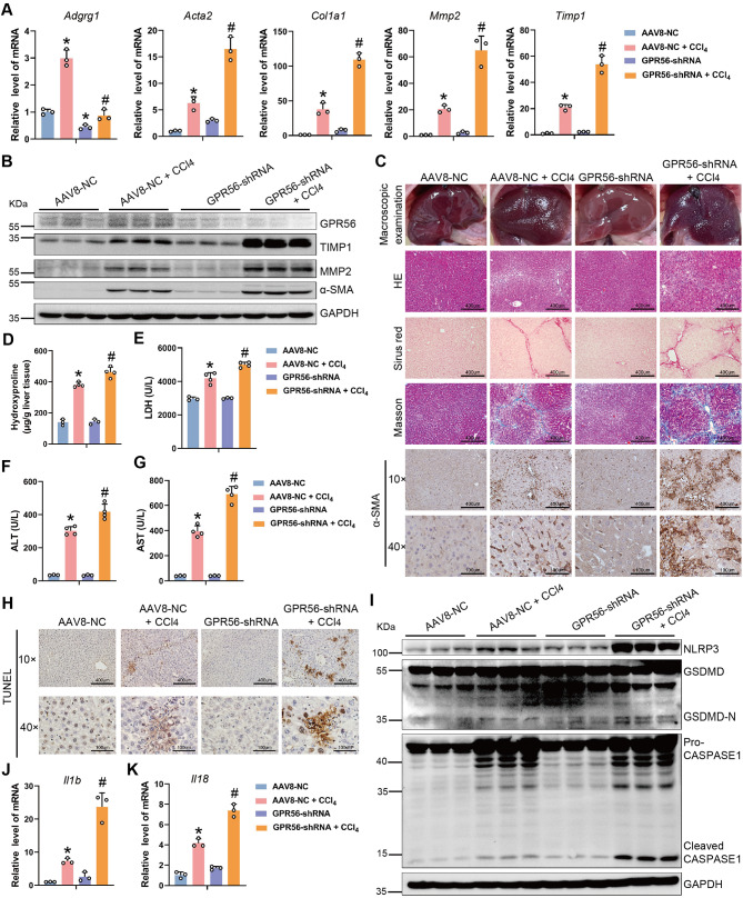
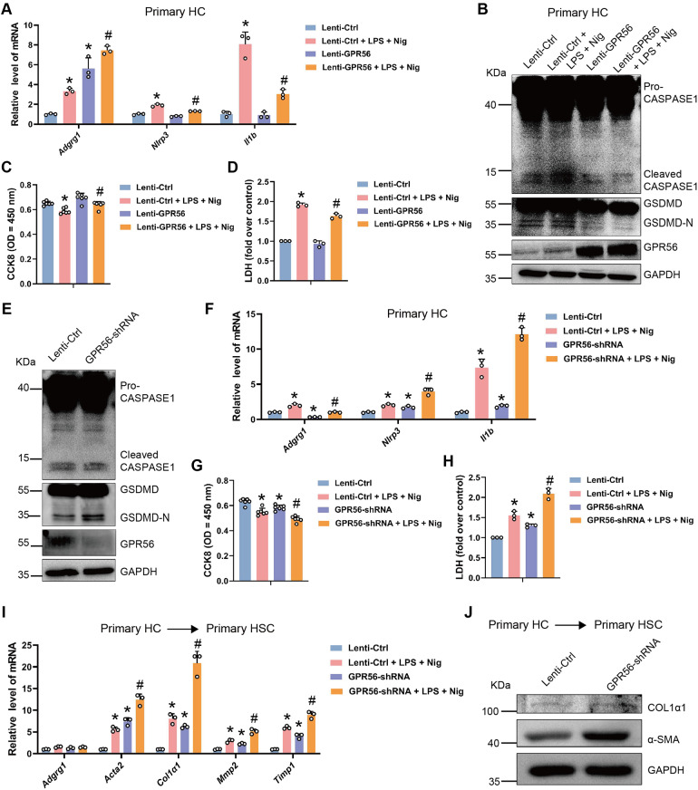
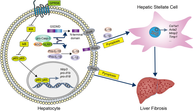
Results: GPR56 was upregulated in both human and mouse fibrotic liver tissues, as well as hepatocytes from CCl4-induced liver fibrosis mice. ROC analysis showed high diagnostic accuracy for cirrhosis (AUC = 0.895, 95% CI: 0.783-1.000). Moreover, WGCNA and GO enrichment analysis speculated that GPR56 was involved in the inflammatory response and extracellular matrix (ECM) synthesis. In vivo assays revealed that hepatocyte-specific overexpression of GPR56 attenuated, while knockdown of GPR56 exacerbated NLRP3 inflammasome-mediated pyroptosis and liver fibrosis. In vitro experiments confirmed that GPR56 inhibited hepatocyte pyroptosis, leading to the inactivation of hepatic stellate cells (HSC). Mechanistic experiments further revealed that GPR56 attenuated hepatocyte pyroptosis via inhibiting the activation of NF-κB pathway.

Conclusions: Our study identify GPR56 as a suppressor of hepatocyte pyroptosis and liver fibrosis, underscoring its potential as a therapeutic and diagnostic target.

Keywords: GPCR; Hepatic stellate cells; Hepatocytes; Liver fibrosis; NF-κB; Pyroptosis.


Conflict of interest statement

Declarations. Ethics approval and consent to participate: The experimental protocol involving mice was formally approved by the Animal Care and Use Committee of Tianjin Medical University to ensure strict adherence to the highest ethical principles and regulatory guidelines. Consent for publication: All authors approved the manuscript and gave their consent for submission and publication. Competing interests: The authors have declared that no conflict of interest exists.

Figures

Fig. 1
Fig. 1
GPR56 is increased in mouse fibrotic liver tissues. (A) Mouse liver macroscopic examination, HE staining, Sirius red staining, Masson staining, and IHC staining of α-SMA and GPR56 in the livers of mice treated with or without CCl4 for 6 weeks. (B) The protein level of TIMP1, MMP2, α-SMA and GPR56 in the livers of mice treated with or without CCl4 was determined by western blot. GAPDH was used as an internal control. (C) The mRNA level of Acta2, Col1a1, Mmp2, Timp1 and Adgrg1 in the livers of mice treated with or without CCl4 was detected by qRT-PCR. (D) Mouse liver macroscopic examination, HE staining, Sirius red staining, Masson staining, and IHC staining of α-SMA and GPR56 in the livers of mice with or without BDL for 21 days. (E) The protein level of TIMP1, MMP2, α-SMA and GPR56 in the livers of mice with or without BDL was determined by western blot. GAPDH was used as an internal control. (F) The mRNA level of Acta2, Col1a1, Mmp2, Timp1 and Adgrg1 in the livers of mice with or without BDL was detected by qRT-PCR. (G) qRT-PCR was used to assess the expression of Adgrg1 in HC, HSC, HM and LSEC isolated from mice treated with or without CCl4. (H) The protein level of GPR56 in HC isolated from mice treated with or without CCl4 was determined by western blot. GAPDH was used as an internal control. *p < 0.05 vs. Control or Sham
Fig. 2
Fig. 2
GPR56 is elevate in human cirrhotic liver tissues. (A-C) The expression of GPR56 in normal human liver tissue (n = 10) and liver tissue from patients with cirrhosis (n = 20) was determined by western blot, qRT-PCR and IHC. GAPDH was used as an internal control. (D) Diagnostic ROC curve of GPR56. ***p < 0.001
Fig. 3
Fig. 3
Hepatocyte-specific overexpression of GPR56 attenuates CCl4-induced liver fibrosis and cell pyroptosis. Balb/c mice were randomly divided into 4 groups: AAV8-TBG-Ctrl group (n = 6), AAV8-TBG-Ctrl + CCl4 group (n = 8), AAV8-TBG-GPR56 group (n = 6) and AAV8-TBG-GPR56 + CCl4 group (n = 8). (A) The mRNA level of Adgrg1, Acta2, Col1a1, Mmp2 and Timp1 was detected by qRT-PCR. (B) The protein level of GPR56, TIMP1, MMP2 and α-SMA was determined by western blot. GAPDH was used as an internal control. (C) Mouse liver macroscopic examination, HE, Sirius red and Masson staining of liver tissues and IHC staining of α-SMA. (D) The content of hepatic hydroxyproline was quantified. (E-G) Serum LDH (E), ALT (F), and AST (G) were examined. (H)TUNEL staining. (I) The protein level of NLRP3, GSDMD and Caspase 1 was determined by western blot. GAPDH was used as an internal control. (J, K) The mRNA level of Il1b and Il18 was detected by qRT-PCR. *p < 0.05 vs. AAV8-TBG-Ctrl, #p < 0.05 vs. AAV8-TBG-Ctrl + CCl4
Fig. 4
Fig. 4
Hepatocyte-specific overexpression of GPR56 attenuates BDL-induced liver fibrosis and cell pyroptosis. C57BL/6J mice were randomly divided into 4 groups: AAV8-TBG-Ctrl group (n = 6), AAV8- TBG-Ctrl + BDL group (n = 10), AAV8-TBG-GPR56 group (n = 6) and AAV8-TBG-GPR56 + BDL group (n = 10). (A) The mRNA level of Adgrg1, Acta2, Col1a1, Mmp2 and Timp1 was detected by qRT-PCR. (B) The protein level of GPR56, TIMP1, MMP2 and α-SMA was determined by western blot. GAPDH was used as an internal control. (C) Mouse liver macroscopic examination, HE, Sirius red and Masson staining of liver tissues and IHC staining of α-SMA. (D) The content of hepatic hydroxyproline was quantified. (E-G) Serum LDH (E), ALT (F), and AST (G) were examined. (H)TUNEL staining. (I) The protein level of NLRP3, GSDMD and Caspase 1 was determined by western blot. GAPDH was used as an internal control. (J, K) The mRNA level of Il1b and Il18 was detected by qRT-PCR. *p < 0.05 vs. AAV8-TBG-Ctrl, #p < 0.05 vs. AAV8-TBG-Ctrl + BDL
Fig. 5
Fig. 5
Knockdown of GPR56 exacerbates CCl4-induced liver fibrosis and cell pyroptosis. Balb/c mice were randomly divided into 4 groups: AAV8-NC group (n = 6), AAV8-NC + CCl4 group (n = 8), GPR56-shRNA group (n = 6) and GPR56-shRNA + CCl4 group (n = 8). (A) The mRNA level of Adgrg1, Acta2, Col1a1, Mmp2 and Timp1 was detected by qRT-PCR. (B) The protein level of GPR56, TIMP1, MMP2 and α-SMA was determined by western blot. GAPDH was used as an internal control. (C) Mouse liver macroscopic examination, HE, Sirius red and Masson staining of liver tissues and IHC staining of α-SMA. (D) The content of hepatic hydroxyproline was quantified. (E-G) Serum LDH (E), ALT (F), and AST (G) were examined. (H)TUNEL staining. (I) The protein level of NLRP3, GSDMD and Caspase 1 was determined by western blot. GAPDH was used as an internal control. (J, K) The mRNA level of Il1b and Il18 was detected by qRT-PCR. *p < 0.05 vs. AAV8-NC, #p < 0.05 vs. AAV8-NC + CCl4
Fig. 6
Fig. 6
GPR56 inhibits LPS/Nigericin-induced hepatocyte pyroptosis. (A-D) Primary HC were infected with lentivirus-mediated control or GPR56 for 72 h, then these cells were stimulated with 100 ng/ml LPS for 4 h and subsequently treated them with 100ng/ml for 2 h. The mRNA level of Adgrg1, Nlrp3 and Il1b was detected by qRT-PCR (A); the protein level of GPR56, GSDMD and Caspase 1 was determined by western blot (B). GAPDH was used as an internal control. Cell proliferation ability was assessed by CCK8 (C); pyroptosis was measured by supernatant LDH activity (D). (E) Primary HC were infected with lentivirus-mediated control or GPR56-shRNA for 72 h, the protein level of GPR56, GSDMD and Caspase 1 was determined by western blot. GAPDH was used as an internal control. (F-H) Primary HC were infected with lentivirus-mediated control or GPR56-shRNA for 72 h, then these cells were stimulated with 100 ng/ml LPS for 4 h and subsequently treated them with 100ng/ml for 2 h. The mRNA level of Adgrg1, Nlrp3 and Il1b was detected by qRT-PCR (F); cell proliferation ability was assessed by CCK8 (G); pyroptosis was measured by supernatant LDH activity (H). (I, J) The conditional medium (CM) from control or GPR56-silenced HC in the presence or absence of LPS/Nig were used to incubate mouse primary HSCs for 24 h. The mRNA level of Adgrg1, Acta2, Col1a1, Mmp2 and Timp1 was detected by qRT-PCR (I); the protein level of COL1α1 and α-SMA was determined by western blot (J). GAPDH was used as an internal control. *p < 0.05 vs. Lenti-Ctrl, #p < 0.05 vs. Lenti-Ctrl + LPS + Nig
Fig. 7
Fig. 7
GPR56 inhibits HC pyroptosis through the NF-κB pathway. (A, B) Primary HC were infected with lentivirus-mediated control or GPR56 for 72 h, then these cells were stimulated with 100 ng/ml LPS for 4 h and subsequently treated them with 100ng/ml for 2 h. The protein level of IKK, phos-IKK, IκB, phos-IκB, p65 and phos-p65 was determined by western blot (A). GAPDH was used as an internal control. The expression and location of p65 was determined by confocal analysis (B); scale bar = 10 μm. (C-F) 5 µmol/L BAY 11-7082 was used to treat GPR56-silenced primary HC for 24 h, the protein level of IKK, phos-IKK, IκB, phos-IκB, p65 and phos-p65 was determined by western blot (C). GAPDH was used as an internal control. The expression and location of p65 was determined by confocal analysis (D); scale bar = 10 μm. The protein level of GSDMD was determined by western blot (E). GAPDH was used as an internal control. The mRNA level of Nlrp3 and Il1b was detected by qRT-PCR (F). *p < 0.05 vs. Lenti-Ctrl, #p < 0.05 vs. GPR56-shRNA
Fig. 8
Fig. 8
Schematic diagram illustrates the role of GPR56 during liver fibrosis

Similar articles

References

    1. Horn P, Tacke F. Metabolic reprogramming in liver fibrosis. Cell Metab. 2024;36:1439–55. - PubMed
    1. Akkiz H, Gieseler RK, Canbay A. Liver fibrosis: from basic science towards clinical progress, focusing on the central role of hepatic stellate cells. Int J Mol Sci 2024, 25. - PMC - PubMed
    1. Wang T, Liu B, Huang J, Zhao Q, Shen H, Bi T, Liu Z, Dai Y, Sun Q. IFN-gamma-mediated Inhibition of JAK/STAT signaling via nano-scutellarin treatment is an efficient strategy for ameliorating liver fibrosis. J Transl Med. 2025;23:195. - PMC - PubMed
    1. Xie Y, Yao J, Yan M, Lin Y, Wei J, Wang H, Mao Y, Liu P, Li X. Pretreatment of UC-MSCs with IFN-alpha2 improves treatment of liver fibrosis by recruiting neutrophils. J Transl Med. 2023;21:832. - PMC - PubMed
    1. Lu Z, Li X, Qi Y, Li B, Chen L. Genetic evidence of the causal relationship between chronic liver diseases and musculoskeletal disorders. J Transl Med. 2024;22:138. - PMC - PubMed

Substances

LinkOut - more resources