

Porphyria cutanea tarda and systemic lupus erythematosus: a case report
- PMID: 40481483
- PMCID: PMC12143034
- DOI: 10.1186/s13256-024-04911-7
Porphyria cutanea tarda and systemic lupus erythematosus: a case report
Abstract
Background: Systemic lupus erythematosus is characterized by multiorgan involvement and the presence of autoantibodies. Porphyria cutanea tarda is a condition that affects the liver and skin by downregulating and inhibiting the enzyme uroporphyrinogen decarboxylase in erythrocytes. The presence of the two diseases simultaneously is rare, so we present this case report and a panoramic review of this uncommon association with scarce description found in the reviewed literature, in a patient with history of kidney transplant who is a user of immunosuppression and immunomodulation medications also required for the management of these diseases, which remains an unknown and unstudied scenario. We believe that it is important to publish this case so that any doctor in clinical practice who is faced with a patient of this complexity can refer to literature as a reference. In addition, articles of this type contribute to the clinical diagnostic and treatment approach.
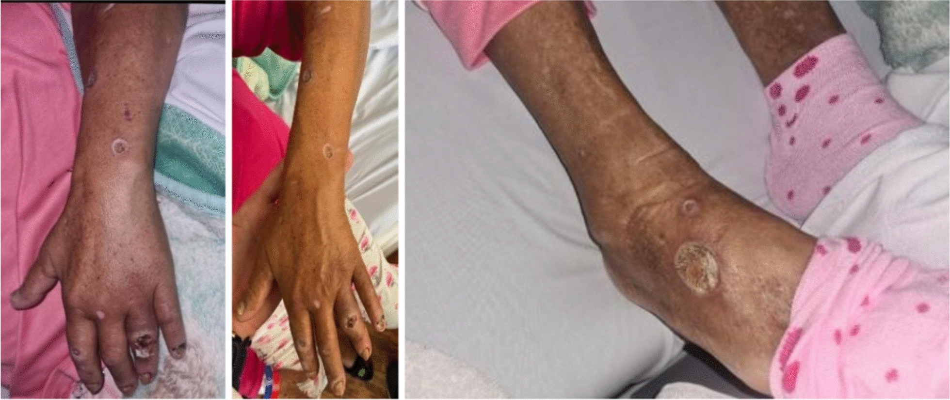
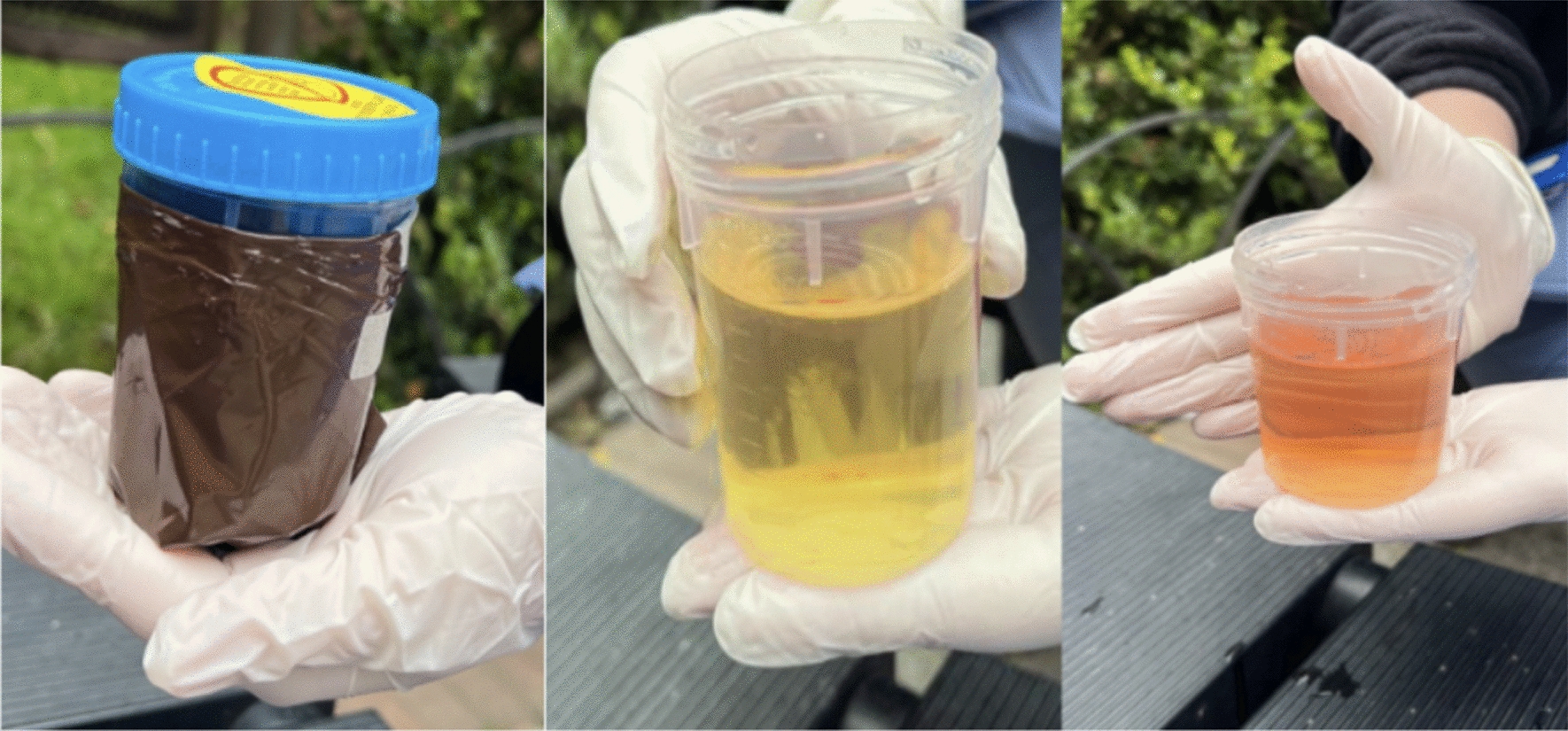
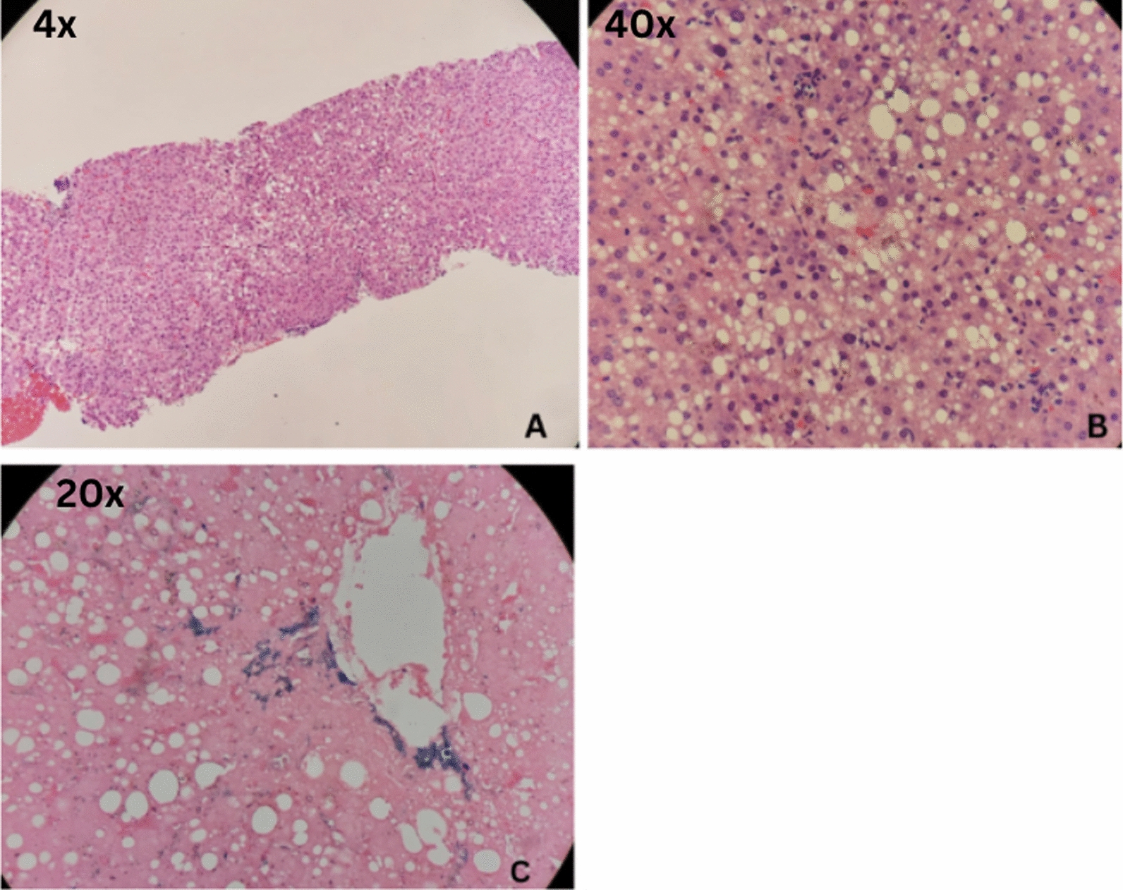
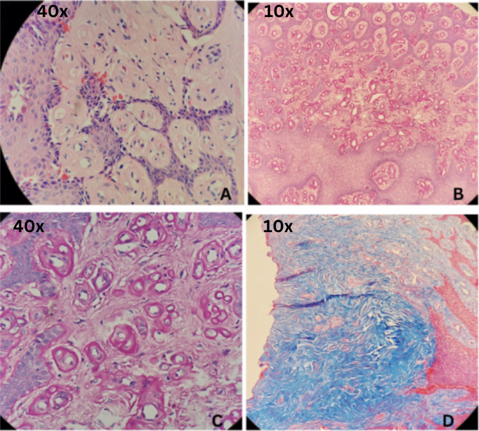
Case presentation: A 62-year-old mestiza woman, immunosuppressed due to a history of renal transplantation, presented with vesicular lesions, hyperpigmentation, and hypertrichosis in photoexposed areas. She also reported changes in urine color upon photoexposure. Laboratory tests, including immune profiling, urine uroporphyrins, and histopathology, confirmed a diagnosis of cutaneous porphyria and systemic lupus erythematosus. The patient was started on low-dose antimalarial treatment with regular liver function monitoring, showing an adequate response.
Conclusions: In the review, 13 articles were found. Porphyria cutanea tarda combined with systemic lupus erythematosus is rarely reported, and the diagnosis of both pathologies becomes a challenge for physicians in diagnosis and treatment, as shown in this case, since porphyria can be the first and only manifestation of underlying systemic lupus erythematosus.
Keywords: Case report; Porphyria cutanea tarda; Systemic lupus erythematosus.
© 2025. The Author(s).
Conflict of interest statement
Declarations. Ethics approval and consent to participate: Not applicable. Consent for publication: Written informed consent was obtained from the patient for publication of this case report and any accompanying images. A copy of the written consent is available for review by the Editor-in-Chief of this journal. Competing interests: Not applicable.
Figures
References
-
- Fritsch S, Wojcik AS, Machota Junior MM, Brenner FM, Paiva ES. Increased photosensitivity? Case report of porphyria cutanea tarda associated with systemic lupus erythematosus. An Bras Dermatol. 2012;87(4):604–7. - PubMed
-
- Sinha A, Dixon N, O’Sullivan MM, Sowden JM. Porphyria cutanea tarda in a patient with systemic lupus erythematosus. Lupus. 1999;8(6):484–6. - PubMed
Publication types
MeSH terms
Substances
LinkOut - more resources
Full Text Sources
Medical
