. 2025 Jun 6;45(1):18.
doi: 10.1186/s41232-025-00385-2.

Genetic deletion of microsomal prostaglandin E synthase-1 promotes imiquimod-induced psoriasis in mice

Affiliations

Genetic deletion of microsomal prostaglandin E synthase-1 promotes imiquimod-induced psoriasis in mice

Fumiaki Kojima et al. Inflamm Regen. .

Abstract

Background: Psoriasis is a chronic inflammatory disease associated with abnormalities in the immune system. Microsomal prostaglandin E synthase-1 (mPGES-1), a terminal enzyme for prostaglandin (PG) E2 biosynthesis, is highly expressed in the skin of psoriasis patients. However, the detailed role of mPGES-1 in psoriasis remains unclear. In the present study, we aimed to investigate the role of mPGES-1 in psoriasis-like skin inflammation induced by imiquimod (IMQ), a well-established model of psoriasis.

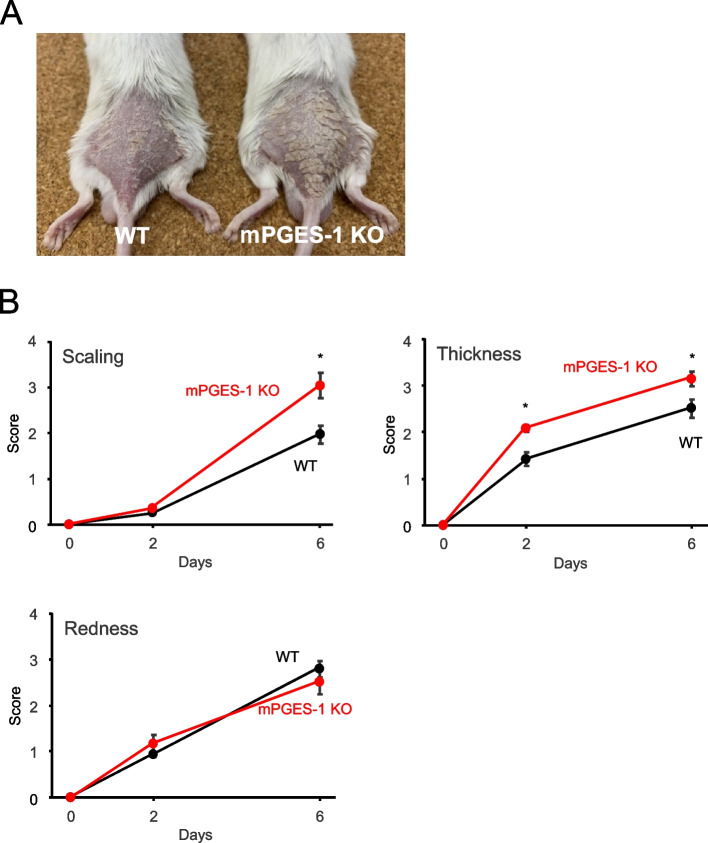
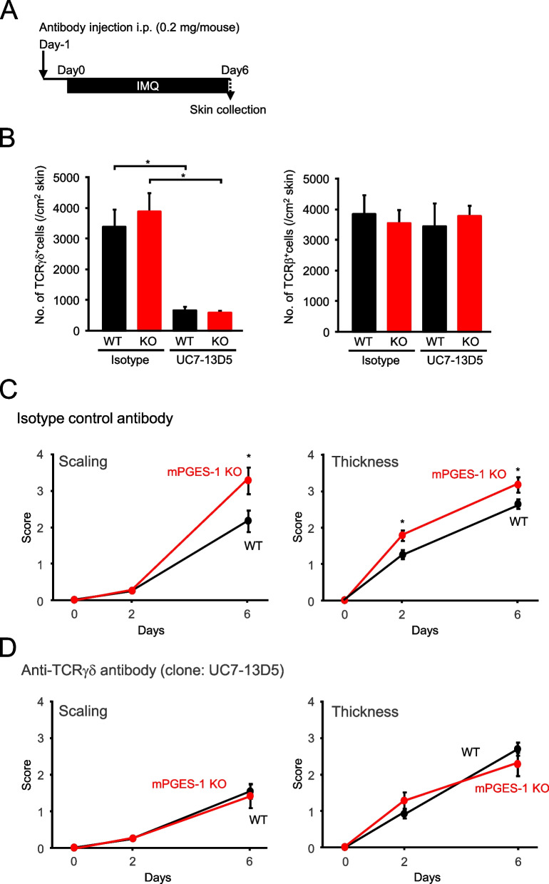
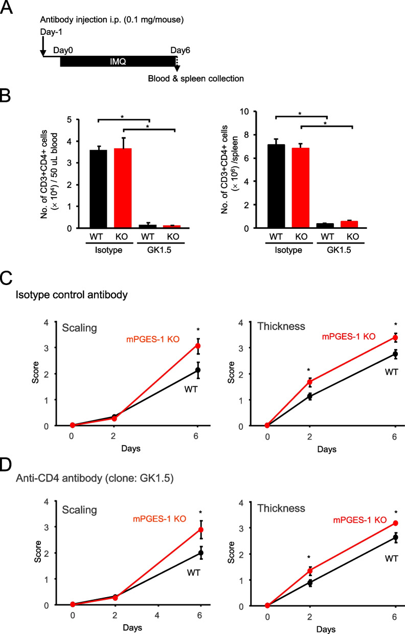
Methods: Psoriasis was induced in mPGES-1-deficient (mPGES-1-/-) and wild-type (WT) mice by administering IMQ for 6 days. Psoriasis was evaluated based on the scores of the macroscopic symptoms, including skin scaling, thickness, and redness, and on the histological features. The skin expression of mPGES-1 was determined by real-time polymerase chain reaction and Western blotting. The impact of mPGES-1 deficiency on T-cell immunity was determined by flow cytometry and γδ T-cell depletion in vivo with anti-T-cell receptor (TCR) γδ antibody.

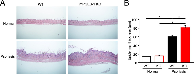
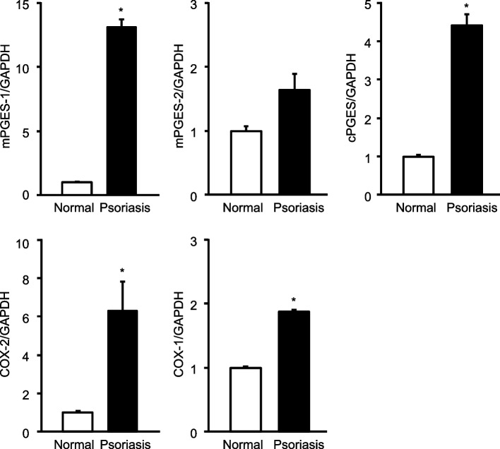
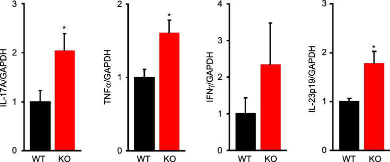
Results: The inflamed skin of mPGES-1-/- mice showed severe symptoms after the administration of IMQ. Histological analysis further showed significant exacerbation of psoriasis in mPGES-1-/- mice. In WT mice, the mPGES-1 expression was highly induced at both mRNA and protein levels in the skin, and PGE2 increased significantly after IMQ administration, while the PGE2 production was largely abolished in mPGES-1-/- mice. These data indicate that mPGES-1 is the main enzyme responsible for PGE2 production in the skin. Furthermore, the lack of mPGES-1 increased the numbers of IL-17A-producing γδ T cells in the skin with IMQ-induced psoriasis, and γδ T-cell depletion resulted in a reduction of the facilitated psoriasis symptoms under the condition of mPGES-1 deficiency.

Conclusions: Our study results demonstrate that mPGES-1 is the main enzyme responsible for skin PGE2 production, and that mPGES-1 deficiency facilitates the development of psoriasis by affecting the development of T-cell-mediated immunity. Therefore, mPGES-1 might impact both skin inflammation and T-cell-mediated immunity associated with psoriasis.

Keywords: Cyclooxygenase; Immunity; Inflammation; Interleukin-17 A; Prostaglandin E synthase; Prostaglandin E2; Psoriasis; γδ T cell.


Conflict of interest statement

Declarations. Ethics approval and consent to participate: All animal experiments were approved by the Animal Research and Ethics Committee of Kitasato University (approval number: Ei-ken 24–06), and all experiments involving the mPGES-1−/− mice were approved by the Safety Committee for Recombinant DNA Experiments of Kitasato University (approval number: 4929). Consent for publication: Not applicable. Competing interests: The authors declare that they have no competing interests.

Figures

Fig. 1
Fig. 1
Clinical course of IMQ-induced psoriasis in mice with mPGES-1 genetic deletion. A Typical examples of WT and mPGES-1−/− mice with psoriasis-like pathology induced by IMQ treatment for 6 days. B Time course of change in the scaling, skin thickness, and redness scores of WT and mPGES-1−/− mice after the indicated days of exposure to IMQ (n = 14 to 16). *P < 0.05; ANOVA followed by Tukey’s multiple comparison test
Fig. 2
Fig. 2
Histological analysis of IMQ-induced psoriasis in mPGES-1−/− mice. A Skin samples of WT and mPGES-1−/− mice were collected on day 6 after the start of exposure to IMQ, and sections were stained with H&E. The results are representative samples of skin tissues from the WT and mPGES-1−/− mice. B Epidermal thickness was assessed on H&E-stained sections (n = 8 to 11). *P < 0.05; ANOVA followed by Tukey’s multiple comparison test
Fig. 3
Fig. 3
Effect of mPGES-1 gene deletion on inflammatory cell infiltration and MPO activity in IMQ-induced psoriasis. A Representative FCM histogram showing leukocytes stained with an anti-CD45 antibody in the skin isolated from WT and mPGES-1−/− mice with psoriasis at days 3 and 6 after the IMQ treatment. The ratio of CD45-positive cells in the skin on days 3 and 6 after the start of exposure to IMQ in both genotypes (n = 3). *P < 0.05 vs WT; t-test. B The MPO activity in the skin samples from the WT and mPGES-1−/− mice treated with IMQ for the indicated days was measured to quantify the inflammatory cell infiltrates in the skin (n = 5 to 12). *P < 0.05; ANOVA followed by Tukey’s multiple comparison test
Fig. 4
Fig. 4
The expressions of mRNA for the PGE2 biosynthetic enzymes in the skin after exposure to IMQ. The expression of mRNA for PGES and COX isozymes in the skin samples obtained from the WT mice treated or not treated with IMQ for 6 days was analyzed by real-time PCR (n = 3 to 6). The levels of mRNA expression are shown as the fold induction relative to the expression in WT mice without treatment with IMQ (assigned the value “1”). *P < 0.05 vs normal; t-test
Fig. 5
Fig. 5
mPGES-1 protein expression and prostanoid production in the skin with psoriasis induced by the administration of IMQ. A The expression of protein for PGES and COX isozymes in the skin samples obtained from mice treated with IMQ for the indicated days was examined by Western blot analysis. The results are representative blots for the WT and mPGES-1−/− mice. B The PGE2 and PGD2 levels in the skin from the mice treated or not treated with IMQ for 3 and 6 days were measured by ELISA (n = 5 to 11). *P < 0.05; ANOVA followed by Tukey’s multiple comparison test
Fig. 6
Fig. 6
The expression of cytokines in the skin of mPGES-1−/− mice with IMQ-induced psoriasis. The expression of mRNA for IL-17 A, TNFα, IFNγ, and IL-23p19 in the skin samples obtained from mice treated with IMQ for 6 days was analyzed by real-time PCR (n = 4 to 6). The levels of mRNA expression are shown as the fold induction relative to that of the WT mice (assigned the value “1”). *P < 0.05 vs WT mice; t-test
Fig. 7
Fig. 7
Profile of T-cell subsets in the skin of mPGES-1−/− mice with IMQ-induced psoriasis. A Representative FCM histogram showing CD3-positive T cells in the skin isolated from the WT and mPGES-1−/− mice with psoriasis at days 3 and 6 after the IMQ treatment. The ratio of CD3-positive cells in the skin on days 3 and 6 after the start of exposure to IMQ in both genotypes (n = 3). B Representative FCM plot of TCRβ- and TCRγδ-positive T cells in the skin isolated from the WT and mPGES-1−/− mice with psoriasis on day 6. The ratio of αβ T and γδ T cells in the skin on day 6 after the start of exposure to IMQ (n = 7 to 9). *P < 0.05 vs WT; t-test
Fig. 8
Fig. 8
Generation of IL-17 A- and IFNγ-producing T-cell subsets in the skin of mPGES-1−/− mice with IMQ-induced psoriasis. A Representative FCM plot and ratio of IL-17 A- and IFNγ-producing αβ T cells in the skin isolated from the WT and mPGES-1−/− mice with psoriasis on day 6. B The number of IL-17 A- and IFNγ-producing αβ T cells per unit area of the skin on day 6 after the start of exposure to IMQ (n = 5). C Representative FCM plot and ratio of IL-17 A- and IFNγ-producing γδ T cells in the skin isolated from the WT and mPGES-1−/− mice with psoriasis on day 6. D The number of IL-17 A- and IFNγ-producing γδ T cells per unit area of the skin on day 6 after the start of exposure to IMQ (n = 5). *P < 0.05 vs WT; t-test
Fig. 9
Fig. 9
Effect of mPGES-1 gene deletion on the population of Vγ4-positive/negative IL-17 A-producing γδ T cells in IMQ-induced psoriasis. A Representative FCM plot of Vγ4-positive/negative IL-17 A-producing γδ T cells in the skin isolated from the WT and mPGES-1−/− mice with psoriasis on day 6. B The ratio of Vγ4-positive IL-17 A-producing T cells and Vγ4-negative IL-17 A-producing T cells among γδ T cells in the skin on day 6 after the start of exposure to IMQ (n = 4). *P < 0.05 vs WT; t-test
Fig. 10
Fig. 10
Effect of γδ T-cell depletion on the exacerbated IMQ-induced psoriasis in mPGES-1−/− mice. A Schematic representation of the experimental plan. B The efficacy of in vivo γδ T-cell depletion was confirmed by flow cytometry analysis of the T-cell population in the skin. C, D Effect of isotype control (C) and anti-TCR γδ (D) antibodies on the scaling and skin thickness scores of the WT and mPGES-1−/− mice after indicated days of exposure to IMQ (n = 9 to 15). *P < 0.05; ANOVA followed by Tukey’s multiple comparison test
Fig. 11
Fig. 11
Effect of CD4-positive T-cell depletion on the exacerbated IMQ-induced psoriasis in mPGES-1−/− mice. A Schematic representation of the experimental plan. B The efficacy of in vivo CD4-positive T-cell depletion was confirmed by flow cytometry analysis of T-cell population in the peripheral blood and spleen. C, D Effect of isotype control antibody (C) and anti-CD4 antibody (D) on scaling and skin thickness score of WT and mPGES-1−/− mice after indicated days of exposure to IMQ (n = 10 to 13). *P < 0.05; ANOVA followed by Tukey multiple comparison test

Similar articles

References

    1. Ahmed A, Leon A, Butler DC, Reichenberg J. Quality-of-life effects of common dermatological diseases. Semin Cutan Med Surg. 2013. 10.12788/j.sder.0009. - PubMed
    1. van der Fits L, Mourits S, Voerman JS, Kant M, Boon L, Laman JD, et al. Imiquimod-induced psoriasis-like skin inflammation in mice is mediated via the IL-23/IL-17 axis. J Immunol. 2009. 10.4049/jimmunol.0802999. - PubMed
    1. Moos S, Mohebiany AN, Waisman A, Kurschus FC. Imiquimod-induced psoriasis in mice depends on the IL-17 signaling of keratinocytes. J Invest Dermatol. 2019. 10.1016/j.jid.2019.01.006. - PubMed
    1. Hawkes JE, Yan BY, Chan TC, Krueger JG. Discovery of the IL-23/IL-17 signaling pathway and the treatment of psoriasis. J Immunol. 2018. 10.4049/jimmunol.1800013. - PMC - PubMed
    1. Kojima F, Kapoor M, Kawai S, Crofford LJ. New insights into eicosanoid biosynthetic pathways: implications for arthritis. Expert Rev Clin Immunol. 2006. 10.1586/1744666X.2.2.277. - PubMed

LinkOut - more resources