Review
. 2025 Jun 7;15(1):81.
doi: 10.1186/s13578-025-01403-z.

Cilia at the crossroad: convergence of regulatory mechanisms to govern cilia dynamics during cell signaling and the cell cycle

Affiliations
Review

Cilia at the crossroad: convergence of regulatory mechanisms to govern cilia dynamics during cell signaling and the cell cycle

Lukáš Čajánek et al. Cell Biosci. .

Abstract

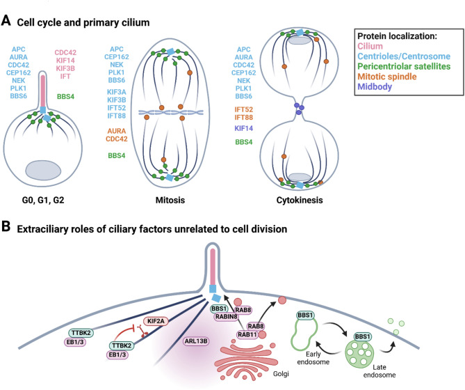
Cilia are versatile, microtubule-based organelles that facilitate cellular signaling, motility, and environmental sensing in eukaryotic cells. These dynamic structures act as hubs for key developmental signaling pathways, while their assembly and disassembly are intricately regulated along cell cycle transitions. Recent findings show that factors regulating ciliogenesis and cilia dynamics often integrate their roles across other cellular processes, including cell cycle regulation, cytoskeletal organization, and intracellular trafficking, ensuring multilevel crosstalk of mechanisms controlling organogenesis. Disruptions in these shared regulators lead to broad defects associated with both ciliopathies and cancer. This review explores the crosstalk of regulatory mechanisms governing cilia assembly, disassembly, and maintenance during ciliary signaling and the cell cycle, along with the broader implications for development, tissue homeostasis, and disease.

Keywords: Cancer; Cell cycle regulation; Cilia; Ciliary dynamics; Ciliary signaling; Ciliopathies; Tissue development.


Conflict of interest statement

Declarations. Ethics approval and consent to participate: Not applicable. Consent for publication: Not applicable. Competing Interests.: The authors declare that they have no competing interests.

Figures

Fig. 1
Fig. 1
Primary cilium structure and mechanism of intracellular ciliogenesis. A Primary cilium originates from the basal body (grey), modified centriole, which possesses two sets of appendages – distal (DAs) and subdistal (SDAs). The central structure is the membrane-enclosed microtubule-based ciliary axoneme (pink). The transition zone (TZ) acts as a diffusion barrier between the basal body and the ciliary axoneme. The ciliary membrane is rich in phosphoinositides (PIP), while phosphatidylinositol 4,5-bisphosphate (PIP2) levels are increased in the periciliary membrane at the cilium base; the phosphatidylinositol (3,4,5)-trisphosphate (PIP3) seems specifically enriched in the membrane surrounding the TZ. Active protein transport is facilitated by intraflagellar transport (IFT) machinery, which works in cooperation with the BBSome – cargo adapter complex. IFT is a bidirectional movement of complexes along the microtubules from the base of the cilium to its tip (anterograde transport) by kinesin motors and from the tip back to the ciliary base (retrograde transport) by dynein motors. B Intracellular ciliogenesis begins with the formation of the ciliary vesicle at the distal appendages, a process facilitated by EHD1/EHD3 and RAB34. TTBK2 phosphorylates CEP83 and other DAs components, enabling the ciliary vesicle to dock to the mother centriole. Ciliary vesicle membrane expansion is regulated by the RAB11/RABIN8/RAB8 cascade, driving the development and elongation of the ciliary membrane. Simultaneously, IFT mediates the growth of the axoneme, coordinating its assembly with the extension of the ciliary membrane. This image was created with BioRender.com
Fig. 2
Fig. 2
Primary cilium dynamics during cell cycle. Primary cilia are disassembled before mitosis and reassembled after cell division during the early G1 phase. Cilium disassembly occurs through several potentially overlapping mechanisms, including gradual resorption mediated by AURA kinase and HDAC6, CDC42 and actin-dependent ectocytosis, and Katanin-dependent ciliary decapitation. Axoneme (light pink), basal body (grey) with distal appendages (DAs; pink). This image was created with BioRender.com
Fig. 3
Fig. 3
Dual roles of cilia regulators in cell cycle and intracellular organization. A Established mitotic regulators such as AURA, NEKs, APC, CDC42, and PLK1 also play critical roles in regulating cilium disassembly. Common ciliary factors such as IFTs (e.g. IFT52 and IFT88), KIF3A/B, KIF14, CEP162, BBS4, and BBS6 also facilitate the formation of the mitotic spindle, chromosome alignment, and the progression of cytokinesis. B The factors involved in ciliogenesis and cilia function are vital to intracellular organization. TTBK2 binds to EB1/3 and promotes microtubule growth by suppressing depolymerizing activity of the KIF2A kinesin. ARL13B regulates the non-canonical Hedgehog (HH) pathway and is key in controlling actin organization. The RAB11/RABIN8/RAB8 cascade controls polarized vesicular trafficking to the plasma and ciliary membrane, where the latter is mediated by the BBS1-RABIN8 module. BBS1 is also involved in the endomembrane vesicular trafficking between the early and late endosomes. This image was created with BioRender.com
Fig. 4
Fig. 4
Interactions between individual modules of primary cilia. A The basal body and pericentriolar satellites are crucial for the growth and maintenance of the ciliary axoneme. TTBK2 at the basal body promotes cilium growth by suppressing the depolymerizing kinesin KIF2A. In C. elegans, the basal body is degraded once the cilium is assembled. B Anterograde and retrograde IFT display functional crosstalk in cilium length control. Inhibition of the heteromeric KIF3A/3B kinesin II disrupts anterograde IFT and indirectly halts retrograde IFT, causing IFT particles to accumulate at the ciliary base. Inhibition of the dynein motor by ciliobrevin D or mutation in IFT-A complexes indirectly abrogates anterograde IFT. C Retrograde IFT is crucial for correct TZ establishment. Mutations in components of retrograde IFT lead to the mislocalization of TZ components inside the cilium, and the shortening of cilia. D Posttranslational modifications (PTMs) of axonemal tubulin are crucial for cilia function. Axoneme acetylation, glutamylation, and glycylation are involved in cilia beating and sperm motility, likely modulating the velocity of the anterograde IFT. PTMs can also compensate for each other; axoneme hyper-glycylation can compensate for axoneme deacetylation in zebrafish cilia, while axoneme hyper-glutamylation can compensate for axoneme deglycylation in murine photoreceptors. This image was created with BioRender.com
Fig. 5
Fig. 5
Adaptive changes to cilia dynamics during cell signaling. A Activation of HH signaling triggers cilia shortening and promotes cell cycle entry, passing on this effect to the subsequent progeny (1). In fibro/adipogenic progenitors (FAPs), the length of cilia modulates muscle regeneration. Short or absent cilia on FAPs block adipogenesis and promote myogenesis, while long cilia favor adipogenesis (2). Similarly, HH signaling induces myoblast differentiation from muscle stem cells, where myoblasts lose cilia to form myofibrils (3). In BBSome deficiency, HH signaling triggers CDC42/actin-dependent ectocytosis of ciliary GPR161, leading to subsequent cilia shortening (4). After neural tube closure, HH-producing cells elongate cilia to attenuate their responsiveness to the HH pathway (5). During differentiation of neural progenitors, apical abscission of the ciliary axoneme from the basal body leads to a temporary cilia dysfunction. B The effects of WNT signaling on primary cilia dynamics are context-dependent. During tissue differentiation, WNT functions together with HH to either stimulate cilia disassembly in myoblasts or sustain cilia dependent signaling in bone marrow mesenchymal stem cells (BM MSCs) during osteogenesis (1). In MSCs, WNT and HH act inhibitory on adipo-commitment, leading to the emergence of adipocytes through IGF1R/FFAR4 cilia-dependent signaling and eventual cilia disassembly (1). In cell culture systems, activation of WNT with WNT3a ligand or pharmacological inhibition of WNT ligands secretion, does not alter number or length of cilia (2). C The effects of TGFβ signaling on cilia dynamics exhibit a context- and cell type-specific dependency. Activation of the TGFβ signaling pathway leads to the shortening of primary cilia in cultured chondrocytes (1), whereas it maintains cilia length in cilia of Xenopus left-right organizer (LRO) (2). D NOTCH signaling is primarily triggered by the fluid flow-induced alterations in cilia dynamics, which in turn promotes cilia function in diverse cellular systems, such as respiratory epithelium (1), zebrafish left-right organizer (LRO) (2) and endothelial cells in the myocardium (3). This image was created with BioRender.com

Similar articles

References

    1. Beales P, Jackson PK. Cilia - the prodigal organelle. Cilia. 2012;1:1–3. - PMC - PubMed
    1. Sorokin SP. Reconstructions of centriole formation and ciliogenesis in mammalian lungs. J Cell Sci. 1968;3:207–30. - PubMed
    1. Gopalakrishnan J, et al. Emerging principles of primary cilia dynamics in controlling tissue organization and function. EMBO J. 2023;42:e113891. - PMC - PubMed
    1. Bangs F, Anderson KV. Primary cilia and mammalian Hedgehog signaling. Cold Spring Harb Perspect Biol. 2017. 10.1101/cshperspect.a028175 - PMC - PubMed
    1. Mill P, Christensen ST, Pedersen LB. Primary cilia as dynamic and diverse signalling hubs in development and disease. Nat Rev Genet. 2023;24:421–41. - PMC - PubMed

LinkOut - more resources