. 2025 Jun 7;16(1):294.
doi: 10.1186/s13287-025-04419-x.

Metabolic markers detect early ostedifferentiation of mesenchymal stem cells from multiple donors

Affiliations

Metabolic markers detect early ostedifferentiation of mesenchymal stem cells from multiple donors

Daniela S C Bispo et al. Stem Cell Res Ther. .

Abstract

Background: Mesenchymal stem cells (MSC) are pivotal bioengineering tools, offering significant promise for applications in bone regeneration. However, their therapeutic potential is limited by inter-donor variability and experimental issues. This study aimed to identify robust metabolic markers of osteodifferentiation applicable across multiple donors, while providing insight into the metabolic pathways actively involved in the process.

Methods: Untargeted nuclear magnetic resonance (NMR) metabolomics was applied to characterize the intra- and extracellular metabolic adaptations of human adipose-derived MSC (hAMSC) undergoing osteogenic differentiation, compared to proliferation alone. Multivariate and univariate statistical analysis was carried out on data from three independent donors, and cross-validation was employed to evaluate the predictive capacity of the proposed markers.

Results: Variations in the levels of selected (nine) intracellular and (seventeen) extracellular metabolites detect osteodifferentiation by day 7 (out of 21), with nearly 100% accuracy. These signatures suggest a metabolic shift from glycolysis/OxPhos to lactic fermentation, fatty acid β-oxidation and phosphocreatine hydrolysis. Intracellular glucose, lactate, citrate and specific amino acids are redirected towards protein synthesis and glycosylation, with some of the secreted metabolites (e.g., citrate) seemingly involved in biomineralization and other extracellular roles. Membrane metabolism, antioxidant mechanisms and adenosine metabolism are also impacted by osteodifferentiation.

Conclusions: These findings reveal effective donor-independent markers of hAMSC osteodifferentiation, with a robust extracellular signature standing out for potential rapid and non-invasive detection of osteocommitted cells.

Keywords: Bone regeneration; Mesenchymal stem cells; Metabolic markers; Metabolomics; Nuclear magnetic resonance (NMR) spectroscopy; Osteogenesis; Osteogenic differentiation.


Conflict of interest statement

Declarations. Ethics approval and consent to participate: Adipose tissue from donor 1 was obtained under an agreement between the University of Aveiro and “Hospital da Luz”, Aveiro, dated 17th February 2023 and upon written consent from the donor. The human adipose-derived stem cells (donors 2 and 3) were purchased from the American Type Culture Collection (Lot 70017032, Ref. ATCC PCS-500-011) and Lonza (Lot 22TL018258, Ref. LSLZPT-5006) having been accompanied by Certificates of Analysis stating that cells were isolated from donated human tissue after obtaining permission for their use in research applications by informed consent or legal authorization. Regarding osteoblasts, these were purchased from Lonza (CC-2538, Lot 19TL217387), certified to have been isolated from donated human tissue after obtaining permission for their use in research applications. All human cells were used within the framework of the BetterBone project (2022.04286.PTDC, doi: https://doi.org/10.54499/2022.04286.PTDC ) Human ethics and consent to participate: Not applicable. Consent for publication: Not applicable. Competing interests: Authors declare that they have no competing interests. Clinical trial number: Not applicable.

Figures

Fig. 1
Fig. 1
Experimental design for hAMSC harvesting and media sampling (from 3 donors) throughout proliferation (control) and osteodifferentiation. For each donor, cell samples were collected (in triplicate) on D0, D1, D4, D7, D14 and D21 (purple) and media samples (also in triplicate) on D1, D4, D7, D11, D14, D18 and D21 (pink). Some figure elements were adapted from Servier Medical Art and licensed under a Creative Commons Attribution 3.0 Unported (CC BY 3.0) license
Fig. 2
Fig. 2
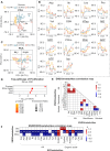
Metabolic changes in proliferating (non-differentiating) hAMSC (control, CTR). (A) PCA scores scatter plot of full NMR spectra of hAMSC polar extracts. (B) Time course evolution of the 14 endometabolites comprised in the donor-independent signature of proliferation (% variation in Table S2, Additional file 1). (C) PCA scores scatter plot of integrals of 7 selected endometabolites: HTau, MG, GPC, PEtn, Ino, ADP and ATP. (D) Top enriched pathways descriptive of hAMSC proliferation for all donors. (E) Endometabolite correlations, all timepoints (ρ >|0.7|, p-value < 0.001). (F) Endometabolite/Exometabolite correlations (ρ >|0.8|, p-value < 0.001). 3-HIBA, 3-hydroxyisobutyrate; ADP, adenosine diphosphate; ATP, adenosine triphosphate; Cho, choline; Cr, creatine; GPC, glycerophosphocholine; GPL, glycerophospholipids; GSH, glutathione (reduced); HTau, hypotaurine; Ino, inosine; MG, methylguanidine; PCho, phosphocholine; PEtn, phosphoethanolamine; Pyr, pyruvate; α-KG, α-ketoglutarate
Fig. 3
Fig. 3
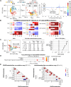
Changes in the endometabolome of osteodifferentiating hAMSC. (A) PCA scores scatter plot (left) obtained with full NMR spectra of hAMSC polar extracts, under control (CTR) and osteoinduced (OI) conditions, pairwise PLS-DA scores scatter plot (middle) comparing CTR to OI, and corresponding LV1 loadings plot (right). (B) Donor-independent (DI) osteogenic signature of 9 endometabolites (heatmap of % variation for OI compared to CTR at each timepoint), *, p-value < 0.05; (*), visually confirmed differences (p-value not calculated as n = 1 for D21 of OI2). (C) PCA scores scatter plot obtained with selected integrals (left), MCCV of PLS-DA models obtained excluding progressively earlier timepoints (middle), and VIP scores corresponding to D21 samples (model v.) (right). (D) Top enriched pathways related to osteodifferentiation at D21, compared to controls, considering all donors. (E) Spearman correlation maps for endometabolites across all timepoints (left) and in D14 and D21 (right), ρ >|0.8| and p-value < 0.001. Etn, ethanolamine; PCr, phosphocreatine; UDP-GalNAc, uridine diphospho-N-acetylgalactosamine; UDP-GlcNAc, uridine diphospho-N-acetylglucosamine; Uδ, unassigned signal at chemical shift δ. Other metabolite abbreviations specified in the caption of Fig. 2. CR, Classification rate (accuracy); Q2, predictive power; sens., sensitivity; spec., specificity
Fig. 4
Fig. 4
Changes in the exometabolome of osteodifferentiating hAMSC. (A) PCA scores scatter plot obtained with the full NMR spectra of the media from hAMSC under control (CTR) and osteoinduced (OI) conditions. (B) 17-exometabolite donor-independent (DI) osteogenic signature, which includes patterns of increased secretion (solid arrow, top right), decreased secretion (dashed arrow, bottom right), and reduced uptake (dashed arrow, bottom left) relative to controls, as well as metabolites with donor-dependent patterns but DI final levels (white double arrow). Heatmaps represent % variation in OI compared to CTR, at each timepoint (p-values were < 0.05). a non-significant variation, bp-value not computed due to n < 3, visual confirmation only. Examples of time-course evolutions are shown in each quadrant (a full account may be found in Figure S9, Additional file 1). (C) PCA scores scatter plot obtained only with the 17-exometabolite DI signature (integrals) (top), MCCV results from PLS-DA progressively excluding early timepoints (middle), and VIP scores corresponding to D21 samples. (bottom). Unit variance (UV) used for all multivariate models
Fig. 5
Fig. 5
Key metabolic fluctuations in the endo- and exometabolomes of osteodifferentiating hAMSC (compared to proliferation alone). Metabolites detected by NMR are shown in bold. Metabolite names in red and blue were shown to increase or decrease in level, respectively, in osteodifferentiating cells compared to controls (this is also shown by colored arrows next to metabolite names, arrows in brackets reflect variations in 2 of the donors). Superscripts indicate the first day of change. Donor-independent metabolite changes are highlighted in yellow, both inside and outside the cells. Changed metabolite fluxes across the cell membrane are represented by solid or dashed arrows, for enhanced or attenuated fluxes, respectively. Some elements of this picture were adapted from Servier Medical Art (smart.servier.com) and are licensed under a Creative Commons Attribution 3.0 Unported (CC BY 3.0) license. 2-HIBA, 2-hydroxyisobutyrate; 3-HBA, 3-hydroxybutyrate; 3-HIBA, 3-hydroxyisobutyrate; α-KG, α-ketoglutarate; Ado, adenosine; ADP, adenosine diphosphate; AMP, adenosine monophosphate; ATP, adenosine triphosphate; BCAAs, branched-chained amino acids; BCKAs, branched-chain α-ketoacids; Cho, choline; Cr, creatine; ER, endoplasmic reticulum; FA, fatty acid; GPC, glycerophosphocoline; GSH, glutathione (reduced form); IMP, inosine monophosphate; Ino, inosine; MG, methylguanidine; Orn, ornithine; PCr, phosphocreatine; PEtn, phosphoethanolamine; Pi, inorganic phosphate; PtdCho, phosphatidylcholine; PtdEtn, phosphatidylethanolamine; ROS, reactive oxygen species; TCA, tricarboxylic acid cycle; UDP-GalNAc, uridine diphospho-N-acetylgalactosamine; UDP-GlcNAc, uridine diphospho-N-acetylglucosamine

References

    1. Iaquinta MR, Mazzoni E, Bononi I, Rotondo JC, Mazziotta C, Montesi M, et al. Adult stem cells for bone regeneration and repair. Front Cell Dev Biol. 2019;7:268. - PMC - PubMed
    1. Česnik AB, Švajger U. The issue of heterogeneity of MSC-based advanced therapy medicinal products–a review. Front Cell Dev Biol. 2024;12:1400347. - PMC - PubMed
    1. Phinney DG, Galipeau J. Manufacturing mesenchymal stromal cells for clinical applications: A survey of good manufacturing practices at U.S. Academic centers. Cytotherapy. 2019;21:782–92. - PubMed
    1. Feng F, Thompson MP, Thomas BE, Duffy ER, Kim J, Kurosawa S, et al. A computational solution to improve biomarker reproducibility during long-term projects. PLoS ONE. 2019;14:e0209060. - PMC - PubMed
    1. Bustin SA. Quantification of mRNA using real-time reverse transcription PCR (RT-PCR): trends and problems. J Mol Endocrinol. 2002;29:23–39. - PubMed

LinkOut - more resources