. 2025 Jul;75(2):100028.
doi: 10.1016/j.jphyss.2025.100028. Epub 2025 Jun 4.

Differences in the autonomic regulation of temperature in the lower lip and tongue during activation of the lingual nerve

Affiliations

Differences in the autonomic regulation of temperature in the lower lip and tongue during activation of the lingual nerve

Syed Taufiqul Islam et al. J Physiol Sci. 2025 Jul.

Abstract

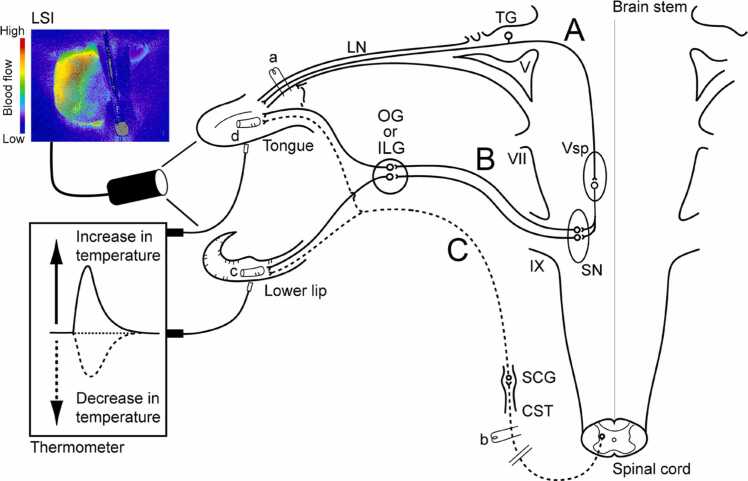
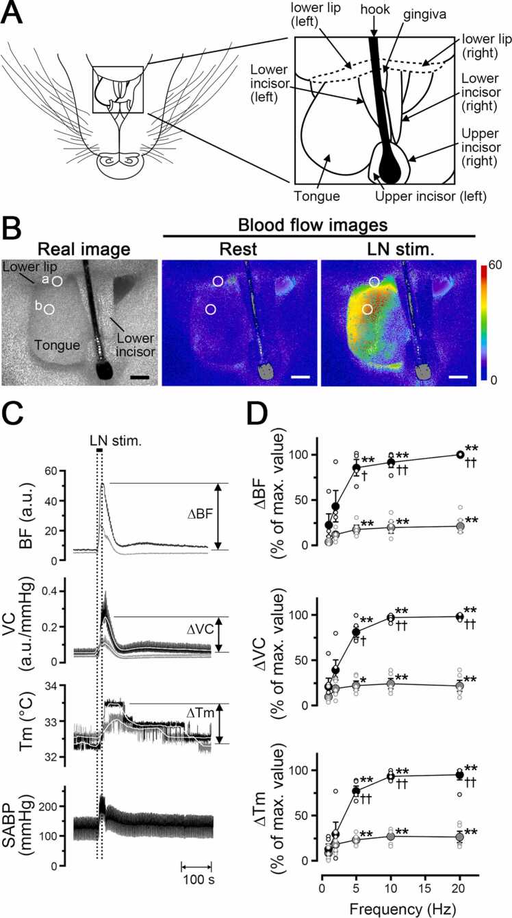
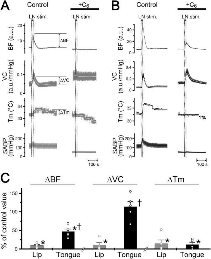
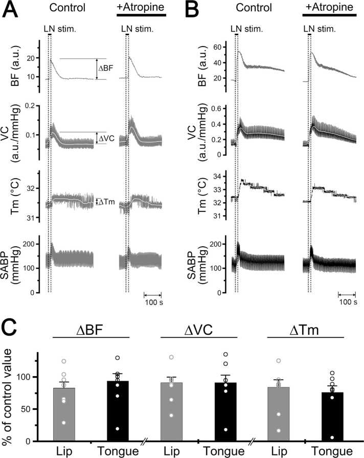
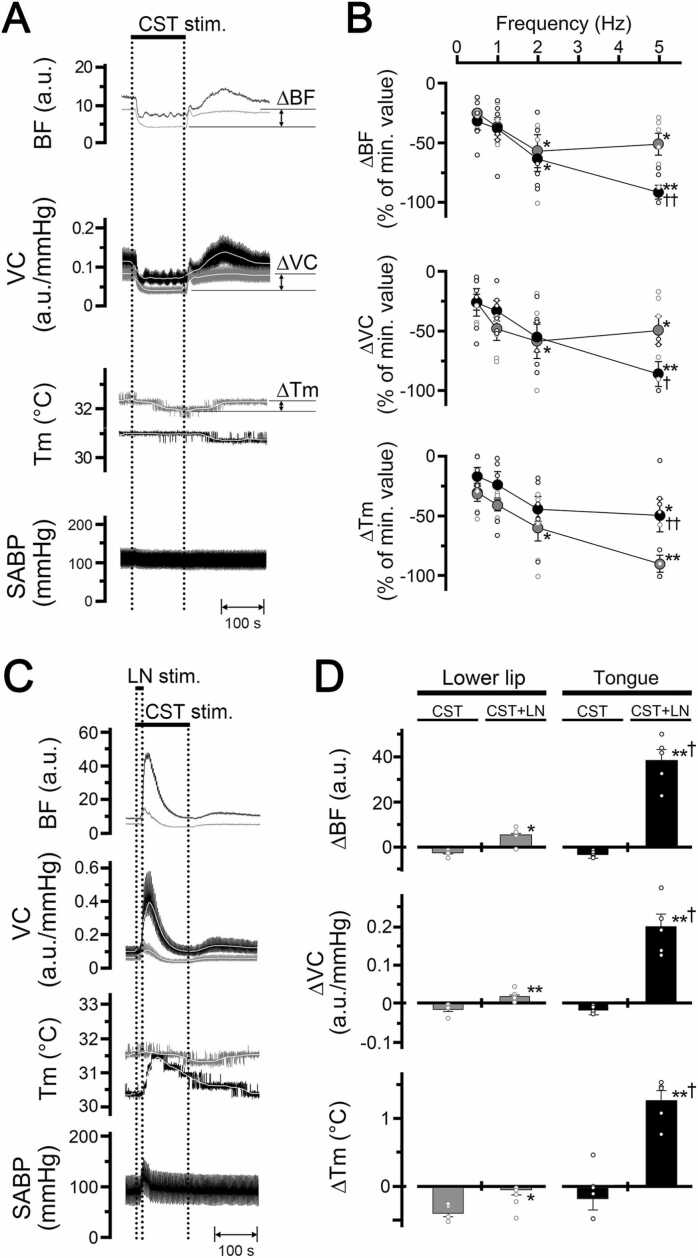
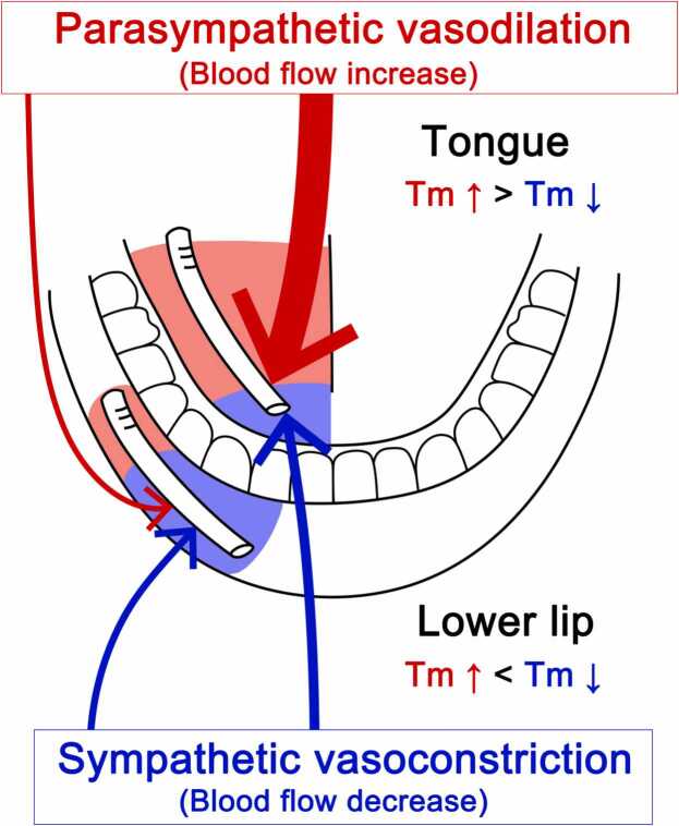
Orofacial temperature influences orofacial functions and is related to hemodynamics mediated by the autonomic nerves. Although the properties of autonomic vasomotor responses differ in orofacial tissues, differences in the autonomic regulation of orofacial temperature are unclear. We examined the differences in blood flow (BF) and temperature (Tm) between the extraoral (lower lip) and intraoral tissues (tongue) of urethane-anesthetized rats. Noncholinergic parasympathetic vasodilation evoked by trigeminal-mediated reflex elicited significant increases in BF and Tm in both tissues, and these increases were larger in the tongue than in the lower lip. Activation of cervical sympathetic nerves significantly decreased BF and Tm in both tissues. These decreases were restored by parasympathetic reflex vasodilation; the effects were larger in the tongue than in the lower lip. Our results suggest that parasympathetic vasodilation is involved in the maintenance of BF and Tm, and that the effects may be greater in intraoral than in extraoral tissues.

Keywords: Cervical sympathetic nerves; Noncholinergic parasympathetic vasodilation; Orofacial temperature; Trigeminal mediated reflex.


Conflict of interest statement

Declaration of Competing Interest The authors declare that they have no known competing financial interests or personal relationships that could have appeared to influence the work reported in this paper.

Figures

Fig. 1
Fig. 1
Schematic representation of the electrical stimulation and measurement sites of both blood flow and local temperature in rats. Stimulation sites: peripheral portion of the lingual nerve (LN) (a), and peripheral cut end of superior cervical sympathetic trunk (CST) (b). Blood flow and local temperature measurement sites: (c) lower lip and (d) tongue, using laser speckle imaging (LSI) and a thermometer, respectively. The continuous lines indicate: (A) trigeminal sensory inputs to the trigeminal spinal nucleus (Vsp) in the brainstem and (B) parasympathetic vasodilator fibers to the lower lip and tongue from the salivatory nuclei (SN). The dashed lines indicate: sympathetic vasoconstrictor fibers to the lower lip and tongue from the superior cervical ganglion (SCG) of the superior cervical sympathetic trunk (C). Other abbreviations: ILG, intralingual ganglion; OG, otic ganglion; TG, trigeminal ganglion; V, trigeminal nerve root; VII, facial nerve root; IX, glossopharyngeal nerve root.
Fig. 2
Fig. 2
Relationships between hemodynamics and local temperature during trigeminal afferent inputs in the orofacial tissues in the rat. A: Illustration of measurement sites of blood flow and temperature in the mandibular area including the lower lip and tongue in a supine rat. B: Typical example of the real image and speckle images of the blood flow in the lower lip and inferior surface of the tongue at the basal level (rest) and produced by left lingual nerve stimulation (LN stim.) for 20 s with a supramaximal intensity (100 µA) at 20 Hz using 2-ms pulses. Scale bars represent 3.5 mm. C: Increases in blood flow (BF, a.u.; arbitrary units), vascular conductance (VC, a.u./mmHg), and local temperature (Tm, °C) in the lower lip (gray traces) and tongue (black traces) extracted from a region of interest (ROI) indicated by the white circles in a and b, and systemic arterial blood pressure (SABP, mmHg) evoked by lingual nerve stimulation (horizontal bar with dashed lines). The white traces indicate the mean vascular conductance and local temperature at the measuring sites. D: Mean ± standard error of the mean (SEM) of ∆BF, ∆VC, and ∆Tm of the lower lip (gray symbols) and tongue (black symbols) evoked by lingual nerve stimulation at 100 µA and various frequencies (1–20 Hz) (n = 6 in each group). Each value is expressed as a percentage of the maximum response. The statistical significance of the differences from base value (at a frequency of 1 Hz) was assessed by ANOVA followed by a post hoc test (Bonferroni/Dunn test). *P < 0.01, ** P < 0.001 vs. base value. P < 0.01, ††P < 0.001, significant differences between the increases in the lower lip and tongue recorded by lingual nerve stimulation (ANOVA followed by a contrast test). Individual data points are indicated by the white markers.
Fig. 3
Fig. 3
Effects of hexamethonium on changes in the hemodynamics and local temperature in the lower lip and tongue evoked by lingual nerve stimulation. A, B: Typical examples of the effects of intravenous administration of hexamethonium (C6) at 10 mg/kg for 10 min (0.1 ml/min) on changes in blood flow (BF, a.u.; arbitrary units), vascular conductance (VC, a.u./mmHg), and local temperature (Tm, °C) of the lower lip (gray traces, A) and tongue (black traces, B) on the left side evoked by left lingual nerve stimulation (20 s, 100 µA, 20 Hz, 2 ms). C: Mean ± SEM of increases in the ∆BF, ∆VC, and ∆Tm of the lower lip (gray bars) and tongue (black bars) evoked by lingual nerve stimulation with administration of C6 (n = 6 in each group). Each value is expressed as a percentage of the control response before treatment. The statistical significance of the differences from the control was assessed by ANOVA followed by a post hoc test (Bonferroni/Dunn test). *P < 0.001 vs. control. P < 0.01, significant differences between the changes evoked by lingual nerve stimulation in the lower lip and tongue (ANOVA followed by a contrast test). Individual data points are indicated by the white markers.
Fig. 4
Fig. 4
Effects of atropine on changes in the hemodynamics and local temperature in the lower lip and tongue evoked by lingual nerve stimulation. A, B: Typical examples of the effects of intravenous administration of atropine at 100 μg/kg for 10 min (0.1 ml/min) on changes in blood flow (BF, a.u.; arbitrary units), vascular conductance (VC, a.u./mmHg), and local temperature (Tm, °C) of the lower lip (gray traces, A) and tongue (black traces, B) on the left side evoked by left lingual nerve stimulation (20 s, 100 µA, 20 Hz, 2 ms). C: Mean ± SEM of increases in the ∆BF, ∆VC, and ∆Tm of the lower lip (gray bars) and tongue (black bars) evoked by lingual nerve stimulation with the administration of atropine (n = 7 in each group). Each value is expressed as a percentage of the control response before treatment. The statistical significance of the differences from the control was assessed by ANOVA followed by a post hoc test (Bonferroni/Dunn test). Individual data points are indicated by white markers.
Fig. 5
Fig. 5
Sympathetic effects on cardiovascular parameters and local temperature in the lower lip and tongue. A: Typical examples of changes in the blood flow (BF, a.u.; arbitrary units), vascular conductance (VC, a.u./mmHg), and local temperature (Tm, °C) in the lower lip (gray traces) and tongue (black traces) on the left side, and SABP evoked by electrical stimulation of the peripheral cut end of the left cervical sympathetic trunk (CST stim.; horizontal bar with dashed lines) for 2 min with a supramaximal intensity (100 µA) at 5 Hz using 2-ms pulses. B: Mean ± SEM of decreases in the ∆BF, ∆VC, and ∆Tm in the lower lip (gray symbols) and tongue (black symbols) evoked by cervical sympathetic trunk stimulation at 100 µA and various frequencies (0.5–5 Hz) (n = 6 in each group). Each value is expressed as a percentage of the minimum response. The statistical significance of the differences from base value (at frequency of 0.5 Hz) was assessed by ANOVA followed by a post hoc test (Bonferroni/Dunn test). *P < 0.01, ** P < 0.001 vs. base value (at the frequency of 0.5 Hz). P < 0.05, ††P < 0.01, significant differences between the decreases evoked by cervical sympathetic trunk stimulation in the lower lip and tongue (ANOVA followed by a contrast test). C: Typical examples of the changes in blood flow (BF), vascular conductance (VC), and local temperature (Tm) of the lower lip (gray traces) and tongue (black traces) evoked by cervical sympathetic trunk stimulation (2 min, 100 µA, 5 Hz, 2 ms) in combination with lingual nerve stimulation (20 s, 100 µA, 20 Hz, 2 ms). D: Mean data ± SEM of the changes in the ∆BF, ∆VC, and ∆Tm of the lower lip (gray bars) and tongue (black bars) evoked by cervical sympathetic trunk stimulation alone (CST), and in combination with lingual nerve stimulation (CST+LN) (n = 6 in each group). *P < 0.05, **P < 0.001 vs. responses evoked by cervical sympathetic trunk stimulation alone at each site (paired t-test). P < 0.001, significant differences between the changes evoked by cervical sympathetic trunk stimulation with lingual nerve stimulation in the lower lip and tongue (unpaired t-test). Individual data points are indicated by white markers.
Fig. 6
Fig. 6
Proposed schema for differences in the regulation of blood flow and local temperature (Tm) in the lower lip (extraoral tissues) and tongue (intraoral tissues) mediated by the autonomic nervous systems. The increases in blood flow and local temperature evoked by parasympathetic vasodilation mediated by trigeminal afferent inputs in the tongue were significantly greater than those in the lower lip. Sympathetic vasoconstriction evoked by excess sympathetic activity reduced blood flow and local temperature in both tissues. These decreases were restored by parasympathetic reflex vasodilation; the effects were larger in the tongue than in the lower lip. Therefore, the local temperature in the tongue may be maintained at a higher level than that in the lower lip through the activity of the parasympathetic nervous system.

Similar articles

References

    1. Ishii H., Niioka T., Sudo E., Izumi H. Evidence for parasympathetic vasodilator fibres in the rat masseter muscle. J Physiol. 2005;569(Pt 2):617–629. doi: 10.1113/jphysiol.2005.087643. - DOI - PMC - PubMed
    1. Izumi H., Karita K. Somatosensory stimulation causes autonomic vasodilatation in cat lip. J Physiol. 1992;450:191–202. doi: 10.1113/jphysiol.1992.sp019123. - DOI - PMC - PubMed
    1. Mizuta K., Karita K., Izumi H. Parasympathetic reflex vasodilatation in rat submandibular gland. Am J Physiol Regul Integr Comp Physiol. 2000;279(2):R677–R683. doi: 10.1152/ajpregu.2000.279.2.R677. - DOI - PubMed
    1. Sato T., Ishii H. Differences in control of parasympathetic vasodilation between submandibular and sublingual glands in the rat. Am J Physiol. 2015;309(11):R1432–R1438. doi: 10.1152/ajpregu.00306.2015. - DOI - PubMed
    1. Izumi H., Karita K. The parasympathetic vasodilator fibers in the trigeminal portion of the distal lingual nerve in the cat tongue. Am J Physiol. 1994;266(5 Pt 2):R1517–R1522. doi: 10.1152/ajpregu.1994.266.5.R1517. - DOI - PubMed

LinkOut - more resources