. 2025 Jun 8;23(1):429.
doi: 10.1186/s12951-025-03471-2.

Tumor microenvironment remodeling with a telomere-targeting agent and its cooperative antitumor effects with a nanovaccine

Affiliations

Tumor microenvironment remodeling with a telomere-targeting agent and its cooperative antitumor effects with a nanovaccine

Jing Bai et al. J Nanobiotechnology. .

Abstract

The nucleoside analogue 6-thio-2'-deoxyguanosine (6-thio-dG, also known as THIO) is a telomere-targeting agent with important clinical potency. It can selectively kill telomerase-positive tumor cells. We previously reported that THIO could successfully induce immunogenic cell death (ICD) in multiple mouse tumor cell lines. In this study, we further explored the potential impact of THIO on remodeling the tumor microenvironment, regulating anti-tumor immune responses, and its possible synergistic effects with other therapeutic methods, such as tumor vaccines. Our results showed that THIO could also induce ICD in various human tumor cell lines. The induction of ICD in tumor cells promoted the migration and maturation of antigen-presenting cells. Administration of THIO significantly inhibited the growth of established CT26 and TC-1 tumors in mice. Meanwhile, it enhanced the anti-tumor CTL response and reduced the levels of immunosuppressive myeloid-derived suppressor cells (MDSCs) in both the spleen and tumor tissues. Additionally, THIO had a direct inhibitory effect on the proliferation and differentiation of MDSCs. Moreover, when combined with bacterial biomimetic vesicles or a nanovaccine, such as THIO with BBV or different Q11-tumor antigen peptide nanofibers, it exhibited enhanced anti-tumor effects and immune responses compared to monotherapy in either "immune hot" TC-1 tumors or "immune cold" B16-F10 tumors. In summary, THIO has the ability to remodel the tumor microenvironment, exert a specific killing effect on tumor cells, and effectively cooperate with tumor vaccines. This broadens the anti-tumor mechanisms of THIO and provides a promising strategy for improving anti-tumor immunotherapies.

Keywords: 6-thio-2′-deoxyguanosine (THIO); Cancer vaccine; Immunogenic cell death (ICD); Tumor immunotherapy; Tumor microenvironment.


Conflict of interest statement

Declarations. Ethics approval and consent to participate: All animal procedures were approved by the Institutional Animal Care and Use Committee of the Institute of Medical Biology, Chinese Academy of Medical Sciences. Consent for publication: All authors agreed to publish this manuscript. Competing interests: The authors declare no competing interests.

Figures

Scheme 1
Scheme 1
Schematic representation of THIO-triggered antitumor immune responses and its synergistic action with a nanovaccine. A Self-assembly process of Q11-E7 and Q11-M30 nanofibers, which serve as vaccines specifically targeting TC-1 and B16 tumors, respectively. B THIO selectively eliminates tumor cells and induces immunogenic cell death (ICD) in tumor cells, thereby generating "find me" and ‘‘eat me’’ immunostimulatory signals, releasing pro-inflammatory molecules, and presenting a wide spectrum of tumor antigens. These events collectively promote the migration, antigen presentation, and maturation of dendritic cells (DCs), subsequently initiating antitumor responses mediated by cytotoxic T lymphocytes (CTLs) and natural killer (NK) cells. Additionally, THIO directly suppresses the functions of myeloid-derived suppressor cells (MDSCs). Treatment with THIO results in the remodeling of the tumor immune microenvironment. This process synergizes with the tumor vaccine, which is designed to evoke antigen-specific effector T cell responses by facilitating the generation, migration, survival, and maintenance of antitumor T cell activity. CTLs cytotoxic T lymphocytes; MDSCs myeloid-derived suppressor cells, NK natural killer cells, ATP adenosine triphosphate, CALR calreticulin, IL-1β interleukin-1, HMGB1 high mobility group protein B1. The scheme was created using BioRender.com
Fig. 1
Fig. 1
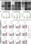
The culture supernatants of THIO-treated mouse tumor cells stimulates the migration and maturation of BMDCs. A Transwell assay for BMDC migration. BMDC were placed into the upper chamber of 24-well Transwell plates, and then the culture supernatants after THIO treatment of CT26, TC-1 and B16-F10 (i.e., ICD Supernatants), untreated cell culture supernatants (i.e., Normal Supernatants), and THIO-containing blank medium control (i.e., Medium + THIO) were placed into the lower chamber and observed and counted the number of cells in the lower chamber with a light microscope at different time points. B Statistical analyses on BMDC migration at different times (n = 3). C Statistical analyses with flow cytometry on BMDC maturation stimulated by the culture supernatants of THIO-treated tumor cells. Maturation markers CD80, CD86, MHC I and MHC II were detected (n = 3). Data are shown as the mean ± standard deviation (SD), repeat experiment 2–3 times independently. Statistical significance was calculated via one-way analysis of variance (ANOVA) followed by Tukey's multiple comparisons. *p < 0.05, **p < 0.01, ***p < 0.001, ****p < 0.0001; ns, not significant
Fig. 2
Fig. 2
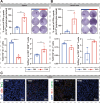
Intraperitoneal treatment of THIO inhibits the growth of subcutaneous CT26 tumors in mice. A Schematic of the treatment schedule: tumor bearing mice received intraperitoneal THIO at 2 mg/kg every other day for a total of five doses. B Representative photographs of excised tumors; dashed circles indicate complete tumor regression. C Tumor growth curves. D Tumor weight summary (n = 8). E Spleen weight summary (n = 8). The mouse experiments were performed in two independent replicates. Data are shown as mean ± standard deviation (SD). Statistical significance for tumor growth curve was calculated by two-way analysis of variance (ANOVA). The comparison between PBS and THIO was conducted by unpaired Student’s t-test. ****p < 0.0001; ns, not significant. Figure A created with BioRender.com
Fig. 3
Fig. 3
Intraperitoneal treatment of THIO promotes anti-tumor immune response and inhibits MDSCs production. A In the spleen, ELISPOT analyzed the spot statistics of IFN-γ secretion by lymphocytes (n = 3), and flow cytometry analyzed the ratio of MDSCs (CD11b+Gr-1+) and CTL (CD3+CD8+IFN-γ+) in the spleen (n = 5). B In lymph nodes, ELISPOT analyzed the spot statistic of IFN-γ secretion by lymphocytes (n = 3) and flow cytometry analyzed the proportion of MDSCs (CD11b+Gr-1+) and CTL (CD3+CD8+IFN-γ+) in lymph nodes (n = 3). C Representative immunofluorescence images of CTL (blue: nucleus, red: CD8, green: IFN-γ) and MDSCs (blue: nucleus, red: Gr-1, green: CD11b) in tumor tissues. Data are shown as mean ± standard deviation (SD). The comparison between PBS and THIO was conducted by unpaired Student’s t-test. *p < 0.05, **p < 0.01, ***p < 0.001, ****p < 0.0001
Fig. 4
Fig. 4
THIO treatment improves anti-tumor immune responses and remodels the tumor microenvironment. A Schematic representation of the response of important immune cells after THIO treatment. B Flow cytometry analysis of lymphocytes isolated from mouse spleen (B, top) (n = 3) and tumor tissues (B, bottom) (n = 3) after two administrations of THIO, including CTLs (CD3+CD8+IFN-γ+), NK (CD3NK1.1+), MDSCs (CD11b+Gr-1+), and Th1 (CD3+CD4+IFN-γ+). (C) Flow cytometry analysis of lymphocytes isolated from mouse spleen (C, top) (n = 3) and tumor tissues (C, bottom) (n = 3) after five administrations, including CTLs (CD3+CD8+IFN-γ+), NK (CD3NK1.1+), MDSCs (CD11b+Gr-1+) and Th1 (CD3+CD4+IFN-γ+). Each treatment group contained five mice, and the experiment was repeated independently twice. Data are shown as mean ± standard deviation (SD). Statistical significance was calculated by unpaired Student’s t-test. *p < 0.05, ** p < 0.01, ***p < 0.001, ****p < 0.0001; ns, not significant. Figure A created with BioRender.com
Fig. 5
Fig. 5
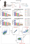
Direct inhibitory effect of THIO on MDSCs. A Schematic representation of MDSCs obtained by isolated stimulation from the bone marrow of hind leg tibia and femur of 8–10 week-old male C57 mice. B Viability of MDSCs after treatment with varying concentrations of THIO for 48 h and 96 h, assessed by the CCK‑8 assay (n = 3). C Flow cytometric analysis of the effect of different THIO concentrations, applied to bone marrow precursor cells for 48 h and 96 h, on subsequent MDSCs differentiation (n = 3). D Flow cytometry gating strategy for identifying MDSCs derived from bone marrow precursor cells. Data are shown as the mean ± standard deviation (SD). Representative results from 2–3 independent experiments. Statistical significance was calculated via one-way analysis of variance (ANOVA) followed by Tukey’s multiple comparisons. ***p < 0.001, ****p < 0.0001; ns, not significant. Figure A created with BioRender.com
Fig. 6
Fig. 6
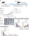
Antitumor immune responses and effects of THIO treatment in combination with BBV against an established TC-1 tumor model. A Flowchart showing the treatment of THIO in combination with BBV. B Pictures of collected tumor masses (n = 8). C Growth curve of tumor (n = 8). D Tumor weight (n = 8). E Flow cytometry analysis of the proportion of MDSCs and CTLs in splenocytes (n = 5), with MDSCs (CD11b+Gr-1+) on the left and CTLs (CD3+CD8+IFN-γ+) on the right. G ELISPOT analysis of IFN-γ-secreting lymphocytes (n = 3). Left panel, statistical graph; right panel, representative images. Data are shown as mean ± standard deviation (SD). The mouse experiments were performed in two independent replicates. Statistical significance was calculated by two-way analysis of variance (ANOVA) and via one-way analysis of variance (ANOVA) followed by Tukey’s multiple comparisons. *p < 0.05, ** p < 0.01, ***p < 0.001, ****p < 0.0001, ns, not significant. Figure A created with BioRender.com
Fig. 7
Fig. 7
Antitumor immune responses and effects of THIO in combination with a nanovaccine Q11-E744-62 in an established TC-1 tumor model. A Flowchart showing the treatment of THIO and Q11-E744-62 nanofibers. B Dynamic surveillance of tumor volume. C Pictures of the collected tumor masses (n = 5). D Tumor weight (n = 5). E Spleen weight (n = 5). F Flowcytometry analyses on the ratios of CTLs (CD3+CD8+IFN-γ+) and MDSCs (CD11b+Gr-1+) in splenocytes (n = 3). G ELISPOT analysis of IFN-γ-secreting lymphocytes in spleen. Left, representative images; right, statistical graph (n = 3). Data are shown as mean ± standard deviation (SD). The mouse experiments were performed in two independent replicates. Statistical significance was calculated by one-way ANOVA. *p < 0.05, **p < 0.01, ***p < 0.001, ****p < 0.0001; ns, not significant. Figure A created with BioRender.com
Fig. 8
Fig. 8
THIO in combination with the nanovaccine Q11-M30 elicits effective antitumor immune responses and effects in an established “immune cold” tumor model of B16-F10 melanoma. A Flowchart showing the treatment of THIO and Q11-M30 nanofibers. B Dynamic monitoring of tumor growth (n = 6). C Pictures of collected tumor masses (n = 6). D Flowcytometry analyses on the ratios of CTLs (CD3+CD8+IFN-γ+) and MDSCs (CD11b+Gr-1+) in splenocytes (n = 3). E Flowcytometry analyses on the ratios of MDSCs (CD11b+Gr-1+) in tumor (n = 3). Data are shown as mean ± standard deviation (SD). The mouse experiments were performed in two independent replicates. Statistical significance was calculated by one-way ANOVA. *p < 0.05, **p < 0.01, ***p < 0.001, ****p < 0.0001; ns, not significant. Figure A created with BioRender.com

Similar articles

References

    1. Sung H, Ferlay J, Siegel RL, Laversanne M, Soerjomataram I, Jemal A, Bray F. Global cancer statistics 2020: GLOBOCAN estimates of incidence and mortality worldwide for 36 cancers in 185 countries. CA Cancer J Clin. 2021;71(3):209–49. - PubMed
    1. Sugarman ET, Zhang G, Shay JW. In perspective: an update on telomere targeting in cancer. Mol Carcinog. 2019;58(9):1581–8. - PMC - PubMed
    1. Mizukoshi E, Kaneko S. Telomerase-targeted cancer immunotherapy. Int J Mol Sci. 2019;20(8):1823. - PMC - PubMed
    1. Man RJ, Chen LW, Zhu HL. Telomerase inhibitors: a patent review (2010–2015). Expert Opin Ther Pat. 2016;26(6):679–88. - PubMed
    1. Mender I, LaRanger R, Luitel K, Peyton M, Girard L, Lai TP, Batten K, Cornelius C, Dalvi MP, Ramirez M, Du W, Wu LF, Altschuler SJ, Brekken R, Martinez ED, Minna JD, Wright WE, Shay JW. Telomerase-mediated strategy for overcoming non-small cell lung cancer targeted therapy and chemotherapy resistance. Neoplasia. 2018;20(8):826–37. - PMC - PubMed

LinkOut - more resources