. 2025 Jul 1;38(3):267-281.
doi: 10.3344/kjp.24419. Epub 2025 Jun 9.

Effect of glucagon-like peptide-1 receptor agonist on paclitaxel induced neurotoxicity in dorsal root ganglion neuronal cells in vitro

Affiliations

Effect of glucagon-like peptide-1 receptor agonist on paclitaxel induced neurotoxicity in dorsal root ganglion neuronal cells in vitro

Younghoon Jung et al. Korean J Pain. .

Abstract

Background: Chemotherapy is the basis of cancer treatment. Chemotherapy-induced peripheral neuropathy (CIPN) is a major adverse effect. CIPN has a high incidence rate and substantially affects the quality of life of cancer survivors. Despite this, no definitive treatment for persistent unmet medical needs is available. Glucagon-like peptide-1 receptor agonists (GLP-1RA), widely used to treat obesity and diabetes, have recently been reported to be efficacious in treating neuropathic pain. The authors aimed to confirm its effects on CIPN and elucidate its underlying mechanisms.

Methods: After differentiation, 50B11 dorsal root ganglion cells were treated with paclitaxel and GLP-1RA to confirm changes in oxidative stress, neuroinflammation, and neuronal damage. Immunofluorescence, flow cytometry, Western blotting, quantitative reverse transcription-polymerase chain reaction, cytokine quantitation by ELISA, and assessments of axonal degeneration and regeneration were performed to evaluate the effect of GLP-1RA on CIPN and confirm the associated mechanisms.

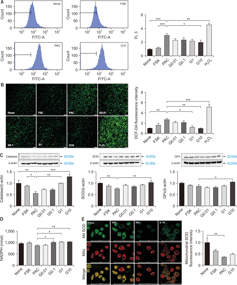
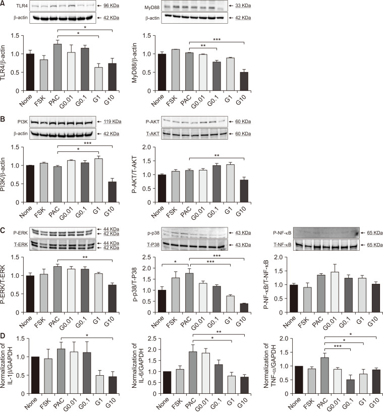
Results: After treatment with paclitaxel, the increased oxidative stress and inflammatory signals decreased with the administration of GLP-1RA. GLP-1RA also led to an increase in β-endorphin and μ-opioid receptors through IL-10. And administration of GLP-1RA had the effect of increasing neurite length. Additionally, the increased expression of the TRP family induced by paclitaxel treatment was restored with GLP-1RA administration.

Conclusions: GLP-1RA reduces oxidative stress and neuroinflammation, thereby alleviating paclitaxel-induced neurotoxicity. Additionally, GLP-1RA increases β-endorphin and μ-opioid receptors and reduces TRP family expression and promotes neuroregeneration, suggesting its effectiveness in mitigating chemotherapy-induced neuropathic pain.

Keywords: Drug Therapy; Exenatide; Ganglia; Glucagon-Like Peptide 1; Nerve Regeneration; Neuroinflammatory Diseases; Oxidative Stress; Paclitaxel; Spinal; beta-Endorphin.


Conflict of interest statement

CONFLICT OF INTEREST

Eunsoo Kim is a section editor of the Korean Journal of Pain. However, he was not involved in the selection of peer reviewers, the evaluation, or the decision-making process for this article.

Figures

Fig. 1
Fig. 1
Cytotoxicity assay of Forskolin + Paclitaxel (uM).
Fig. 2
Fig. 2
None treated cell for 24 hr. ROS: reactive oxygen species, FITC: fluorescein isothiocyanate.
Fig. 3
Fig. 3
Paclitaxel treated cell for 24 hr. ROS: reactive oxygen species, PAC: paclitaxel, FITC: fluorescein isothiocyanate.
Fig. 4
Fig. 4
Effect of GLP-1RA on oxidative stress in paclitaxel-treated 50B11 DRG neuronal cell lines. Flow cytometry (A) and fluorescence microscopy (B) assessment of ROS production. Wild-type 50B11 cells were generated and left unstained or stained with DCF-DA. A single gate was generated by plotting FITC (x-axis) vs. SSC (y-axis). ROS production was significantly increased following paclitaxel inhibition by GLP-1RA in 10 ng/mL concentration. (C) Western blot analysis of antioxidant enzymes. GPx, SOD, and catalase are prominent antioxidant enzymes. The protein levels represent the density ratio of each protein band to the β-actin band. (D) Evaluation of intracellular NADPH using the glutathione peroxidase assay. GPx activity was dose-dependently increased by GLP-1RA treatment in paclitaxel-treated neuronal cell lines. (E) Fluorescence microscopy analysis of mitochondria and mitochondrial superoxide levels. Data are shown as mean ± standard deviation. n = 3. *P < 0.05, **P < 0.01, ***P < 0.001. Scale bar = 100 um. FSK: Forskolin, PAC: Forskolin + Paclitaxel, G0.01: Forskolin + Paclitaxel + GLP-1RA 0.01 µg/mL (treated 24 hr after paclitaxel treatment), G0.1: Forskolin + Paclitaxel + GLP-1RA 0.1 µg/mL (treated 24 hr after paclitaxel treatment), G1: Forskolin + Paclitaxel + GLP-1RA 1 µg/mL (treated 24 hr after paclitaxel treatment), G10: Forskolin + Paclitaxel + GLP-1RA 10 µg/mL (treated 24 hr after paclitaxel treatment), GLP-1RA: glucagon like peptide-1 receptor agonist, DRG: dorsal root ganglion, ROS: reactive oxygen species, DCF-DA: 2,7-dichlorofluorescein diacetate, FITC: fluorescein isothiocyanate, SSC: side scatter, GPx: glutathione peroxidase, SOD: superoxide dismutase, NADPH: nicotinamide adenine dinucleotide phosphate.
Fig. 5
Fig. 5
The effect of GLP-1RA on the expression of proteins related to cell signaling and cytokines related to inflammation in paclitaxel-treated 50b11 DRG neuronal cell line. Western blot analysis of TLR4 and MyD88 (A), phospho ERK, p38 and NF-κB (B), and PI3K and phosphor AKT (C). Protein levels represented a density ratio of each protein band to the β-actin bands. (D) QPCR analysis of pro-inflammatory cytokines. GLP-1RA significantly inhibited IL-1β, IL-6, and TNF-α in paclitaxel-treated cells. Data are shown as mean ± standard deviation. n = 3. *P < 0.05, **P < 0.01, ***P < 0.001. Scale bar = 100 um. FSK: Forskolin, PAC: Forskolin + Paclitaxel, G0.01: Forskolin + Paclitaxel + GLP-1RA 0.01 µg/mL (treated 24 hr after paclitaxel treatment), G0.1: Forskolin + Paclitaxel + GLP-1RA 0.1 µg/mL (treated 24 hr after paclitaxel treatment), G1: Forskolin + Paclitaxel + GLP-1RA 1 µg/mL (treated 24 hr after paclitaxel treatment), G10: Forskolin + Paclitaxel + GLP-1RA 10 µg/mL (treated 24 hr after paclitaxel treatment), GLP-1RA: glucagon like peptide-1 receptor agonist, DRG: dorsal root ganglion, TLR4: Toll-like receptor 4, MyD88: myeloid differentiation primary response 88, ERK: extracellular signal regulated kinase, NF-κB: nuclear factor kappa-light chain-enhancer of activated B cells, PI3K: phosphoinositide 3-kinase, AKT: protein kinase B, IL-1β: interleukin-1β, IL-6: interleukin-6, TNF-α: tumor necrosis factor-α, GAPDH: glyceraldehyde 3-phosphate dehydrogenase.
Fig. 6
Fig. 6
The effect of GLP-1RA on IL-10 and β-endorphin in paclitaxel-treated 50B11 DRG neuronal cell line. (A) QPCR analysis for IL-10 mRNA level and ELISA assay for secreted IL-10. IL-10 mRNA expression and secreted IL-10 were significantly decreased by paclitaxel, which was restored by GLP-1RA in the 10 ng/mL concentration. (B) QPCR analysis and immunofluorescence assay of POMC and β-endorphin. (C) QPCR analysis and immunofluorescence assay of μ-opioid receptor. Data are shown as mean ± standard deviation. n = 3. *P < 0.05, **P < 0.01, ***P < 0.001. Scale bar = 100 um. FSK: Forskolin, PAC: Forskolin + Paclitaxel, G0.01: Forskolin + Paclitaxel + GLP-1RA 0.01 µg/mL (treated 24 hr after paclitaxel treatment), G0.1: Forskolin + Paclitaxel + GLP-1RA 0.1 µg/mL (treated 24 hr after paclitaxel treatment), G1: Forskolin + Paclitaxel + GLP-1RA 1 µg/mL (treated 24 hr after paclitaxel treatment), G10: Forskolin + Paclitaxel + GLP-1RA 10 µg/mL (treated 24 hr after paclitaxel treatment), GLP-1RA: glucagon like peptide-1 receptor agonist, IL-10: interleukin-10, DRG: dorsal root ganglion, POMC: pro-opiomelanocortin, MOR: μ-opioid receptor, GAPDH: glyceraldehyde 3-phosphate dehydrogenase.
Fig. 7
Fig. 7
The effect of GLP-1RA on the expression of pain receptor, level of intracellular calcium and cAMP, and length of neurite in paclitaxel-treated 50b11 DRG neuronal cell line. (A) Western blot analysis of TRPV1 and TRPA1. Pain receptor expression was significantly increased by paclitaxel, inhibited by GLP-1RA in 10 ng/mL. Protein levels represented a density ratio of each protein band to the β-actin bands. (B) Calcium detection assay in the cytosol. The level of intracellular calcium concentration was increased by paclitaxel, which GLP-1RA inhibited. (C) Cyclic AMP ELISA. The cyclic AMP ELISA is a competitive immunoassay for quantitatively determining extracellular cyclic AMP in culture supernatant. The level of secreted cAMP was increased by paclitaxel, inhibited by GLP-1RA in the concentration of 10 ng/mL. (D) Immunofluorescence assay of beta III tubulin (constitutively expressed in the central and peripheral nervous systems) with GAP43 (a nervous system-specific, growth-associated protein enriched in growth cones). Data are shown as mean ± standard deviation. n = 3. *P < 0.05, ***P < 0.001. Scale bar = 100 um. FSK: Forskolin, PAC: Forskolin + Paclitaxel, G0.01: Forskolin + Paclitaxel + GLP-1RA 0.01 µg/mL (treated 24 hr after paclitaxel treatment), G0.1: Forskolin + Paclitaxel + GLP-1RA 0.1 µg/mL (treated 24 hr after paclitaxel treatment), G1: Forskolin + Paclitaxel + GLP-1RA 1 µg/mL (treated 24 hr after paclitaxel treatment), G10: Forskolin + Paclitaxel + GLP-1RA 10 µg/mL (treated 24 hr after paclitaxel treatment), GLP-1RA: glucagon like peptide-1 receptor agonist, DRG: dorsal root ganglion, TRPV1: transient receptor potential vanilloid 1, TRPA1: transient receptor potential ankyrin 1, GAP43: growth associated protein 43.
Fig. 8
Fig. 8
The shematic illustration of the effect of GLP-1RA in the paclitaxel-induced neurotoxicity model. GLP-1R: glucagon like peptide-1 receptor, TRPV1: transient receptor potential vanilloid 1, TRPA1: transient receptor potential ankyrin 1, PKC: protein kinase C, PI3K: phosphoinositide 3-kinase, AKT: protein kinase B, MAPK: mitogen-activated protein kinase, NF-κB: nuclear factor kappa-light chain-enhancer of activated B cells, IL-1β: interleukin-1β, IL-6: interleukin-6, TNF-α: tumor necrosis factor-α, NADPH: nicotinamide adenine dinucleotide phosphate, ROS: reactive oxygen species, IL-10: interleukin-10, POMC: pro-opiomelanocortin, STAT3: signal transducer and activator of transcription 3.

Similar articles

References

    1. Kang MJ, Won YJ, Lee JJ, Jung KW, Kim HJ, Kong HJ, et al. Community of Population-Based Regional Cancer Registries, author. Cancer Statistics in Korea: incidence, mortality, survival, and prevalence in 2019. Cancer Res Treat. 2022;54:330–44. doi: 10.4143/crt.2022.128. - DOI - PMC - PubMed
    1. Mols F, Beijers T, Vreugdenhil G, van de Poll-Franse L. Chemotherapy-induced peripheral neuropathy and its association with quality of life: a systematic review. Support Care Cancer. 2014;22:2261–9. doi: 10.1007/s00520-014-2255-7. - DOI - PubMed
    1. Zajączkowska R, Kocot-Kępska M, Leppert W, Wrzosek A, Mika J, Wordliczek J. Mechanisms of chemotherapy-induced peripheral neuropathy. Int J Mol Sci. 2019;20:1451. doi: 10.3390/ijms20061451. - DOI - PMC - PubMed
    1. Starobova H, Vetter I. Pathophysiology of chemotherapy-induced peripheral neuropathy. Front Mol Neurosci. 2017;10:174. doi: 10.3389/fnmol.2017.00174. - DOI - PMC - PubMed
    1. Pearce A, Haas M, Viney R, Pearson SA, Haywood P, Brown C, et al. Incidence and severity of self-reported chemotherapy side effects in routine care: a prospective cohort study. PLoS One. 2017;12:e0184360. doi: 10.1371/journal.pone.0184360. - DOI - PMC - PubMed

LinkOut - more resources