. 2025 Jun 8:19476035251344478.
doi: 10.1177/19476035251344478. Online ahead of print.

Mir-450a-5p Ameliorates IL-1β-Induced Chondrocyte Apoptosis, Inflammation, and Extracellular Matrix Degradation by Down-Regulating LITAF

Affiliations

Mir-450a-5p Ameliorates IL-1β-Induced Chondrocyte Apoptosis, Inflammation, and Extracellular Matrix Degradation by Down-Regulating LITAF

Guo-Feng Jia et al. Cartilage. .

Abstract

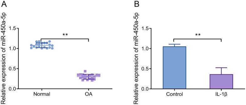
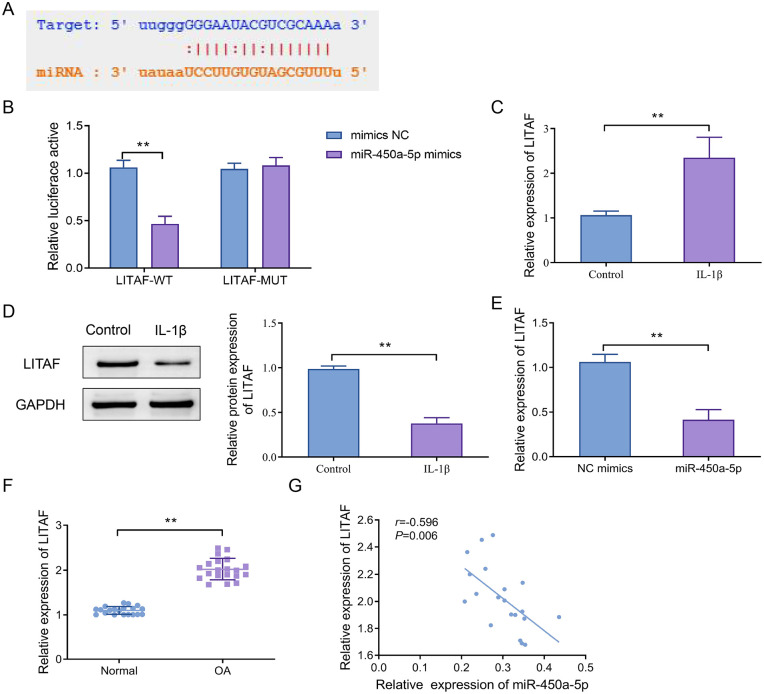
ObjectiveOsteoarthritis (OA) is a degenerative joint disease characterized by cartilage degradation, causing severe pain and disability. Recent studies suggest that miR-450a-5p may regulate inflammatory pathways in OA. This study aimed to elucidate the role of miR-450a-5p in OA, providing a potential therapeutic target for the clinical treatment.MethodsCartilage tissues were collected from OA patients undergoing knee replacement surgery, and CHON-001 cells were treated with interleukin (IL)-1β to induce an OA model in vitro. Real-time quantitative polymerase chain reaction was used to detect the miR-450a-5p expression, and Western blot determined the lipopolysaccharide-induced tumor necrosis factor (TNF)-α factor (LITAF) expression. The targeting relationship between LITAF and miR-450a-5p was verified by dual-luciferase reporter assay. Cell proliferation and apoptosis were assessed using the Cell Counting Kit-8 assay and flow cytometry, respectively. Levels of IL-6, IL-10, and TNF-α were measured via enzyme-linked immunosorbent assay. In addition, Western blot was employed to detect the expressions of matrix metalloproteinase-3 (MMP-3), collagen III, and aggrecan in extracellular matrix (ECM).ResultsMiR-450a-5p expression was significantly down-regulated in OA tissues and IL-1β-induced CHON-001 cells (~60%), while LITAF expression was markedly increased (~1.8-fold). There was a negative correlation between miR-450a-5p and LITAF in OA tissues (r = -0.596, P < 0.01). MiR-450a-5p directly targeted and inhibited LITAF expression. Its overexpression promoted chondrocyte proliferation, reduced apoptosis and inflammatory cytokines, and mitigated ECM degradation.ConclusionsMiR-450a-5p inhibited LITAF expression, thereby attenuating apoptosis, inflammation, and ECM degradation in chondrocytes. It may serve as a promising therapeutic target for OA.

Keywords: CHON-001; IL-1β; LITAF; chondrocytes; miR-450a-5p; osteoarthritis.


Conflict of interest statement

The author(s) declared no potential conflicts of interest with respect to the research, authorship, and/or publication of this article.

Figures

Figure 1.
Figure 1.
MiR-450a-5p expression is significantly elevated in cartilage tissues and IL-1β-induced chondrocytes. (A) RT-qPCR was used to detect the miR-450a-5p expression level of cartilage tissues in OA patients and non-OA patients, **P < 0.01, normal group versus OA group. (B) RT-qPCR was performed to measure the miR-450a-5p expression level in IL-1β-treated CHON-001 cells, **P < 0.01. IL-1β = interleukin-1β; OA = osteoarthritis; RT-qPCR = real-time quantitative polymerase chain reaction.
Figure 2.
Figure 2.
MiR-450a-5p inhibits IL-1β-induced apoptosis, inflammation, and extracellular matrix degradation in chondrocytes. (A) The mRNA levels of miR-450a-5p were examined by RT-qPCR, **P < 0.01, miR-450a-5p group versus NC mimics group; (B) cell proliferation at 0, 24, 48, and 72 hours was detected by CCK-8 assay. (C) Flow cytometry was performed to assess the apoptosis level of CHON-001 cells; (D) ELISA was used to determine the levels of IL-6, IL-18, and TNF-α; (E) Western blot was utilized to detect the protein expression levels of MMP-3, collagen III, and aggrecan; **P < 0.01, IL-1β group versus control group; ##P < 0.01, IL-1β + miR-450a-5p group versus IL-1β + NC mimics group. IL-1β = interleukin-1β; ECM = extracellular matrix; RT-qPCR = real-time quantitative polymerase chain reaction; CCK-8 = cell counting kit-8; ELISA = enzyme-linked immunosorbent assay; IL = interleukin; TNF-α = tumor necrosis factor-α; MMP-3 = matrix metalloproteinase-3.
Figure 3.
Figure 3.
LITAF serves as a direct target of miR-450a-5p. (A) The target sequences for miR-450a-5p binding to LITAF were predicted; (B) a dual-luciferase reporter assay was used to validate the binding of miR-450a-5p to LITAF, **P < 0.01; (C) RT-qPCR was utilized to detect the LITAF mRNA level in CHON-001 cells; (D) Western blot was adopted to determine the protein level of LITAF in CHON-001 cells, **P < 0.01; (E) the expression level of LITAF was measured after transfection with miR-450a-5p, **P < 0.01; (F) RT-qPCR was used to detect the LITAF mRNA level in cartilage tissues of OA patients, **P < 0.01; (G) the correlation between the expression of miR-450a-5p and LITAF was assessed by the Pearson analysis. LITAF = lipopolysaccharide-induced tumor necrosis factor-α factor; RT-qPCR = real-time quantitative polymerase chain reaction; OA = osteoarthritis.
Figure 4.
Figure 4.
LITAF reduces the protective effect of miR-450a-5p on IL-1β-induced chondrocytes. (A) Western blot was used to detect the changes in LITAF protein expression; (B) the proliferation ability of CHON-001 cells was measured by CCK-8; (C–D) the apoptosis level of CHON-001 cells; (E) ELISA was adopted to determine the levels of IL-6, IL-18, and TNF-α; (F–G) Western blot and quantitative statistical analysis for the protein expression levels of MMP-3, collagen III, and aggrecan, **P < 0.01 versus NC mimics + vector group, ##P < 0.01, versus miR-450a-5p + vector group. TNF-α = tumor necrosis factor-α; LITAF = lipopolysaccharide-induced TNF-α factor; IL = interleukin; CCK-8 = cell counting kit-8; ELISA = enzyme-linked immunosorbent assay; MMP-3 = matrix metalloproteinase-3.
Figure 5.
Figure 5.
The diagram of potential mechanism by which miR-450a-5p targets and inhibits LITAF in OA.

Similar articles

References

    1. Cross M, Smith E, Hoy D, Nolte S, Ackerman I, Fransen M, et al. The global burden of hip and knee osteoarthritis: estimates from the global burden of disease 2010 study. Ann Rheum Dis. 2014;73(7):1323-30. doi: 10.1136/annrheumdis-2013-204763. - DOI - PubMed
    1. Ye H, He Y, Zheng C, Wang F, Yang M, Lin J, et al. Type 2 diabetes complicated with heart failure: research on therapeutic mechanism and potential drug development based on insulin signaling pathway. Front Pharmacol. 2022;13:816588. doi: 10.3389/fphar.2022.816588. - DOI - PMC - PubMed
    1. Vina ER, Kwoh CK. Epidemiology of osteoarthritis: literature update. Curr Opin Rheumatol. 2018;30(2):160-7. doi: 10.1097/BOR.0000000000000479. - DOI - PMC - PubMed
    1. Yunus MHM, Nordin A, Kamal H. Pathophysiological perspective of osteoarthritis. Medicina (Kaunas). 2020;56(11):614. doi: 10.3390/medicina56110614. - DOI - PMC - PubMed
    1. Liu-Bryan R, Terkeltaub R. Emerging regulators of the inflammatory process in osteoarthritis. Nat Rev Rheumatol. 2015;11(1):35-44. doi: 10.1038/nrrheum.2014.162. - DOI - PMC - PubMed

LinkOut - more resources