

Outcomes and Surgical Approaches for Pineal Region Tumors in Adults: A Retrospective Study of a Single-Center Over 12 Years
- PMID: 40485791
- PMCID: PMC12136960
- DOI: 10.1055/s-0044-1801372
Outcomes and Surgical Approaches for Pineal Region Tumors in Adults: A Retrospective Study of a Single-Center Over 12 Years
Abstract
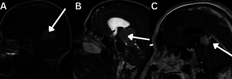
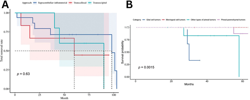
Background Pineal region tumors are considered rare, deeply located, and very difficult to resect. They can cause various symptoms by compressing and obstructing different structures. Contradictory data have been reported regarding various aspects of surgical outcomes in different patient positioning. Objectives This retrospective study aimed to describe the variety of pineal region tumors and patient positioning in pineal region surgeries and compare the neurological outcomes during different approaches. Materials and Methods From January 1, 2010, to December 31, 2022, 61 patients with pineal area tumors were hospitalized at the National Center for Neurosurgery. Thirty-five patients' histology examinations were available. Twenty-nine patients had open surgical excision. Regarding approaches, supracerebellar infratentorial, posterior transfalcine interhemispheric, and occipital transtentorial approaches were employed. Results Among 35 patients, 17 had hydrocephalus and required ventricular drainage to address third ventricle obstruction. Complete tumor resection was achieved in 55% of patients. The mortality rate was 13.7% in the open surgical group and 15.625% in the endoscopic third ventriculostomy (ETV) group. Conclusion Proper patient positioning and selecting the optimal approach are crucial for a successful outcome.
Keywords: germ cell tumor; occipital transtentorial approach; oncology; pineal biopsy; pineal region tumor; supracerebellar approach.
Asian Congress of Neurological Surgeons. This is an open access article published by Thieme under the terms of the Creative Commons Attribution-NonDerivative-NonCommercial License, permitting copying and reproduction so long as the original work is given appropriate credit. Contents may not be used for commercial purposes, or adapted, remixed, transformed or built upon. ( https://creativecommons.org/licenses/by-nc-nd/4.0/ ).
Conflict of interest statement
Conflict of Interest None declared.
Figures


Similar articles
-
Neurosurgical application of pineal region tumor resection with 3D 4K exoscopy via infratentorial approach: a retrospective cohort study.Int J Surg. 2023 Dec 1;109(12):4062-4072. doi: 10.1097/JS9.0000000000000707. Int J Surg. 2023. PMID: 37755386 Free PMC article.
-
Good clinical outcomes and the necessity of CSF drainage in patients undergoing simultaneous biopsy and endoscopic third ventriculostomy in the region of pineal tumors: A systematic review and meta-analysis.J Clin Neurosci. 2024 Aug;126:234-244. doi: 10.1016/j.jocn.2024.07.001. Epub 2024 Jul 5. J Clin Neurosci. 2024. PMID: 38970969
-
Supracerebellar highway-fast and safe road to pediatric pineal tumor resection.Childs Nerv Syst. 2025 Aug 13;41(1):260. doi: 10.1007/s00381-025-06919-w. Childs Nerv Syst. 2025. PMID: 40802062
-
Endoscopic third ventriculostomy (ETV) for idiopathic normal pressure hydrocephalus (iNPH).Cochrane Database Syst Rev. 2015 Jul 29;2015(7):CD010033. doi: 10.1002/14651858.CD010033.pub2. Cochrane Database Syst Rev. 2015. PMID: 26222251 Free PMC article.
-
Prescription of Controlled Substances: Benefits and Risks.2025 Jul 6. In: StatPearls [Internet]. Treasure Island (FL): StatPearls Publishing; 2025 Jan–. 2025 Jul 6. In: StatPearls [Internet]. Treasure Island (FL): StatPearls Publishing; 2025 Jan–. PMID: 30726003 Free Books & Documents.
References
-
- Abecassis I J, Hanak B W, Ellenbogen R G. Philadelphia, PA: Elsevier;; 2018. Pineal region tumors; pp. 602–62100.
-
- Simon E, Afif A, M'Baye M, Mertens P.Anatomy of the pineal region applied to its surgical approach Neurochirurgie 201561(2–3):70–76. - PubMed
-
- Feigl G C, Britz G, Staribacher D, Kuzmin D. The minimally invasive lateral occipital infracortical supra-/transtentorial approach in surgery of lesions of the pineal region: a possible alternative to the standard approaches. World Neurosurg. 2023;172:e151–e164. - PubMed
-
- Ferrer E, Santamarta D, Garcia-Fructuoso G, Caral L, Rumià J.Neuroendoscopic management of pineal region tumours Acta Neurochir (Wien) 19971390112–20., discussion 20–21 - PubMed
LinkOut - more resources
Full Text Sources