

Brain Abscess Mimicking Brain Tumors: A Systematic Review of Individual Patient's Data
- PMID: 40485794
- PMCID: PMC12136936
- DOI: 10.1055/s-0045-1802623
Brain Abscess Mimicking Brain Tumors: A Systematic Review of Individual Patient's Data
Abstract
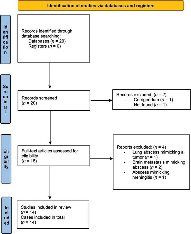
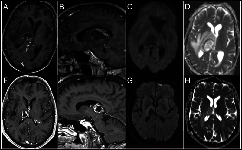
Objectives Brain abscess is a worrisome condition with a 1-year mortality rate of 21% and a 32% rate of new-onset epilepsy. Brain magnetic resonance imaging (MRI) is strongly recommended as a screening modality with contrast-enhanced T1-weighted images, diffusion-weighted imaging (DWI), and attenuated diffusion coefficient. However, there is a 10% rate of false negative, which could potentially impact management and prognosis. Our systematic review aims at identifying risk factors for false negative. Materials and Methods A database search of our institutions plus a systematic literature review was conducted using MEDLINE/PubMed, including studies of brain abscesses misdiagnosed as brain tumors. Data on patient demographics, clinical presentations, imaging findings, pathogens, treatments, and outcomes were extracted and analyzed. We present a case of a 59-year-old male with HIV, who developed a brain abscess misdiagnosed as a tumor. Initial symptoms included left-side weakness and weight loss. Imaging showed a ring-enhancing lesion in the right thalamus. The abscess was caused by T. gondii , and the patient was treated with sulfadiazine, pyrimethamine, ceftriaxone, and metronidazole, achieving a GOS-E score of 8 at 1 year. Results The review included 14 studies, with 1 additional illustrative case, encompassing a total of 15 cases. Patients ranged from 39 to 77 years, with a mean age of 59 years. Comorbidities included human immunodeficiency virus (HIV), glioblastoma, breast cancer, arthritis, gastric cancer, and nephrotic syndrome. Common symptoms were hemiparesis, generalized seizures, headache, and confusion. Imaging often revealed ring-enhancing lesions with restricted diffusion on DWI. Lesions were located in various brain regions. Pathogens identified included 40% Nocardia species, Toxoplasma gondii , Mycobacterium tuberculosis , Aggregatibacter aphrophilus , Rickettsia typhi , Arcanobacterium haemolyticum , Aspergillus terreus , and Providencia rettgeri . Treatments involved antibiotics and, in some cases, surgical intervention. Outcomes measured by the Glasgow Outcome Scale-Extended (GOS-E) at 1 year indicated good recovery in most cases. Conclusion Despite the high sensitivity and specificity of brain MRI in diagnosing brain abscesses, the standard protocol used for the past two decades still results in a 10% false-negative rate. Such inaccuracies can significantly impact the patient's management, potentially delaying antibiotic therapy and impacting the surgical planning, hence affecting the outcome. Immunocompromised patients are particularly vulnerable to misdiagnoses of brain abscesses as brain tumors. To improve diagnostic accuracy, new imaging techniques and computational tools are currently under investigation.
Keywords: MRI; brain abscess; case reports; mimicking; nocardia; systematic review; toxoplasmosis; tumor.
Asian Congress of Neurological Surgeons. This is an open access article published by Thieme under the terms of the Creative Commons Attribution-NonDerivative-NonCommercial License, permitting copying and reproduction so long as the original work is given appropriate credit. Contents may not be used for commercial purposes, or adapted, remixed, transformed or built upon. ( https://creativecommons.org/licenses/by-nc-nd/4.0/ ).
Conflict of interest statement
Conflict of Interest None declared.
Figures
Similar articles
-
Folic acid supplementation and malaria susceptibility and severity among people taking antifolate antimalarial drugs in endemic areas.Cochrane Database Syst Rev. 2022 Feb 1;2(2022):CD014217. doi: 10.1002/14651858.CD014217. Cochrane Database Syst Rev. 2022. PMID: 36321557 Free PMC article.
-
Vesicoureteral Reflux.2024 Apr 30. In: StatPearls [Internet]. Treasure Island (FL): StatPearls Publishing; 2025 Jan–. 2024 Apr 30. In: StatPearls [Internet]. Treasure Island (FL): StatPearls Publishing; 2025 Jan–. PMID: 33085409 Free Books & Documents.
-
Spinal Epidural Abscess.J Educ Teach Emerg Med. 2020 Jan 15;5(1):S26-S52. doi: 10.21980/J8T938. eCollection 2020 Jan. J Educ Teach Emerg Med. 2020. PMID: 37465609 Free PMC article.
-
Case report: Cryptogenic giant brain abscess caused by Providencia rettgeri mimicking stroke and tumor in a patient with impaired immunity.Front Neurol. 2022 Sep 23;13:1007435. doi: 10.3389/fneur.2022.1007435. eCollection 2022. Front Neurol. 2022. PMID: 36212658 Free PMC article.
-
Diffusion-weighted MR imaging in the preoperative assessment of brain abscesses.Surg Neurol. 2002 Dec;58(6):395-402; discussion 402. doi: 10.1016/s0090-3019(02)00929-1. Surg Neurol. 2002. PMID: 12517619 Review.
References
-
- Bodilsen J, Dalager-Pedersen M, van de Beek D, Brouwer M C, Nielsen H. Incidence and mortality of brain abscess in Denmark: a nationwide population-based study. Clin Microbiol Infect. 2020;26(01):95–100. - PubMed
-
- Bodilsen J, Dalager-Pedersen M, van de Beek D, Brouwer M C, Nielsen H. Long-term mortality and epilepsy in patients after brain abscess: a nationwide population-based matched cohort study. Clin Infect Dis. 2020;71(11):2825–2832. - PubMed
-
- Reddy J S, Mishra A M, Behari Set al.The role of diffusion-weighted imaging in the differential diagnosis of intracranial cystic mass lesions: a report of 147 lesions Surg Neurol 20066603246–250., discussion 250–251 - PubMed
-
- ESCMID Study Group for Infections of the Brain (ESGIB) . Bodilsen J, D'Alessandris Q G, Humphreys H et al.European Society of Clinical Microbiology and Infectious Diseases guidelines on diagnosis and treatment of brain abscess in children and adults. Clin Microbiol Infect. 2024;30(01):66–89. - PubMed
-
- Newman-Toker D E, Peterson S M, Badihian S . Rockville, MD: Agency for Healthcare Research and Quality (AHRQ); 2022. Diagnostic Errors in the Emergency Department: A Systematic Review [Internet] - PubMed
LinkOut - more resources
Full Text Sources
Miscellaneous