

doi: 10.1016/j.gendis.2025.101590.
eCollection 2025 Sep.
Otof gene transfer in DFNB9 mice carrying human founder non-truncating alleles
Affiliations
- PMID: 40485982
- PMCID: PMC12142502
- DOI: 10.1016/j.gendis.2025.101590
Item in Clipboard
Otof gene transfer in DFNB9 mice carrying human founder non-truncating alleles
Genes Dis.
.
No abstract available
Conflict of interest statement
Y.C., Q.A., S.K., N.P., T.G., L.B., J.G., M.K., M.B., L.S., M.D., and V.V. are or were employees of Decibel Therapeutics or Regeneron Pharmaceuticals at the time of contribution.
Figures
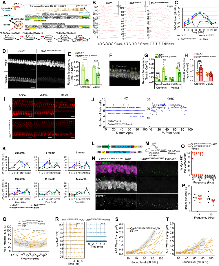
Audiological phenotype of Otofp.R1934Q/p.R1934Q KI mice and gene transfer therapy for treatment of auditory neuropathy spectrum disorder due to the Otof p.R1934Q. (A) Schematic depiction of the targeting strategy for the generation of Otofp.R1934Q/p.R1934Q KI mice. (B)Otofp.R1934Q/p.R1934Q mice did not show any meaningful ABR, while Otof+/+ and Otof+/p.R1934Q mice still showed normal ABR threshold at 3 months of age. (C)Otofp.R1934Q/p.R1934Q mice exhibited comparable DPOAE responses to that of Otof+/+ and Otof+/p.R1934Q mice at 5 weeks of age. (D) Z-projected confocal images of cochlear IHCs of indicated genotypes of Otof+/+ and Otofp.R1934Q/p.R1934Q mice were analyzed for comparison of normalized fluorescence intensity of otoferlin and Vglut3. (E) The level of otoferlin and Vglut3 protein expression was significantly reduced in Otofp.R1934Q/p.R1934Q mice. (G) The ratio of apical and basal protein levels. The ratios of otoferlin in Otofp.R1934Q/p.R1934Q mice versus Otof+/+ controls were significantly decreased. The decreased level of otoferlin was more pronounced at the basal part of IHCs. (H) The ratio of apical/basal protein levels of otoferlin and Vglut3. The ratio of apical otoferlin in Otofp.R1934Q/p.R1934Q mice was significantly increased compared with Otof+/+ controls, indicating that remaining otoferlin shifted apically. (D–H)Otof+/+, 8 IHCs from n = 2; Otofp.R1934Q/p.R1934Q, 16 IHCs from n = 4; scale bar = 20 μm; the data were presented as mean ± standard error; two-way analysis of variance with Bonferroni posttest; ∗∗∗p < 0.001. (I) Representative images of hair cells in apical (10%–20% from the apex), middle (45%–55% from the apex), and basal turns (75%–85% from the apex) from Otof+/+ and Otofp.R1934Q/p.R1934Q mice. IHCs and OHCs were immunolabeled with anti-myosin 6 (red). Scale bar = 10 μm. (J) Numbers of hair cells in a segment spanning 1% of the whole cochlear length. No hair cell loss of either IHCs or OHCs is shown in Otofp.R1934Q/p.R1934Q mice at 4 months of age. n = 5 for Otof+/+ and Otofp.R1934Q/p.R1934Q each group. (K) DPOAEs over time from each DFNB9 genotype. At 2 months of age, OAE responses still existed except those from 16 to 19 kHz. A substantial portion of OAE responses from Otofp.R1934Q/p.R1934Q mice was preserved until 5 months of age. The levels of the signals were L1 = 65 dB and L2 = 55 dB. The stars indicate significant differences between Otofp.R1934Q/p.R1934Q and Otof+/+; one-way ANOVA with Tukey's multiple comparison test; ∗p < 0.05 and ∗∗p < 0.01. (L) Schematic of the dual hybrid vector system with mOtof coding sequences. (M) Dual vectors were injected through the round window in mature mice (4–6 weeks of age). (N) Confocal immunofluorescence imaging of IHCs in AAV- and vehicle-treated Otofp.R1934Q/p.R1934Q mice with anti-Myc tag and Homer co-labeling. Scale bar = 10 μm. (O) Percentage of Myc+ IHCs across frequency regions. Most animals showed > 50% of IHCs with Myc signal. (P) Homer puncta count per IHC. The horizontal bars indicate the median, and the open symbols reflect treated animals without Myc-signal for (O) and (P). The open symbols indicate animals with no Myc-otoferlin detected. (Q) ABR thresholds of all individual animals treated with dual AAV vectors (n = 8) and vehicle (n = 4). All animals treated with AAV vectors showed detectable ABR thresholds, with most animals in normal hearing range (the grey shaded areas indicate the 90% interval of observed thresholds in normal hearing C57BL/6 mice; the black line indicates the mean). In contrast, animals treated with the vehicle only show no detectable ABRs below the equipment limit. (R) ABR at 4 weeks post-treatment in AAV- and vehicle-treated Otofp.R1934Q/p.R1934Q mice. ABR waveforms in response to 16 kHz tone bursts were restored in AAV-treated mice, whereas vehicle-treated mice showed no detectable ABRs. (S) ABR wave I amplitude growth curves of individual animals. (T) ABR wave II amplitude growth curves of individual animals. The grey shaded area in panels (R–T) indicates the mean and standard deviation of wave amplitudes in Otof+/+ mice. Wave I amplitudes were smaller than those of normal hearing mice, whereas wave II amplitudes were comparable to those of normal hearing mice. The dashed lines indicate mice (n = 3) with no Myc-otoferlin detection. ABR threshold and amplitude recovery in these mice were poorer than the mice with robust expression. gRNA, guide RNA; KI, knock-in; mRNA, messenger RNA; mOtof, mouse otoferlin; ssODNs, single-stranded oligodeoxynucleotides; ABR, auditory brainstem response; DPOAE, distortion product otoacoustic emission; SPL, sound pressure level; IHC, inner hair cell; SE, standard error; Vglut3, vesicular glutamate transporter 3; OHC, outer hair cell; OAE, otoacoustic emission; AAV, adeno-associated virus.
References
-
- Roux I., Safieddine S., Nouvian R., et al. Otoferlin, defective in a human deafness form, is essential for exocytosis at the auditory ribbon synapse. Cell. 2006;127(2):277–289. - PubMed
LinkOut - more resources
Full Text Sources
