. 2025 Feb 22;7(6):101366.
doi: 10.1016/j.jhepr.2025.101366. eCollection 2025 Jun.

Altered liver sinusoidal endothelial cells in MASLD and their evolution following lanifibranor treatment

Affiliations

Altered liver sinusoidal endothelial cells in MASLD and their evolution following lanifibranor treatment

Pierre-Emmanuel Rautou et al. JHEP Rep. .

Abstract

Background & aims: Data on changes in liver sinusoidal endothelial cells (LSECs) in patients with metabolic dysfunction-associated steatotic liver disease (MASLD) and their response to treatment are limited. This study aimed at determining (i) features associated with LSEC capillarisation in patients with MASLD; (ii) whether LSEC changes can regress with the pan-peroxisome proliferator-activated receptor (PPAR) agonist lanifibranor; (iii) the role of the different PPAR isotypes on LSEC changes in MASLD.

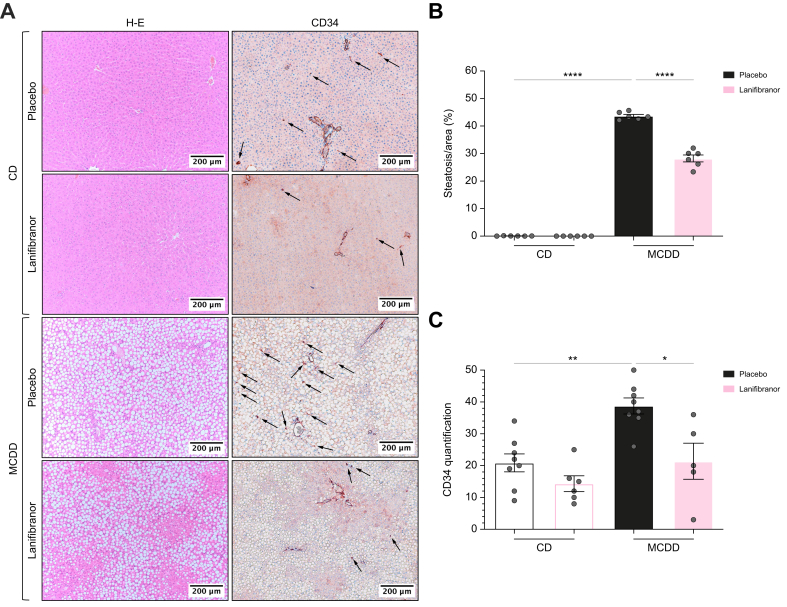
Methods: We analysed CD34 expression, a marker of LSEC capillarisation, on liver biopsies from patients considered for inclusion in the NATIVE trial at baseline (n = 249), and after 24 weeks of placebo or lanifibranor (n = 173). Two rat models of MASLD were used to investigate the effect of lanifibranor or of mono-PPAR agonists on LSECs.

Results: Lobular CD34 staining was more intense in patients with isolated steatosis than in those with no MASLD (52% vs. 10%; p = 0.03). In the overall cohort, this staining was more intense in patients with metabolic dysfunction-associated steatohepatitis (MASH) than in those without (63% vs. 41%; p = 0.01) and strongly correlated with liver fibrosis and to a lesser extent with liver inflammation. Lanifibranor treatment was associated with more common improvement in CD34 periportal staining (p = 0.025), and less frequent worsening of lobular staining (p = 0.028). Compared with healthy rats, rats with MASLD had higher CD34 staining, portal venous pressure, intrahepatic vascular resistance, and impaired liver endothelial function. Lanifibranor normalised or strongly improved these abnormalities, whereas mono-PPAR agonists caused partial improvements.

Conclusions: In patients, LSEC capillarisation was increased at the earliest stages of MASLD and was associated with liver fibrosis and inflammation. In both patients and rats with MASLD, lanifibranor treatment was associated with improvement in liver endothelial phenotype.

Impact and implications: Data on changes in liver sinusoidal endothelial cells (LSECs) in patients with metabolic dysfunction-associated steatotic liver disease (MASLD) and their response to treatment are limited. This study demonstrates that LSEC capillarisation is already present in the lobular zone of the liver of patients and rats at the stage of isolated steatosis, before metabolic dysfunction-associated steatohepatitis (MASH) onset, and progresses with liver fibrosis, and to a lesser extent with liver inflammation. Lanifibranor treatment, a pan-peroxisome proliferator-activated receptor agonist currently tested in a phase III clinical trial, improves LSEC capillarisation but also intrahepatic vascular resistance and portal pressure in MASLD. Targeting LSECs appears to be a promising approach to improve MASH.

Keywords: CD34; Intrahepatic vascular resistance; LSECs; Liver fibrosis; Liver inflammation; MASH; PPAR; Vascular biology; lanifibranor.


Conflict of interest statement

P-ER has received research funding from Terrafirma and acted as consultant for Mursla, Genfit, Boehringer Ingelheim, Cook, Jazz, and Abbelight, and received speaker fees from AbbVie. SMF has been lecturer for AbbVie, Allergan, Bayer, Eisai, Genfit, Gilead Sciences, Janssens Cilag, Intercept, Inventiva, Merck Sharp & Dome, Novo Nordisk, Promethera, Siemens. He has acted as consultant for AbbVie, Actelion, Aelin Therapeutics, AgomAb, Aligos Therapeutics, Allergan, Astellas, Astra Zeneca, Bayer, Boehringer Ingelheim, Bristoll-Meyers Squibb, CSL Behring, Coherus, Echosens, Eisai, Enyo, Galapagos, Galmed, Genetech, Genfit, Genflow Bio, Gilead Sciences, Intercept, Inventiva, Janssens Pharmaceutica, Julius Clinical, Madrigal, Medimmune, Merck Sharp & Dome, NGM Bio, Novartis, Novo Nordisk, PRO.MED.CS, Promethera, Roche. His institution has received grants from Astellas, Falk Pharma, Genfit, Gilead Sciences, GlympsBio, Janssens Pharmaceutica, Inventiva, Merck Sharp & Dome, Pfizer, Roche. WJK received lecturer fees for the PanNASH initiative and received travel grants from Ipsen and Norgine. He is a co-inventor of a patent on the use of lipopigment imaging for disease (filed by MGH/MIT: US 20190307390). MFA has acted as an advisor for 89Bio, Boehringer Ingelheim, Hanmi, Intercept, Inventiva, Madrigal, and Novo Nordisk. She has received grants (paid to her institution) from 89Bio, Akero, Hamni, Inventiva, Madrigal and Novo Nordisk. She has served as a speaker for MedScape, Chronic Liver Disease Foundation, Clinical Care Options, and Fishawack, Inc. Please refer to the accompanying ICMJE disclosure forms for further details.

Figures

Image 1
Graphical abstract
Fig. 1
Fig. 1
CD34 staining is more pronounced in patients with MASH than in patients without MASH (classified according to the SAF algorithm26), particularly in the lobular area. Density of CD34-positive vessels, periportal score, and lobular score were available in 248, 246 and 245 patients, respectively, as detailed in Fig. S3. (A,B) Representative images of CD34 staining with corresponding periportal and lobular score of patients with no MASH and MASH, respectively. (C) Density of CD34-positive vessels is displayed for patients without MASH and with MASH. Percentage of patients with, respectively, periportal (D) and lobular (E) score of 1 or 2 is displayed. Lobular areas are circled. For violin plots, the bars represent the median ± IQR, otherwise bars represent 95% CIs. ∗p <0.05. The Wilcoxon-Mann-Whitney U test, the Χ2 test, or Fisher test was used when appropriate. Patients’ numbers vary between graphs because vessel density, periportal score, and lobular score were unavailable for technical and staining quality reasons for, respectively, one, three, and four patients out of 249. MASH, metabolic dysfunction-associated steatohepatitis; PT, portal tracts; SAF, Steatosis-Activity-Fibrosis score.
Fig. 2
Fig. 2
Comparison of CD34 staining between patients with isolated steatosis (MASL, but no MASH) and without MASLD (according to the SAF algorithm26). Density of CD34-positive vessels, periportal score, and lobular score were available in 40, 38, and 37 patients without MASH, respectively, as detailed in Fig. S3. (A,B) Representative images of CD34 staining with corresponding periportal and lobular score of patients with no MASLD and MASL, respectively. (C) Density of CD34 positive vessels is displayed for patients without MASLD and with MASL. Percentage of patients with, respectively, periportal (D) and lobular (E) score of 1 or 2 is displayed. Density of CD34-positive vessels (F), periportal score (G), and lobular score (H) is displayed according to the level of liver inflammation (CRN-I stages). The Wilcoxon-Mann-Whitney U test was used. Lobular areas are circled. For violin plots, the bars represent the median ± IQR, otherwise bars represent 95% CIs. Patients’ numbers vary between graphs because vessel density, periportal score, and lobular score were unavailable for technical and staining quality reasons for, respectively, one, three, and four patients out of 249. ∗p <0.05. CRN, Clinical Research Network; MASL, metabolic dysfunction-associated steatotic liver; MASLD, metabolic dysfunction-associated steatotic liver disease; MASH, metabolic dysfunction-associated steatohepatitis; PT, portal tracts; SAF, Steatosis-Activity-Fibrosis score.
Fig. 3
Fig. 3
Relationship between CD34 staining level and localisation and histological features of MASLD in 249 patients with a suspicion of MASH. Density of CD34-positive vessels, periportal score, and lobular score were available in 248, 246, and 245 patients, respectively, as detailed in Fig. S3. Baseline density of CD34-positive vessels is displayed according to fibrosis (CRN-F grade) (A) and according to inflammation (CRN-I grade) (B). Percentage of patients with periportal score for CD34 staining of 1 or 2 is displayed according to fibrosis (CRN-F grade) (C) and according to inflammation (CRN-I grade) (D). Percentage of patients with lobular score for CD34 staining of 1 or 2 is displayed according to fibrosis (CRN-F grade) (E) and according to inflammation (CRN-I grade) (F). When appropriate, Kruskal–Wallis, and post hoc Dunn’s tests were performed between all columns, with ∗p <0.05; ∗∗p <0.01; ∗∗∗p <0.001. For violin plots, the bars represent the median ± IQR, otherwise bars represent 95% CIs. Patients’ numbers vary between graphs because vessel density, periportal score, and lobular score were unavailable for technical and staining quality reasons for, respectively, one, three, and four patients out of 249. CA, Cochran–Armitage; CRN, Clinical Research Network; KW, Kruskal–Wallis; MASL, metabolic dysfunction-associated steatotic liver; MASLD, metabolic dysfunction-associated steatotic liver disease; MASH, metabolic dysfunction-associated steatohepatitis; PT, portal tracts; SAF, Steatosis-Activity-Fibrosis score.
Fig. 4
Fig. 4
Association of CD34 staining in the lobular area with clinical features. CD34 lobular score was available 245 patients, as detailed in Fig. S3. On the analysis population (screening failure and randomised patients), serum AST (A), serum ALT (B), cytokeratin 18 M65 fragments (C) and FIB-4 score (D) were significantly increased in patients with CD34 lobular staining score of 1 or 2. The Wilcoxon-Mann-Whitney U test, the Χ2 test, or Fisher test was used when appropriate. For violin plots, the bars represent the median ± IQR, otherwise bars represent 95% CIs. ∗p <0.05; ∗∗p <0.01. AST, aspartate aminotransferase; ALT, alanine aminotransferase; FIB-4, Fibrosis-4.
Fig. 5
Fig. 5
Effect of lanifibranor treatment on CD34 staining. (A) Representative images of CD34 staining of a patient treated with 1,200 mg lanifibranor at baseline (left) and after 24 weeks of treatment (right). (B) Relative change of density of CD34 positive vessels according to treatment group in randomised patients. (C,D) Percentage of patients with improved (decreased by minimum 1 stage) periportal score (C) and lobular score (D) at week 24. (E,F) Percentage of patients with worsened (increased by minimum 1 stage) periportal score (E) and lobular score (F) at week 24. For violin plots, the bars represent the median ± IQR, otherwise bars represent 95% CIs. CA, Cochran–Armitage; KW, Kruskal–Wallis.
Fig. 6
Fig. 6
Assessment of steatosis and LSEC capillarisation in early MASLD. Eight-week-old male Wistar Han rats (n = 6–8/group) were either fed a chow diet (CD) or a methionine-choline-deficient diet (MCDD) for 4 weeks and preventively treated with either placebo or lanifibranor (100 mg/kg) daily QD via oral gavage. (A) Images of H&E-stained and CD34-stained liver tissue sections (Olympus BX43, microscope lens 10 × /0.45 NA Plan Apo; resolution 1 pixel = 0.442 μm). (B) Steatosis quantification defined as fraction of macrovesicular fat droplets per area (%). (C) Blinded CD34 semiquantification. Data were analysed using two-way ANOVA followed by the post hoc Tukey test and presented as mean ± standard error of the mean with ∗p <0.05; ∗∗p <0.01; ∗∗∗∗p <0.0001. Arrows indicate CD34-positive staining. LSEC, liver sinusoidal endothelial cell; MASLD, metabolic dysfunction-associated steatotic liver disease.
Fig. 7
Fig. 7
In vivo haemodynamics and pressures assessment, and in situ ex vivo liver perfusions in two preclinical models of MASLD. Eight-week-old male Wistar Han rats (n = 7–12/group per experiment) were either fed a chow diet (CD) or a methionine-choline-deficient diet (MCDD) for 4 weeks and preventively treated with either placebo or lanifibranor (100 mg/kg) daily QD via oral gavage. In vivo parameters: (A) PVP, (B) MABP. Ex vivo parameters: (E) THPG, (G) dose–response Mx, (I) dose–response ACh. Model 2: 8-week-old male Zucker fatty rats (n = 7–8/group per experiment) fed a high-fat high-fructose diet (HFHFD) and 8-week-old male Zucker lean rats (n = 8/group per experiment) fed a chow diet (CD) were concomitantly treated with either placebo or lanifibranor (100 mg/kg) daily QD via oral gavage during the complete period of 8 weeks of diet. In vivo parameters: (C) PVP, (D) MABP. Ex vivo parameters: (F) THPG, (H) dose–response Mx, (J) dose–response ACh. Pooled in vivo data (n = 16–43/group) were analysed using two-way ANOVA (C,D) followed by the post hoc Tukey test and presented as mean ± standard error of the mean or Kruskal–Wallis test (A,B) followed by the Dunn test and presented as median (IQR). The THPG and vascular relaxation data were analysed using a generalised estimating equation model followed by least significant difference post hoc testing. Data are presented as mean ± SEM. ∗p <0.05; ∗∗p <0.01; ∗∗∗p <0.001; ∗∗∗∗p <0.0001. For clarity only the comparisons with MCDD-placebo are shown in perfusion graphs. ACh, acetylcholine; Log M, logarithmic concentration in mol/L; MABP, mean arterial blood pressure; MASLD, metabolic dysfunction-associated steatotic liver disease; Mx, methoxamine; PVP, portal venous pressure; QD, once per day; THPG, transhepatic pressure gradient.

References

    1. Rinella M.E., Lazarus J.V., Ratziu V., et al. A multisociety Delphi consensus statement on new fatty liver disease nomenclature. J Hepatol. 2023;79:1542–1556. - PubMed
    1. EASL–EASD–EASO Clinical Practice Guidelines for the management of non-alcoholic fatty liver disease. J Hepatol. 2016;64:1388–1402. - PubMed
    1. Rinella M.E., Neuschwander-Tetri B.A., Siddiqui M.S., et al. AASLD Practice Guidance on the clinical assessment and management of nonalcoholic fatty liver disease. Hepatology. 2023;77:1797–1835. - PMC - PubMed
    1. Qi X., Li J., Caussy C., et al. Epidemiology, screening, and co-management of type 2 diabetes mellitus and metabolic dysfunction-associated steatotic liver disease. Hepatology. 2024 doi: 10.1097/HEP.0000000000000913. - DOI - PubMed
    1. Francque S., Szabo G., Abdelmalek M.F., et al. Nonalcoholic steatohepatitis: the role of peroxisome proliferator-activated receptors. Nat Rev Gastroenterol Hepatol. 2021;18:24–39. - PubMed

LinkOut - more resources