

Endovascular treatment of cirsoid aneurysms: A stepwise approach to successful embolization
- PMID: 40486152
- PMCID: PMC12145821
- DOI: 10.1016/j.radcr.2025.04.070
Endovascular treatment of cirsoid aneurysms: A stepwise approach to successful embolization
Abstract
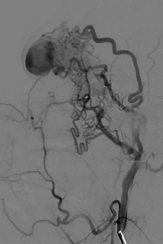
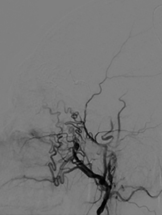
Cirsoid aneurysms, a subtype of arteriovenous fistulas (AVF), of the scalp are rare pathological lesions characterized by abnormal fistulous connections between superficial arteries and draining veins without intervening capillary beds. We present a case report of a cirsoid aneurysm located on the scalp, treated at our tertiary care center using percutaneous endovascular intervention with injection embolics. This report highlights the challenges posed by complex vascular anatomy and high shunt flow in the treatment of such lesions involving the head and neck. We discuss the rationale for selecting the treatment approach, emphasizing the importance of a patient-specific strategy to achieve successful obliteration of the abnormal vascular connections. Our experience underscores the efficacy of transarterial and/or transvenous embolization using appropriate embolic materials in the management of cirsoid aneurysms. This case report contributes to the existing literature on treatment options for scalp AVFs, providing insights into optimizing outcomes in these rare but clinically significant lesions.
Keywords: Arteriovenous fistula; Cirsoid aneurysm; Endovascular embolization.
© 2025 The Authors. Published by Elsevier Inc. on behalf of University of Washington.
Figures


References
-
- Tiwary S.K., Khanna R., Khanna AK. Craniofacial cirsoid aneurysm: 2-stage treatment. J Oral Maxillofac Surg. 2007;65(3):523–525. - PubMed
-
- Sofela A., Osunronbi T., Hettige S. Scalp Cirsoid Aneurysms: case illustration and systematic review of literature. Neurosurgery. 2020;86(2):E98–e107. - PubMed
-
- Shenoy S.N., Raja A. Scalp arteriovenous malformations. Neurol India. 2004;52(4):478–481. - PubMed
-
- Visser A., FitzJohn T., Tan S.T. Surgical management of arteriovenous malformation. J Plast Reconstr Aesthet Surg. 2011;64(3):283–291. - PubMed
Publication types
LinkOut - more resources
Full Text Sources
