Case Reports
. 2025 May 14;20(8):3729-3732.
doi: 10.1016/j.radcr.2025.04.019. eCollection 2025 Aug.

Thoracic aorta aneurysm tracheal compression and anatomical variant of the right subclavian artery: A case report

Affiliations
Case Reports

Thoracic aorta aneurysm tracheal compression and anatomical variant of the right subclavian artery: A case report

Francesca Cianfrone et al. Radiol Case Rep. .

Abstract

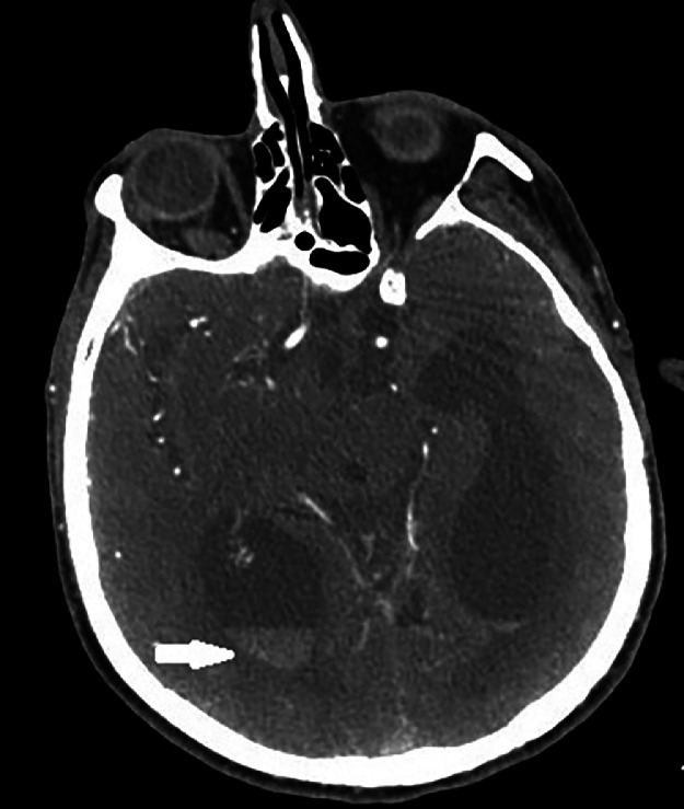
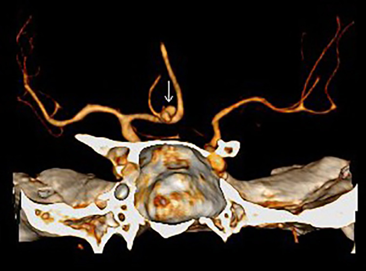
Tracheo-bronchial compression is a complication of vascular congenital and acquired anomalies, usually associated with double aortic arch, aberrant subclavian artery, pulmonary artery sling, Kommerell's diverticulum, and with aneurysms of the aortic arch and thoracic aorta. In this report we present a case of a 75-year-old male with incidental diagnosis of tracheal compression by a thoracic aorta aneurysm combined with anatomical variant of the right subclavian artery, that came up to our attention because of the onset of a subarachnoid hemorrhage (SAH) caused by a bilobar shaped anterior cerebral artery aneurysm rupture.

Keywords: Aberrant subclavian artery; Thoracic aorta aneurysm; Tracheal compression; Vascular anomalies.


Figures

Fig 1
Fig. 1
Pretreatment cranial CT scan image. Unenhanced Axial MDCT Scan showing blood-CSF levels in both lateral ventricles which is consistent with emoventricle. Blood-like hyperdensity in right side subharchnoid spaces of the convexity of fronto-temporal-parietal lobes, cisterna magna and third ventricle are consistent with SAH.
Fig 2
Fig. 2
Post-treatment cranial CT scan image. Enhanced Axial MDCT Scan showing blood-CSF levels in both lateral ventricles which are consistent with emventricle.
Fig 3
Fig. 3
Volume rendered image of vascular structures. Ruptured aneurysm of anterior communicating artery is clearly visible.
Fig 4
Fig. 4
Sagittal chest CT scan image. Unhenanced MDCT of the thorax oblique view. A retro-tracheal and retro-esophageal aneurysm of the aortic arch is well detected (white arrow).
Fig 5
Fig. 5
Axial chest CT scan image. CE-MDCT of the thorax axial view. A huge thoracic aorta aneurysm abutting the esophagus and trachea is visible. Note the tracheal cannula indwell.
Fig 6
Fig. 6
Fiberoptic evaluation that showed a protruding mass at the posterior wall of the trachea with partial occlusion of the portion of the lumen.
Fig 7
Fig. 7
Fiberoptic evaluation corresponding to the distal part of the tracheostomy cannula, with sufficient respiratory space revealed.

Similar articles

References

    1. Kumar A., Dutta V., Negi S., Puri G.D. Vascular airway compression management in a case of aortic arch and descending thoracic aortic aneurysm. Ann Card Anaesth. 2016;19(3):568–571. - PMC - PubMed
    1. Komarov R., Ismailbaev A., Kurasov N., Ivashov I., Saliba M., Danachev A. Aortic arch aneurysm. Tracheobronchial compression as a vital indication for emergency surgery: a case report. Int J Surg Case Rep. 2021;85 - PMC - PubMed
    1. Charrette E.J., Winton T.L., Salerno T.A. Acute respiratory insufficiency from an aneurysm of the descending thoracic aorta. J Thorac Cardiovasc Surg. 1983;85(3):467–470. - PubMed
    1. Malik T.H., Bruce I.A., Kaushik V., Wright N.B., Rothera M.P. The role of magnetic resonance imaging in the assessment of suspected extrinsic tracheobronchial compression due to vascular anomalies. Arch Dis Child. 2006;91(1):52–55. - PMC - PubMed
    1. Lincoln J.C., Deverall P.B., Stark J., Aberdeen E., Waterston D.J. Vascular anomalies compressing the oesophagus and trachea. Thorax. 1969;24(3):295–306. - PMC - PubMed

Publication types

LinkOut - more resources