. 2025 May 23:13:1546483.
doi: 10.3389/fbioe.2025.1546483. eCollection 2025.

hiPSC-derived cardiac fibroblasts dynamically enhance the mechanical function of hiPSC-derived cardiomyocytes on an engineered substrate

Affiliations

hiPSC-derived cardiac fibroblasts dynamically enhance the mechanical function of hiPSC-derived cardiomyocytes on an engineered substrate

Mitchell Josvai et al. Front Bioeng Biotechnol. .

Abstract

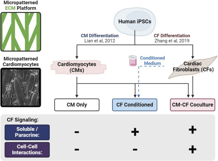
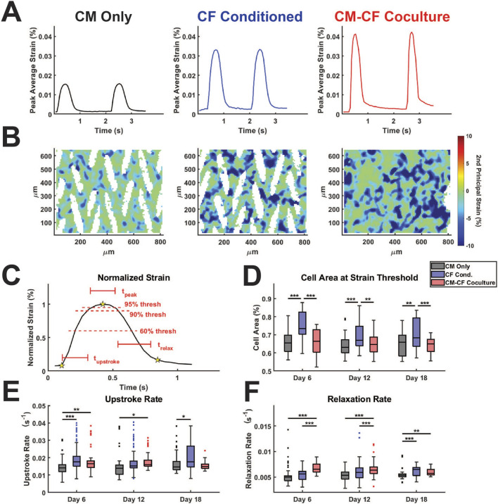
Introduction: Cardiac fibroblasts deposit and turnover the extracellular matrix in the heart, as well as secrete soluble factors that play critical roles in development, homeostasis, and disease. Coculture of CFs and human induced pluripotent stem cell (hiPSC)-derived cardiomyocytes (CMs) enhances CM mechanical output, yet the mechanism remains unclear.

Methods: Here, we use an in vitro engineered platform to compare the effects on CM mechanical function of direct CM-CF Coculture and soluble signaling alone through CF Conditioned Medium to a CM Only monoculture. Mechanical analysis is performed using digital image correlation and custom software to quantify the coordination and organization of CM contractile behavior.

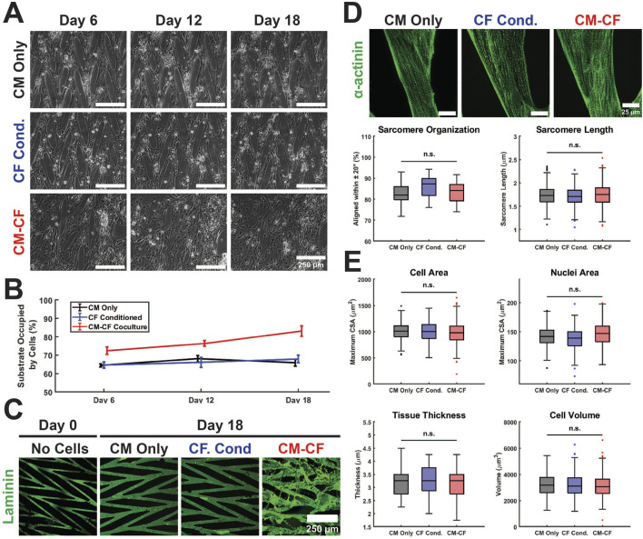
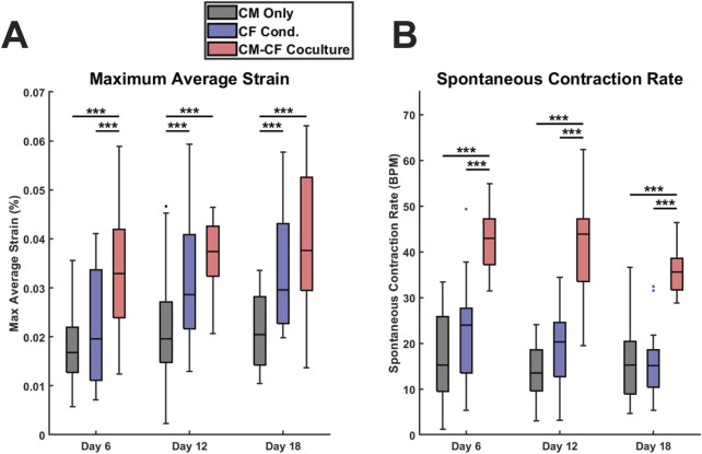
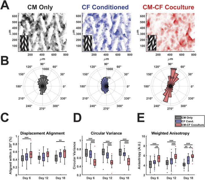
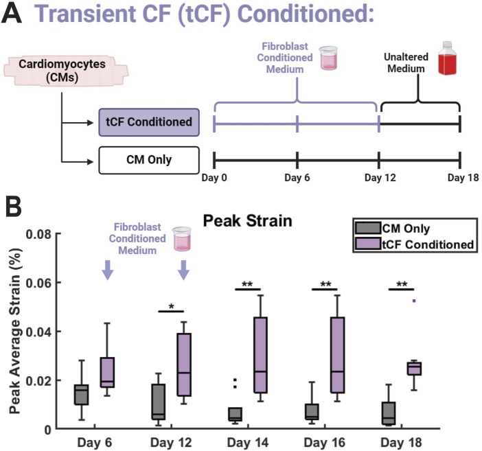
Results: CM-CF Coculture induces larger CM contractile strains, and an increased rate of spontaneous contraction compared to CM Only. Additionally, CM-CF Cocultures have increased contractile anisotropy and myofibril alignment and faster kinetics. The paracrine effects of fibroblast conditioned medium (FCM) are sufficient to induce larger contractile strains and faster contraction kinetics with these effects remaining after the removal of FCM. However, FCM does not influence CM spontaneous rate, contractile alignment, anisotropy, or relaxation kinetics compared to CM Only control.

Discussion: These data suggest that hiPSC-CFs exert dynamic and multifactorial effects on the mechanical function of hiPSC-CMs and highlight the importance of CFs in both the native heart and in vitro cardiac models. Further, this work demonstrates the applicability of the coculture-conditioned medium-monoculture paradigm to decouple the effects of paracrine factor and cell-cell signaling on hiPSC-CM mechanical function and maturation.

Keywords: cardiac fibroblasts; cardiomyocytes; hiPSC; mechanical function; tissue engineering.


Conflict of interest statement

TK is a consultant for Fujifilm Cellular Dynamics Incorporated. The remaining authors declare that the research was conducted in the absence of any commercial or financial relationships that could be construed as a potential conflict of interest.

Figures

FIGURE 1
FIGURE 1
Graphical representation of experimental culture groups. hiPSCs are differentiated into separate populations of CMs and CFs and split among three experimental groups: CM monocultures supplied with basal medium (CM Only), CM monocultures supplied with medium conditioned by a CF monoculture (CF Conditioned), and cocultures of CMs and CFs together (CM-CF Coculture).
FIGURE 2
FIGURE 2
Culture conditions do not alter hiPSC-CM structural characteristics on a micropatterned platform. (A) Representative Day 6 (left), Day 12 (center column), and Day 18 (right) images of CM Only (top), CF Conditioned (center row), and CM-CF Coculture (bottom) groups. Scale bars = 250 µm. (B) CM-CF Cocultures demonstrate progressive remodeling and increases in substrate occupation, while CM Only and CF Conditioned remain unchanged. (C) Representative images of day 0 (right) and day 18 (left) samples stained for the ECM component laminin (green). Day 0 samples are prior to cell seeding and represent only the micropatterned ECM and no cellular modifications. Scale bar = 250 µm. (D) Representative images stained for sarcomeric alpha actinin (top). The day 18 properties of the cardiac sarcomere, including length and organization, are unaltered across conditions (bottom). Scale bars = 25 µm. N = 20 (5 samples, 4 locations each). (E) No changes are observed in the day 18 cell area, nuclei area, tissue thickness, or cell volume across conditions. N = 20 (5 samples, 4 locations each). A minimum of 5 unique hiPSC-CM differentiations were used for each experimental group.
FIGURE 3
FIGURE 3
CF Conditioned medium and CM-CF Coculture exert differential effects on contractile behavior. (A) The maximum contractile strain achieved by each group on days 6, 12, and 18. The maximum contractile strain is the peak of the second principal strain averaged across the full field of imaging as calculated by DIC. (B) The spontaneous rate of contraction for each condition at each experimental timepoint. N = CM Only: d6 = 47, d12 = 43, d18 = 28; CF Conditioned: d6 = 38, d12 = 35, d18 = 23; CM-CF Coculture: d6 = 20, d12 = 23, d18 = 17. A minimum of five unique hiPSC-CM differentiations were used for each experimental group.
FIGURE 4
FIGURE 4
CM-CF Coculture promotes displacement trajectory alignment. (A) Representative displacement vector maps for CM Only (left), CF Conditioned (center) and CM-CF Coculture (right). Inset denotes the orientation of the original micropattern design. Vector maps correspond to the day 18 samples in Figure 2A. (B) Representative polar histograms (corresponding to the vector maps in Figure 4A) for each experimental group, demonstrating the alignment of displacement trajectories. Polar histograms account only for displacement trajectory and not magnitude, though displacements with a magnitude less than 75% of the median for an individual sample were excluded from summary measurements in Figures 4C–E. (C) Displacement trajectory alignment (defined as the percentage of displacements aligned within 20 degrees of the most predominant principal strain orientation), (D) circular variance of displacement angles (ranges from 0 to 1), and (E) weighted anisotropy (defined as the sum of all vector magnitudes aligned within 20 degrees of the predominant orientation divided by the sum of all unaligned vector magnitudes). N = CM Only: d6 = 47, d12 = 43, d18 = 28; CF Conditioned: d6 = 38, d12 = 35, d18 = 23; CM-CF Coculture: d6 = 20, d12 = 23, d18 = 17. A minimum of 5 unique hiPSC-CM differentiations were used for each experimental group.
FIGURE 5
FIGURE 5
CF Conditioned Medium and CM-CF Coculture alter the contractile and relaxation kinetics of hiPSC-CMs. (A) Representative strain traces and (B) strain maps, corresponding to the day 18 samples in Figures 2A, 4A. Representative strain maps are from the peak of the first contraction shown in Figure 5A. (C) Normalized strain schematic demonstrating initiation (first start), peak (second star) and rest (third star) points used for kinetic calculations and to determine the 60% and 90% threshold values. (D) Cell area reaching the 60% strain threshold at the maximum strain state, used as a metric to approximate the percentage of cells achieving considerable contraction states. (E) Contraction upstroke strain rate and (F) relaxation strain rate. N = CM Only: d6 = 47, d12 = 43, d18 = 28; CF Conditioned: d6 = 38, d12 = 35, d18 = 23; CM-CF Coculture: d6 = 20, d12 = 23, d18 = 17. A minimum of 5 unique hiPSC-CM differentiations were used for each experimental group.
FIGURE 6
FIGURE 6
Transient CF (tCF) Conditioned medium experimental results. (A) Experimental design for tCF Conditioned. Samples are provided with fibroblast conditioned medium from days 0 to 10, then provided with only basal medium until day 18. CM Only samples were provided with only basal medium from days 0 through 18. (B) The maximum contractile strain achieved on days 6, 12, 14, 16, and 18 as calculated by DIC. N = 10 for both groups at all time points. A minimum of 3 unique hiPSC-CM differentiations were used for each experimental group.

Similar articles

References

    1. Abecasis B., Gomes-Alves P., Rosa S., Gouveia P. J., Ferreira L., Serra M., et al. (2019). Unveiling the molecular crosstalk in a human induced pluripotent stem cell-derived cardiac model. Biotech and Bioeng. 116, 1245–1252. 10.1002/bit.26929 - DOI - PubMed
    1. Acevedo-Acevedo S., Crone W. C. (2015). Substrate stiffness effect and chromosome missegregation in hIPS cells. J. Negat. Results Biomed. 14, 22. 10.1186/s12952-015-0042-8 - DOI - PMC - PubMed
    1. Ahmed R. E., Anzai T., Chanthra N., Uosaki H. (2020). A brief review of current maturation methods for human induced pluripotent stem cells-derived cardiomyocytes. Front. Cell Dev. Biol. 8, 178. 10.3389/fcell.2020.00178 - DOI - PMC - PubMed
    1. Aigha I., Raynaud C. (2016). Maturation of pluripotent stem cell derived cardiomyocytes: the new challenge. Glob. Cardiol. Sci. Pract. 2016, e201606. 10.21542/gcsp.2016.6 - DOI - PMC - PubMed
    1. Avazmohammadi R., Soares J. S., Li D. S., Raut S. S., Gorman R. C., Sacks M. S. (2019). A contemporary look at biomechanical models of myocardium. Annu. Rev. Biomed. Eng. 21, 417–442. 10.1146/annurev-bioeng-062117-121129 - DOI - PMC - PubMed

LinkOut - more resources