Review
. 2025 May 23:19:1604435.
doi: 10.3389/fnins.2025.1604435. eCollection 2025.

Brain organoid model systems of neurodegenerative diseases: recent progress and future prospects

Affiliations
Review

Brain organoid model systems of neurodegenerative diseases: recent progress and future prospects

Saniyah Shaikh et al. Front Neurosci. .

Abstract

Neurological diseases are a leading cause of disability, morbidity, and mortality, affecting 43% of the world's population. The detailed study of neurological diseases, testing of drugs, and repair of site-specific defects require physiologically relevant models that recapitulate key events and dynamic neurodevelopmental processes in a highly organized fashion. As an evolving technology, self-organizing and self-assembling brain organoids offer the advantage of modeling different stages of brain development in a 3D microenvironment. Herein, we review the utility, advantages, and limitations of the latest breakthroughs in brain organoid endeavors in the context of modeling three of the most prevalent neurodegenerative diseases-Alzheimer's, Parkinson's, and Huntington's disease. We conclude the review with a perspective on the future prospects of brain organoid models with their myriad possible applications in translational medicine.

Keywords: Alzheimer’s disease; Huntington’s disease; Parkinson’s disease; brain; disease modeling; neurodegenerative disorders; organoids; stem cells.


Conflict of interest statement

The authors declare that the research was conducted in the absence of any commercial or financial relationships that could be construed as a potential conflict of interest.

Figures

Figure 1
Figure 1
Advanced techniques enhancing brain organoid research. Demonstration of cutting-edge techniques such as single-cell omics, high-resolution imaging, and bioengineering tools (such as scaffolds and microfluidics). These methods enhance the accuracy of disease models, organoid development, and functionality. Created in BioRender. LiTe, R. (2025) https://BioRender.com/zb8h1lw.
Figure 2
Figure 2
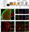
(a) Schematic representation of the procedures for developing cerebral organoids using hiPSCs. (b) Representative fluorescent images in organoids showed the ventricular zone (VZ)-like generated by new-born neurons (Tuj1, green) and neural progenitor cells (Sox2, red) after differentiation (at week 4). (c,d) High resolution confocal images displayed the cortical layer formation (Ctip2 stained deep cortical layer marker and Satb2 stained superficial cortical layer marker) at different developmental periods [week 4 (c) and week 12 (d), respectively]. (e,f) Proliferation and migration differentiation pattern of in organoids; the differentiation pattern of astrocytes in organoids were monitored by GFAP immunostaining (astrocytic marker) at different time points [week 4 (e) and week 12 (f), respectively]. Scale bar: 100 μm. Adapted from Zhao et al. (2020), with copyright permission under the terms of the CC-BY-NC-ND 4.0 license.
Figure 3
Figure 3
Organoid modeling of AD. This diagram illustrates patient-derived FAD and APOE4 organoids’ ability to represent actual disease pathology. Created in BioRender. Yaqinuddin, A. (2025) https://BioRender.com/vo2gq0k.
Figure 4
Figure 4
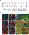
Generation and characterization of iPSC-derived midbrain organoids from Parkinson’s disease (PD) patients and healthy controls. (A) Schematic showing the differentiation process when utilizing induced pluripotent stem cells (iPSCs) to produce brain organoids. (B) Immunohistochemical evaluation following 4 weeks in culture demonstrating expression of markers specific to the midbrain: neural progenitors (SOX2), TH (dopaminergic neurons), TUJ1 and MAP2 (neuronal markers), FOXA2 and LMX1A (midbrain identity), and Ki67 (proliferation). Adapted from Patikas et al. (2023) with copyright permission under the terms of the CC-BY-NC-ND 4.0 license.
Figure 5
Figure 5
Pathway-to-phenotype mapping in PD organoid models and key molecular pathways implicated in PD. Created in BioRender. Yaqinuddin, A. (2025) https://BioRender.com/vo2gq0k.
Figure 6
Figure 6
Organoid-based modeling of HD. This flowchart maps how mutant huntingtin (muHTT) expression in hiPSC-derived organoids leads to altered gene expression profiles. Created in BioRender. Yaqinuddin, A. (2025) https://BioRender.com/vo2gq0k.

References

    1. Akula S. K., Exposito-Alonso D., Walsh C. A. (2023). Shaping the brain: the emergence of cortical structure and folding. Dev. Cell 58, 2836–2849. doi: 10.1016/j.devcel.2023.11.004, PMID: - DOI - PMC - PubMed
    1. Albanese A., Swaney J. M., Yun D. H., Evans N. B., Antonucci J. M., Velasco S., et al. . (2020). Multiscale 3D phenotyping of human cerebral organoids. Sci. Rep. 10:21487. doi: 10.1038/s41598-020-78130-7, PMID: - DOI - PMC - PubMed
    1. An J., Shin M., Beak G., Yoon J., Kim S., Cho H. Y., et al. . (2024). Drug evaluation of Parkinson’s disease patient-derived midbrain organoids using mesoporous au Nanodot-patterned 3D concave electrode. ACS Sens. 9, 3573–3580. doi: 10.1021/acssensors.4c00476, PMID: - DOI - PubMed
    1. Arber C., Lovejoy C., Harris L., Willumsen N., Alatza A., Casey J. M., et al. . (2021). Familial Alzheimer’s disease mutations in PSEN1 lead to premature human stem cell neurogenesis. Cell Rep. 34:108615. doi: 10.1016/j.celrep.2020.108615 - DOI - PMC - PubMed
    1. Bagley J. A., Reumann D., Bian S., Lévi-Strauss J., Knoblich J. A. (2017). Fused cerebral organoids model interactions between brain regions. Nat. Methods 14, 743–751. doi: 10.1038/nmeth.4304, PMID: - DOI - PMC - PubMed

LinkOut - more resources