. 2025 Apr;15(4):1938-1955.
doi: 10.1016/j.apsb.2025.02.014. Epub 2025 Feb 18.

CDK1-mediated phosphorylation of USP37 regulates SND1 stability and promotes oncogenesis in colorectal cancer

Affiliations

CDK1-mediated phosphorylation of USP37 regulates SND1 stability and promotes oncogenesis in colorectal cancer

Liang Wu et al. Acta Pharm Sin B. 2025 Apr.

Abstract

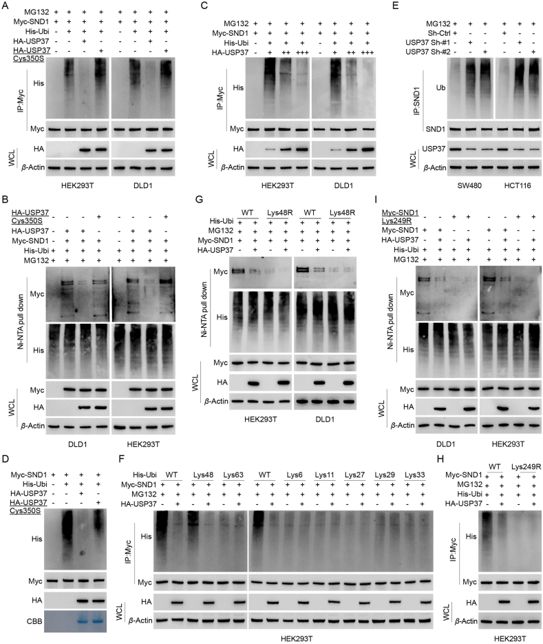
Colorectal cancer (CRC) poses a severe global health challenge with high incidence and mortality rates. USP37 has been identified as the bona fide deubiquitinase of SND1, playing a critical role in stabilizing SND1, thereby augmenting its oncogenic potential. The interaction between USP37 and SND1 was confirmed through extensive proteomics, ubiquitinomics, and interactomics, underscoring their synergistic effects on CRC proliferation and metastasis. Additionally, CDK1 has emerged as a pivotal regulator of USP37, phosphorylating it at threonine 631 rather than serine 628, enhancing its deubiquitinase activity, and consequently stabilizing SND1 to drive CRC malignancy further. Histological analyses of human CRC samples linked the upregulation of CDK1 and USP37 with increased SND1 levels and poor patient prognosis. High-throughput virtual screening and subsequent experimental validation identified Dacarbazine as a pharmacological inhibitor of USP37, and its inhibition disrupted SND1 stability, hindering CRC cell proliferation and metastasis. This study reveals a novel and promising molecular mechanism driving CRC progression through the CDK1-USP37-SND1 axis, highlighting the clinical importance of targeting this pathway to improve patient outcomes.

Keywords: CDK1; Colorectal cancer; Dacarbazine; Deubiquitination; Oncogenesis; Phosphorylation; SND1; USP37.


Conflict of interest statement

The authors have declared that no conflict of interest exists.

Figures

Image 1
Graphical abstract
Figure 1
Figure 1
Specific interaction of USP37, which plays an oncogenic role in CRC tissues, with SND1. (A) IHC staining of USP37 was performed on 180 human CRC specimens. Representative consecutive sections from the samples are depicted. Scale is as indicated. The Wilcoxon matched-pairs test was employed to compare cancerous tissues (CA) with matched adjacent tissues (AD). (B) Kaplan–Meier survival curves for CRC patients with high (n = 115) and low (n = 65) USP37 levels were delineated. (C) A Venn diagram depicts proteins upregulated, ubiquitin-modified proteins downregulated, and USP37 binding proteins in DLD1 cells overexpressing USP37 (USP37 vs. vector), as determined by LC–MS/MS. (D) LC–MS/MS analysis of SND1 peptides within USP37 immunoprecipitates was conducted. (E) IP analysis using USP37 and SND1 antibodies was performed on cell lysates from SW480 and HCT116, followed by IB analysis. (F) HEK293T cells were co-transfected with HA-USP37 and Myc-SND1. Cell lysates were then subjected to IP using anti-HA or anti-Myc antibodies. (G) Representative images of fixed SW480, HCT116, and DLD1 cells after immunofluorescent staining with antibodies against USP37 (green) and SND1 (red). Nuclei were counterstained with DAPI (blue). Scale bar, 10 μm. (H) Representative images of in situ PLA for USP37/SND1 using anti-USP37 and anti-SND1 antibodies in fixed SW480 and HCT116 cells. The PLA-detected proximity complexes are denoted by fluorescent rolling circle products (red dots). Scale bar, 10 μm. (I) Purified HA-USP37 WT or USP37 Cys350S were incubated with GST or GST-SND1 coupled to glutathione-sepharose beads. HA-tagged proteins retained on sepharose were detected using IB. Recombinant GST-SND1 purified from bacteria was analyzed using SDS-PAGE and Coomassie Brilliant Blue staining. (J, K) Schematic representations of HA-tagged full-length (FL) USP37, Myc-tagged FL SND1, and their various deletion mutants. (L, M) In HEK293T cells, Myc-SND1 and HA-tagged FL USP37 or its deletion mutants were co-transfected and subjected to IP analysis using HA or Myc magnetic beads, followed by IB analysis with Myc and HA antibodies. (N) In HEK293T cells, HA-USP37 and Myc-tagged FL SND1 or its deletion mutants were co-transfected and subjected to IP analysis using Myc magnetic beads, followed by IB analysis with antibodies against HA and Myc.
Figure 2
Figure 2
USP37 maintains SND1 stability. (A) USP37 was knocked down in SW480 and HCT116 cells using two independent shRNAs. Subsequently, IB analysis was performed to determine the SND1 level. (B, C) USP37 WT or Cys350S mutant was overexpressed in DLD1 (B) and HEK293T (C) cells. (D, E) Increasing amounts of USP37 were transfected into HEK293T (C) and DLD1 (D) cells, and SND1 level was detected. (F) DLD1 cells were transduced with HA-USP37 WT alone or in combination with USP37 shRNA, and SND1 levels were analyzed by IB analysis. (G, H) The qRT-PCR analysis was conducted to assess SND1 mRNA in SW480 and HCT116 cells with depleted endogenous USP37 via two independent shRNAs. (I) SW480 cells transfected with two independent USP37 shRNAs were treated with or without the proteasome inhibitor MG132 (20 μmol/L, 8 h), followed by an analysis of USP37 and SND1. (J) IB analysis of SND1 levels was conducted in SW480 and HCT116 cells transduced with USP37 shRNA and shRNA-resistant (Sh-Res) HA-USP37. (K) DLD1 cells transfected with USP37 or USP37 Cys350S were treated with 100 μg/mL CHX, harvested at indicated times, and then subjected to IB with antibodies against SND1 and USP37. Quantification of SND1 levels relative to β-actin is shown. (L, M) HCT116 (L) and SW480 (M) cells stably expressing control shRNA (shRNA-Ctrl) or USP37 shRNA were treated with 100 μg/mL CHX, followed by IB with antibodies against SND1 and USP37. Quantification of SND1 levels relative to β-actin is displayed. (G, H, K, L, and M) Results are quantified and represented as mean ± SD from three independent experiments. ∗∗∗P<0.001 indicates a statistically significant difference, ‘ns’ indicates no statistical significance, determined by one-way ANOVA (G, K, L, and M) and two-tailed Student's t-test (H).
Figure 3
Figure 3
USP37 deubiquitinates SND1. (A) HEK293T or DLD1 cells were co-transfected with Myc-SND1, His-Ubi, and HA-USP37 WT or USP37 Cys350S. Cell lysates were subjected to IP with Myc beads, followed by IB with anti-His and anti-Myc antibodies. Cells were harvested after treatment with 20 μmol/L MG132 for 8 h. (B) Cells co-transfected with the specified plasmids were processed to pull down His-Ubi with Ni-NTA beads, and IB was performed to detect polyubiquitinated SND1. (C) Increasing amounts of USP37 were transfected into HEK293T (C) and DLD1 (D) cells, and treated with MG132 for 8 h before collection. Cell lysates were subjected to IP with Myc beads, followed by IB with anti-His and anti-Myc antibodies. (D) In vitro deubiquitination assays were conducted. Ubiquitinated Myc-SND1 was treated with USP37 WT or USP37 Cys350S. Myc-SND1 was immunoprecipitated with Myc beads, followed by IB with anti-HA and anti-His antibodies. Recombinant USP37 WT or USP37 Cys350S was analyzed by SDS-PAGE and Coomassie Brilliant Blue staining. (E) Cells stably expressing shRNA-Ctrl or two independent USP37 shRNAs were transfected with the described plasmids and treated with MG132 for 8 h. Cell lysates were subjected to IP with SND1 antibody and the ubiquitination of SND1 was detected by IB. (F) HEK293T cells were co-transfected with Myc-SND1, HA-USP37, and His-Ubi WT, Lys6 (specifically Lys6-only), Lys11, Lys27, Lys29, Lys33, Lys48, or Lys63 plasmids to analyze the ubiquitination linkage of SND1. (G) HEK293T cells were co-transfected with Myc-SND1, HA-USP37, and His-Ubi WT or Lys48R (specifically, only Lys48 is mutated to Arg). His-Ubi was pulled down with Ni-NTA beads to analyze the ubiquitination linkage of SND1. (H) HEK293T cells were co-transfected with HA-USP37, His-Ubi, and either Myc-SND1 or Myc-SND1 Lys249R. Cells were collected after treatment with 20 μmol/L MG132 for 8 h. Cell lysates were subjected to IP with Myc beads, followed by IB with anti-His and anti-Myc antibodies. (I) HEK293T and DLD1 cells were co-transfected with the specified plasmids. His-Ubi was pulled down with Ni-NTA beads, and IB was conducted to detect polyubiquitinated Myc-SND1 or Myc-SND1 Lys249R protein.
Figure 4
Figure 4
USP37 promotes tumor progression of CRC through stabilizing SND1. (A–F) Cell growth and migration were assessed in SW480 and HCT116 cells transduced with shRNA-Ctrl or USP37 shRNA, and either vector control or SND1 reconstitution. The number of colonies formed and cells migrated were quantitatively analyzed. Representative images are shown in (A) and (D). Scale bar, 1 cm (A); 200 μm (D). (G–I) Cells as indicated (5 × 10⁶ per mouse) were subcutaneously injected into nude mice (n = 7). Tumor volume (H) and weight (I) were calculated. Scale: 1 cm. (J, K) Cells as indicated (2 × 10⁶ per mouse) were injected via the tail vein into nude mice to establish a lung metastasis model (n = 6). Representative images of lung tissue and H&E-stained sections are presented in (J), along with the calculation of metastatic nodules per group (K). (L) Cells as indicated (2 × 10⁶ per mouse) were injected via the tail vein into nude mice to establish a lung metastasis model (n = 6). Representative images of lung tissue and H&E-stained sections are presented in (J). (M) SND1 was knocked down in DLD1 cells with overexpression of the USP37 gene. Cells, as indicated (5 × 10⁶ per mouse), were subcutaneously injected into nude mice to form xenograft tumors (n = 7), and gross photographs are shown. (N) Cells as indicated (2 × 10⁶ per mouse) were injected via the tail vein into nude mice to establish a lung metastasis model (n = 6). Representative images of lung tissue and H&E-stained sections are presented in (N). (O) Cells as indicated (2 × 10⁶ per mouse) were injected beneath the splenic capsule to establish a liver metastasis model (n = 6). Representative images of liver tissue and H&E-stained sections are shown in (O). (B, C, E, F, H, I, and K) The results are quantified and represented as mean ± SD. For (B, C, E, and F) n = 3 biological replicates. ∗∗∗P<0.001, one-way ANOVA (B, C, E, F, H, I, and K).
Figure 5
Figure 5
CDK1 binds and phosphorylates USP37. (A) LC–MS/MS identified phosphosite data indicating that the threonine residue corresponding to Thr631 of the USP37 (depicted in blue within the peptide sequence) was phosphorylated. (B) The phosphorylation site at Thr631 of the USP37 is highly conserved throughout evolution. The amino acid sequence of the CDK1-targeted USP37 substrate exhibits conservation across different species. (C, D) IP of DLD1 cell lysates was performed with IgG, anti-USP37 (C), or anti-CDK1 (D) antibodies. The immunoprecipitates were then subjected to IB with designated antibodies. (E) Purified Flag-CDK1 was incubated with GST or GST-USP37 coupled to glutathione-sepharose beads. Proteins retained on the Sepharose were detected by IB with Flag. Recombinant GST-USP37 purified from bacteria was analyzed using SDS-PAGE and Coomassie blue staining. (F) DLD1 cells were transfected with designated plasmids and treated with vehicle or CDK1 inhibitor (RO-3306). IP was performed on cell lysates using an anti-HA antibody, and phosphorylation of USP37 was detected using a phospho-CDK substrate antibody. (G, H) The indicated plasmids and CDK1 siRNA were transfected into DLD1 and HEK293T cells. IP was performed on cell lysates using an anti-HA antibody, and phosphorylation of USP37 was detected using a phospho-CDK substrate antibody. (I) DLD1 cells were transfected with vector or HA-USP37 (WT or Thr631A mutant). IP was performed on cell lysates using an anti-HA antibody, and phosphorylation of USP37 was detected using a phospho-CDK substrate antibody. (J) In vitro kinase assays were conducted with GST-USP37 (WT or Thr631A) affinity-purified from HEK293T cells and cyclin B/CDK1. Coomassie staining of USP37 was used as a loading control. (K) SW480 cells were transfected with vector or Myc-SND1. Cell lysates were subjected to IP using anti-Myc antibody, followed by immunoblotting with designated antibodies. (L) SW480 cells were transfected with vector or Myc-SND1. Cell lysates were subjected to IP using an anti-Myc antibody, and phosphorylation of SND1 was detected using a phospho-CDK substrate antibody. (M, N) A cycloheximide pulse-chase assay was performed in cells, and results were quantified in the right panel (N). The results represent mean ± SD from three independent experiments. (O) SW480 cells were pretreated with RO-3306 (2 μmol/L) for 24 h followed by a cycloheximide pulse-chase assay, and results were quantified. The results are quantified and represented as mean ± SD. from three independent experiments. (P) A cycloheximide pulse-chase assay was performed in cells, and results were quantified. (Q) SW480 and HCT116 cells were transfected with siRNA-Ctrl or two independent CDK1 siRNAs, and IP was performed on cell lysates with anti-SND1 antibodies. IB analysis was conducted to detect the ubiquitination of SND1. (R) Cells were treated with vehicle or RO-3306 for 24 h in the presence of MG-132 (20 μmol/L), and cell lysates were subjected to IP with IgG or anti-SND1, followed by IB to detect the ubiquitination of SND1. (S, T) Cell migration and growth were assessed in SW480 and HCT116 cells transduced with siRNA-Ctrl or CDK1 siRNAs and transfected with vector or SND1. The number of migrated cells (S) and colony formation (T) were quantitatively analyzed. (U) SW480 cells transfected with vector or SND1 were treated with vehicle or RO-3306 for 24 h, and the migration of cells (U) was quantitatively analyzed. (N, O, P, S, T, and U) The results are quantified and represented as mean ± SD. from three independent experiments. ∗∗∗P<0.001, one-way ANOVA (N, S, T, and U), two-tailed Student's t-test (O and P).
Figure 6
Figure 6
CDK1-mediated phosphorylation activates USP37 towards SND1. (A) USP37 was knocked down in SW480 and HCT116 cells using two independent shRNAs and treated with vehicle or RO-3306. IB analysis was performed to assess the SND1 level. (B) The indicated plasmids were transfected into HEK293T and DLD1 cells, and treated with vehicle or RO-3306. Cell lysates were subjected to IP with an anti-Myc antibody, followed by IB analysis of ubiquitinated SND1. (C) The indicated plasmids and siRNAs were transfected into SW480 cells. IP with anti-SND1 antibody was performed on cell lysates, followed by IB analysis of ubiquitinated SND1. (D) The indicated plasmids were transfected into HEK293T and DLD1 cells. Cell lysates were subjected to IP with an anti-Myc antibody, followed by IB analysis of ubiquitinated SND1. (E) SW480 cells stably expressing USP37 shRNA were transfected with the specified plasmids, followed by IB analysis of SND1 expression levels. (F, G) HCT116 (F) and SW480 (G) cells with stable knockdown of USP37 were transfected with the indicated plasmids. The cells were then injected into nude mice to form xenograft tumors, with representative gross images shown. Vehicle or RO-3306 treatment was administered at predetermined time points. (H, I) Quantitative analysis of tumor volume for the constructed xenografts from HCT116 (H) and SW480 (I) cells. (J, K) Quantitative analysis of tumor weight for the constructed xenografts from HCT116 (H) and SW480 (I) cells. (L–N) A lung metastasis model was established using cells from F and G, with the number of metastatic foci quantitatively analyzed (M and N). (H, I, J, K, M, and N) Results are quantified and represented as mean ± SD. ∗∗∗P<0.001, ns indicates no statistical significance, one-way ANOVA (H, I, J, K, M, and N).
Figure 7
Figure 7
Pharmacological inhibition of USP37 attenuates SND1 stability and inhibits CRC cell proliferation and metastasis. (A) The workflow for HTVS utilized for the identification of USP37 inhibitors. (B) The chemical structure and in silico docking of Dacarbazine into the human USP37. (C) IB analysis of SND1 and β-actin in HCT116 cells pretreated with 20 μmol/L MG132 for 1 h, followed by overnight treatment with 15 μmol/L Dacarbazine in the presence of MG132. (D) HEK293T cells co-transfected with Myc-SND1 and His-Ubi, pretreated with 20 μmol/L MG132 for 1 h, then treated overnight with 15 μmol/L Dacarbazine in the presence of MG132. IP was performed using anti-Myc beads, followed by immunoblotting with antibodies against His and SND1. (E) SW480 cells were pretreated with Dacarbazine (15 μmol/L) for 24 h followed by a cycloheximide pulse-chase assay. The results are quantified and represented as mean ± SD from three independent experiments. (F) IB analysis of SND1 expression in control and USP37 gene knockout HCT116 cells treated overnight with 15 μmol/L Dacarbazine. (G) The deubiquitination of SND1 by USP37 at different concentrations of Dacarbazine. HA-USP37 was exposed to 5, 10, and 15 μmol/L Dacarbazine, then incubated with ubiquitinated SND1. The ubiquitination level of SND1 was measured. (H) HEK293T cells co-transfected with Myc-SND1, HA-USP37, and His-Ubi, then treated with 15 μmol/L Dacarbazine for 24 h. After pre-treatment with MG132 (20 μmol/L) for 6 h, IP was performed using anti-Myc beads, followed by IB with antibodies against Myc, HA, and His. (I) DLD1 cells transfected with vector or HA-USP37 were treated with vehicle or Dacarbazine (15 μmol/L) for 24 h, then at set time points, CHX (100 μg/mL) was added, and USP37 and SND1 were analyzed by IB. (J, K) DLD1 cells transfected with vector or USP37 were injected into nude mice to form xenograft tumors and are presented in gross photographs. Intraperitoneal injection of Dacarbazine (50 mg/kg) or vehicle was administered at scheduled time points. Quantification of the weight of the xenograft tumors constructed from the cells shown (K). Scale bar, 1 cm. (L, M) A lung metastasis model was constructed using the cells shown in J, and representative images of HE-stained sections are presented. The number of metastatic foci was quantified (M). (N, O) HCT116 cells stably transfected with control or USP37 sgRNA were used to construct xenograft tumors, presented in gross photographs. Intraperitoneal injection of Dacarbazine (50 mg/kg) or vehicle was administered at scheduled time points. Quantification of the weight of the xenograft tumors constructed from the cells shown (O). Scale bar, 1 cm. (P, Q) A lung metastasis model was constructed using the cells shown in N, and representative images of HE-stained sections are presented. The number of metastatic foci was quantified (Q). (E, K, M, O, and Q) The results are quantified and represented as mean ± SD. ∗∗∗P < 0.001, ns indicates no statistical significance, one-way ANOVA (K, M, O, and Q), two-tailed Student's t-test (E).
Figure 8
Figure 8
CDK1, USP37, and SND1 expression is upregulated in CRC and positively correlates with poor patient prognosis. (A, B) IB analysis was performed on cell lysates from 42 pairs of CRC tumor tissues and matched adjacent tissues to detect the expression of USP37, SND1, and CDK1. The correlation between USP37 and SND1 in CRC tumor tissues and matched adjacent tissues (B) was analyzed statistically using the chi-square test; Pearson r denotes the correlation coefficient. (C, D) Representative images of IHC staining of CDK1, USP37, and SND1 in CRC tumor samples. (E–G) A positive correlation of SND1 expression with USP37 (E) and CDK1 (F) was demonstrated. Additionally, a positive correlation of USP37 expression with CDK1 was shown (G). (H) Kaplan–Meier survival curves for CRC patients with high (n = 132) and low (n = 48) expression of SND1. (I) Kaplan–Meier survival curves for CRC patients with high (n = 127) and low (n = 53) expression of CDK1 protein. (J) The working model illustrates that CDK1-mediated phosphorylation of USP37 regulates SND1 stability and promotes oncogenesis in CRC.

References

    1. Morgan E., Arnold M., Gini A., Lorenzoni V., Cabasag C.J., Laversanne M., et al. Global burden of colorectal cancer in 2020 and 2040: incidence and mortality estimates from GLOBOCAN. Gut. 2023;72:338–344. - PubMed
    1. Biller L.H., Schrag D. Diagnosis and treatment of metastatic colorectal cancer: a review. JAMA. 2021;325:669–685. - PubMed
    1. Cheng C., Yao H., Li H., Liu J., Liu Z., Wu Y., et al. Blockade of the deubiquitinating enzyme USP48 degrades oncogenic HMGA2 and inhibits colorectal cancer invasion and metastasis. Acta Pharm Sin B. 2024;14:1624–1643. - PMC - PubMed
    1. Dewson G., Eichhorn P.J.A., Komander D. Deubiquitinases in cancer. Nat Rev Cancer. 2023;23:842–862. - PubMed
    1. Chen Y., Zhao Y., Yang X., Ren X., Huang S., Gong S., et al. USP44 regulates irradiation-induced DNA double-strand break repair and suppresses tumorigenesis in nasopharyngeal carcinoma. Nat Commun. 2022;13:501. - PMC - PubMed

LinkOut - more resources