. 2025 May 23:13:1585987.
doi: 10.3389/fcell.2025.1585987. eCollection 2025.

SNHG12 downregulation induces follicular dysplasia by modulating the glycolysis of granulosa cell in polycystic ovary syndrome

Affiliations

SNHG12 downregulation induces follicular dysplasia by modulating the glycolysis of granulosa cell in polycystic ovary syndrome

Sisi Yan et al. Front Cell Dev Biol. .

Abstract

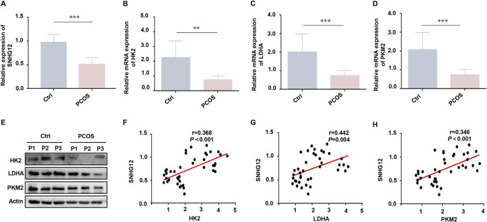
Introduction: Polycystic ovary syndrome (PCOS) is characterized by follicular dysplasia, with granulosa cells (GCs) glycolysis playing a pivotal role in this pathology. Although the involvement of long noncoding RNAs (lncRNAs) in diverse biological processes of PCOS has been well documented, the molecular mechanism of lncRNA small nucleolar RNA host gene 12 (SNHG12) in PCOS remains unclear.

Methods: In this study, we measured SNHG12 expression in GCs of PCOS patients and healthy controls using RT-PCR and performed correlation analysis between SNHG12 expression and glycolytic markers. Using granulosa-like tumor (KGN) cells, we investigated glycolytic capacity and examined the relationship among SNHG12, PTEN and HMGB1 through RNA immunoprecipitation (RIP) and chromatin immunoprecipitation (ChIP) assays. Finally, DHEA-induced PCOS mice was constructed using SNHG12 adenovirus to explore its role in PCOS.

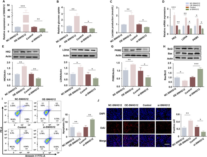
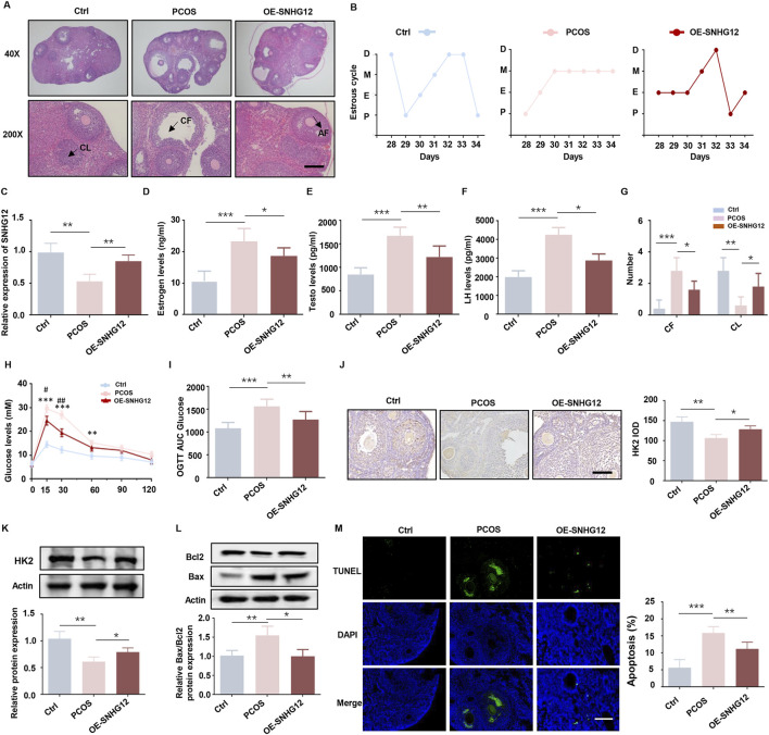
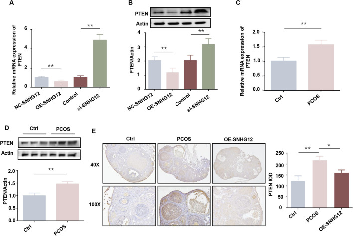
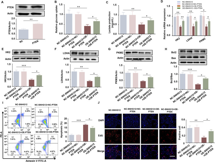
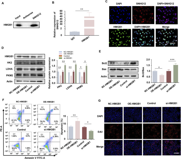
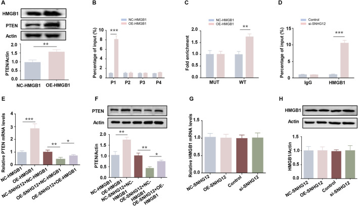
Results: SNHG12 expression was significantly downregulated in GCs from PCOS patients compared with healthy controls, and showed positive correlation with glycolytic markers. Functional studies demonstrated that SNHG12 knockdown impaired glycolysis in KGN cells, while SNHG12 overexpression partially restored glycolysis. Furthermore, SNHG12-induced glycolysis affected apoptosis of KGN cells, which mediated follicular dysplasia through lactate production and apoptotic pathways. In vivo, adenovirus-mediated SNHG12 overexpression alleviated the symptoms of PCOS mice. Mechanistically, RIP and ChIP assays revealed that SNHG12 interacts with HMGB1 and inhibits PTEN transcription by preventing HMGB1 from binding to the PTEN promoter, thereby promoting glycolysis in KGN cells.

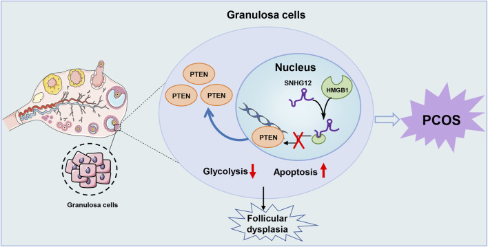
Conclusion: Our findings collectively demonstrate that the SNHG12/HMGB1/PTEN axis serves as a novel regulatory mechanism in PCOS by modulating glycolytic-mediated follicular dysplasia in GCs, offering a potential therapeutic target for PCOS.

Keywords: HMGB1; PCOS; SNHG12; follicular dysplasia; glycolysis; granulosa cells.


Conflict of interest statement

The authors declare that the research was conducted in the absence of any commercial or financial relationships that could be construed as a potential conflict of interest.

Figures

FIGURE 1
FIGURE 1
SNHG12 is downregulated in GCs of PCOS patients and associates with glycolytic dysfunction. (A) Relative mRNA expression of SNHG12 in GCs from PCOS patients (n = 20) and healthy controls (n = 20). (B–D) RT-PCR analysis of HK2, LDHA and PKM2 mRNA levels in GCs from PCOS patients and controls. (E) Western blot analysis of HK2, LDHA and PKM2 protein levels in GCs from PCOS patients and controls. (F–H) Correlation analysis between the SNHG12 expression and key glycolytic enzymes (HK2, LDHA and PKM2) in GCs. * P < 0.05, ** P < 0.01, and *** P < 0.001.
FIGURE 2
FIGURE 2
SNHG12 exerts a significant effect on glycolysis of KGN cells. (A) mRNA levels of SNHG12 in KGN cells transfected with OE-SNHG12 or si-SNHG12. (B,C) Glucose uptake and lactate production in KGN cells transfected with OE-SNHG12 or si-SNHG12. (D) Relative mRNA expression of HK2, LDHA and PKM2 in KGN cells treated with SNHG12 overexpression or knockdown. (E–G) Protein levels of HK2, LDHA and PKM2 in KGN cells treated with SNHG12 overexpression or knockdown. (H) Western blot analysis of the Bax/Bcl2 ratio in KGN cells transfected with OE-SNHG12 or si-SNHG12. (I) Apoptosis of KGN cells in each group was detected by flow cytometry. (J) Representative images of EdU assay results in different groups. Scale bar = 50 μm. n = 3, * P < 0.05, ** P < 0.01, and *** P < 0.001.
FIGURE 3
FIGURE 3
SNHG12 alleviates metabolic disorders and improves ovarian follicle development in PCOS mice. (A) Representative HE staining of ovarian sections from control (n = 6), PCOS (n = 6) and OE-SNHG12 groups (magnification: 40× and 200×). Scale bar: 50 µm. (B) Cytological analysis of vaginal smears in control, PCOS and OE-SNHG12 groups. (C) Relative mRNA expression of SNHG12 in ovarian tissues in the indicated groups. (D–F) Serum levels of estradiol (E2), testosterone (T), and luteinizing hormone (LH) in the indicated groups. (G) The number of cystic follicles (CF) in the ovaries of the mice in the indicated groups. (H,I) Glucose tolerance tests (GTT) and area under the curve (AUC) analysis in the experimental mice. (J) IHC staining of HK2 in ovarian tissues from the indicated groups (magnification: 400×). Scale bar: 50 µm. (K) Western blot analysis of HK2 expression in ovarian tissues from each group. (L) Western blot analysis of the Bax/Bcl2 ratio in ovarian tissues from each group. (M) Representative images of TUNEL assay results in mouse ovaries from the indicated groups. Scale bar: 50 µm * P < 0.05, ** P < 0.01, and *** P < 0.001.
FIGURE 4
FIGURE 4
PTEN is a key downstream target of SNHG12. (A) mRNA levels of PTEN in KGN cells transfected with OE-SNHG12 or si-SNHG12. (B) Protein levels of PTEN in KGN cells transfected with OE-SNHG12 or si-SNHG12. (C,D) mRNA and protein levels of PTEN in GCs from PCOS patients (n = 20) and healthy controls (n = 20). (E) IHC staining of PTEN in ovarian tissues from control and PCOS mice (magnification: 40× and 100×). Scale bar: 50 µm * P < 0.05, ** P < 0.01, and *** P < 0.001.
FIGURE 5
FIGURE 5
SNHG12 regulates glycolysis in KGN cells partially through PTEN. (A) Western blot analysis confirming efficiency of PTEN overexpression in KGN cells. (B,C) Glucose uptake and lactate production in KGN cells transfected with OE-SNHG12 or PTEN. (D) Relative mRNA expression of HK2, LDHA and PKM2 in the indicated KGN cells. (E–G) Protein levels of HK2, LDHA and PKM2 in the indicated KGN cells. (H) Western blot analysis of the Bax/Bcl2 ratio in the indicated KGN cells. (I) Apoptosis of KGN cells in each group was detected by flow cytometry. (J) Representative images of EdU assay results in the indicated KGN cells. Scale bar = 50 μm. n = 3, * P < 0.05, ** P < 0.01, and *** P < 0.001.
FIGURE 6
FIGURE 6
SNHG12 promotes glycolysis in KGN cells by interacting with HMGB1. (A,B) RNA pull-down experiment and RIP assays validating the interaction between SNHG12 and HMGB1. (C) Immunofluorescence analysis showing co-location of SNHG12 with HMGB1 in the nucleus of KGN cells. Scale bar: 20 µm. (D) Protein levels of HK2, LDHA and PKM2 in KGN cells with HMGB1 overexpression or knockdown. (E) Western blot analysis of the Bax/Bcl2 ratio in the aforementioned KGN cells. (F) Flow cytometry analysis of apoptosis in the aforementioned KGN cells. (G) Representative images of EdU assay results in the aforementioned KGN cells. Scale bar = 50 μm. n = 3, * P < 0.05, ** P < 0.01, and *** P < 0.001.
FIGURE 7
FIGURE 7
SNHG12 suppresses PTEN transcription by competitively binding to HMGB1. (A) Western blot analysis of the PTEN in HMGB1-overexpressing KGN cells. (B) ChIP-PCR assay of binding of HMGB1 to PTEN promoter region in KGN cells. (C) Luciferase reporter assays of KGN cells overexpressing HMGB1 and transfected with reporter plasmids containing WT and MUT PTEN promoter. (D) ChIP assays performed in KGN cells using HMGB1 antibodies or IgG. (E,F) RT-PCR and Western blot analyses of PTEN expression levels after HMGB1 upregulation in SNHG12-knockdown KGN cells. (G,H) mRNA and protein levels of HMGB1 in KGN cells were determined by RT-PCR and Western blot when treated with OE-SNHG12 or si-SNHG12. n = 3, * P < 0.05, ** P < 0.01, *** P < 0.001.
FIGURE 8
FIGURE 8
Schematic illustration of the role of SNHG12 in GCs during PCOS. Our study demonstrates that SNHG12 regulates glycolysis in GCs by disrupting the interaction between HMGB1 and the PTEN promoter, thereby modulating PTEN transcription and contributing to the pathophysiology of PCOS.

Similar articles

References

    1. Aldakheel F. M., Abuderman A. A., Alduraywish S. A., Xiao Y., Guo W. W. (2021). MicroRNA-21 inhibits ovarian granulosa cell proliferation by targeting SNHG7 in premature ovarian failure with polycystic ovary syndrome. J. Reprod. Immunol. 146, 103328. 10.1016/j.jri.2021.103328 - DOI - PubMed
    1. Cao J., Huo P., Cui K., Wei H., Cao J., Wang J., et al. (2022). Follicular fluid-derived exosomal miR-143-3p/miR-155-5p regulate follicular dysplasia by modulating glycolysis in granulosa cells in polycystic ovary syndrome. Cell Commun. Signal 20, 61. 10.1186/s12964-022-00876-6 - DOI - PMC - PubMed
    1. Chatterjee N., Pazarentzos E., Mayekar M. K., Gui P., Allegakoen D. V., Hrustanovic G., et al. (2019). Synthetic essentiality of metabolic regulator PDHK1 in PTEN-deficient cells and cancers. Cell Rep. 28, 2317–2330. 10.1016/j.celrep.2019.07.063 - DOI - PMC - PubMed
    1. Cui X., Lei X., Huang T., Mao X., Shen Z., Yang X., et al. (2024). Follicular fluid-derived extracellular vesicles miR-34a-5p regulates granulosa cell glycolysis in polycystic ovary syndrome by targeting LDHA. J. Ovarian Res. 17, 223. 10.1186/s13048-024-01542-w - DOI - PMC - PubMed
    1. Duan R., Zhai Y., Wang Q., Zhao L., Wang Y., Yu N., et al. (2024). LINC01764 promotes colorectal cancer cells proliferation, metastasis, and 5-fluorouracil resistance by regulating glucose and glutamine metabolism via promoting c-MYC translation. MedComm 5, e70003. 10.1002/mco2.70003 - DOI - PMC - PubMed

LinkOut - more resources