Approach to Residual Anterolateral Rotatory Knee Instability After Anterior Cruciate Ligament Reconstruction
- PMID: 40487140
- PMCID: PMC12136665
- DOI: 10.2106/JBJS.OA.25.00002
Approach to Residual Anterolateral Rotatory Knee Instability After Anterior Cruciate Ligament Reconstruction
Abstract
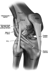
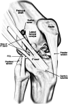
» Arthroscopic anterior cruciate ligament (ACL) reconstruction is widely regarded for its excellent results in restoring tibiofemoral anterior laxity to near-normal levels.» However, some operated patients may still experience anterolateral rotatory instability, leading to dissatisfaction and feelings of instability. After ruling out injuries to the posteromedial corner, lateral collateral ligament, and posterolateral corner, the focus should shift to the anterolateral ligament (ALL) and Kaplan fibers.» For ALL injuries causing internal rotatory instability at around 30 degrees knee flexion, a modified deep Lemaire tenodesis is recommended.» Kaplan fiber injuries leading to internal rotatory instability at angles greater than 30 degrees knee flexion can be treated with a modified superficial Lemaire surgery and iliotibial band strap fixation in the distal Kaplan fiber anatomical position.
Copyright © 2025 The Authors. Published by The Journal of Bone and Joint Surgery, Incorporated. All rights reserved.
Conflict of interest statement
The authors declare that there is no conflict of interest that could be perceived as prejudicing the impartiality of the research reported. All co-authors have agreed with the final manuscript's contents, and no financial interest remains to be announced. Disclosure: The Disclosure of Potential Conflicts of Interest forms are provided with the online version of the article (http://links.lww.com/JBJSOA/A816).
Figures


References
-
- Halewood C, Amis AA. Clinically relevant biomechanics of the knee capsule and ligaments. Knee Surg Sports Traumatol Arthrosc. 2015;23(10):2789-96. - PubMed
-
- Grassi A, Nitri M, Moulton S, Marcheggiani Muccioli GM, Bondi A, Romagnoli M, Zaffagnini S. Does the type of graft affect the outcome of revision anterior cruciate ligament reconstruction?: a meta-analysis of 32 studies. Bone Joint J. 2017;99-B(6):714-23. - PubMed
-
- Grassi A, Zicaro JP, Costa-Paz M, Samuelsson K, Wilson A, Zaffagnini S, Condello V, ESSKA Arthroscopy Committee. Good mid-term outcomes and low rates of residual rotatory laxity, complications and failures after revision anterior cruciate ligament reconstruction (ACL) and lateral extra-articular tenodesis (LET). Knee Surg Sports Traumatol Arthrosc. 2020;28(2):418-31. - PubMed
Publication types
LinkOut - more resources
Full Text Sources
