. 2025 May 8:32:101833.
doi: 10.1016/j.mtbio.2025.101833. eCollection 2025 Jun.

Synchronous bacterial barrier and exudate absorption: A novel dual-function dressing strategy for pin-site infection prevention

Affiliations

Synchronous bacterial barrier and exudate absorption: A novel dual-function dressing strategy for pin-site infection prevention

Bing Liang et al. Mater Today Bio. .

Abstract

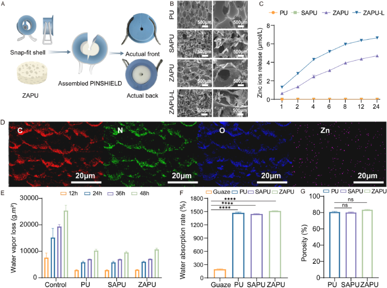
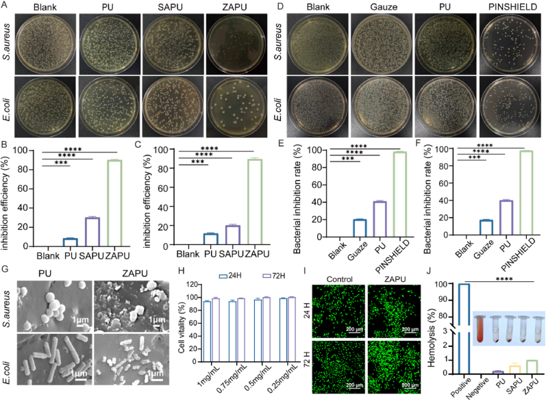
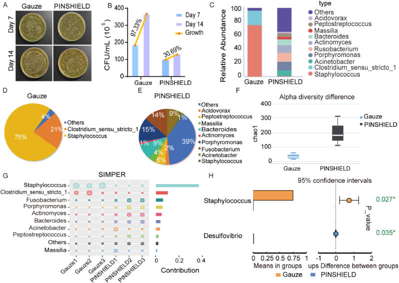
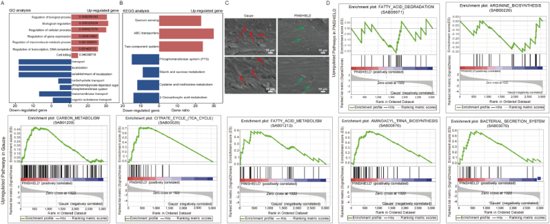
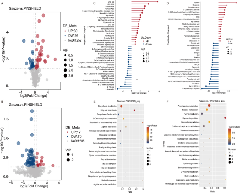
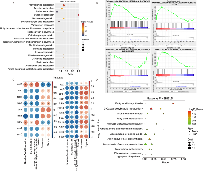
Open pin-site wounds, with infection rates of 11 %-100 %, pose significant clinical challenges, affecting millions globally and often leading to life-threatening complications. Current dressings fail to simultaneously block bacterial invasion and manage internal wound infection, necessitating innovative solutions. This study introduces PINSHIELD, a dual-functional dressing that externally seals wounds while efficiently managing exudate to mitigate pin-site infections (PSI). The external shell provides a physical barrier, while the embedded zinc alginate-polyurethane (ZAPU) layer combines active antibacterial properties with passive bacterial adhesion. The optimized ZAPU structure absorbs exudate and regulates the wound microenvironment, inhibiting bacterial proliferation and limiting infection spread. In vitro studies demonstrated that PINSHIELD inhibited S. aureus and E. coli by 90 %, with a bacterial blocking efficiency exceeding 95 %, significantly outperforming traditional gauze. In vivo results showed reduced inflammation, bacterial loads, and Staphylococcus abundance, while enhancing microbial diversity and enriching health-associated bacteria. Transcriptomic and metabolomic analyses revealed that PINSHIELD downregulated key S. aureus virulence genes (cna, SSL family, aur) and disrupted essential metabolic pathways (e.g., fatty acid biosynthesis, aminoacyl-tRNA synthesis), impairing bacterial adhesion, immune evasion, and biofilm formation. By synchronizing bacterial barrier formation with exudate management, PINSHIELD addresses the complex pathological needs of PSI, enhancing therapeutic efficacy and wound healing. This innovative design provides a versatile platform for infection control and personalized wound care, with broad implications for treating open wounds in orthopedic and other invasive device scenarios.

Keywords: Exudate absorption; Osteomyelitis; Pin-site infection; Staphylococcus aureus, transcriptomic and metabolomic analyses.


Conflict of interest statement

The authors declare that they have no known competing financial interests or personal relationships that could have appeared to influence the work reported in this paper.

Figures

Image 1
Graphical abstract
Fig. 1
Fig. 1
Design and Characterization of PINSHIELD and ZAPU. A) Schematic illustration and physical image of PINSHIELD assembly. B) SEM images of PU, SAPU, ZAPU, and ZAPU-L at 50X and 200× magnifications. C) Zinc ion release profile of ZAPU. D) Elemental mapping of ZAPU. E) Water vapor transmission loss of PU, SAPU, and ZAPU at 12, 24, 36, and 48 h. F) Water absorption rate of Gauze, PU, SAPU, and ZAPU. G) Porosity of PU, SAPU, and ZAPU (∗∗∗∗p < 0.0001).
Fig. 2
Fig. 2
Antibacterial Efficacy and Biocompatibility Evaluation of PINSHIELD. A) Colony count of S.aureus and E.coli on PU, SAPU, and ZAPU dressings. B) Antibacterial rate on S.aureus (ZAPU vs Blank, ∗∗∗p < 0.001, ∗∗∗∗p < 0.0001). C) Antibacterial rate on E.coli (ZAPU vs Blank, ∗∗∗p < 0.001, ∗∗∗∗p < 0.0001). D) Colony count of unblocked S.aureus and E.coli on Gauze, PU, and PINSHIELD. E) Inhibition rate of S.aureus (PINSHIELD vs Blank, ∗∗∗p < 0.001, ∗∗∗∗p < 0.0001). F) Inhibition rate of E.coli (PINSHIELD vs Blank, ∗∗∗p < 0.001, ∗∗∗∗p < 0.0001). G) Representative SEM images (scale bar = 1 μm) showing the morphology and surface adherence of S. aureus and E. coli after 2-h co-culture with PU and ZAPU. H) Cell viability of L929 cells after treatment with ZAPU. I) Live/dead cell staining results of L929 cells treated with ZAPU at 24h and 72h. J) Hemolysis evaluation of PU, SAPU, and ZAPU (ZAPU vs Positive control, ∗∗∗∗p < 0.0001).
Fig. 3
Fig. 3
Infection and Inflammation Control in Pin-Site Wounds by PINSHIELD. A) Schematic representation of pin-site infection (PSI) progression and treatment strategies with PINSHIELD and gauze. B) Gross observations of pin-site wounds on Day 7 and Day 14. C) Peripheral blood was collected from the ear vein of animals on Day 7 for white blood cell (WBC) and neutrophil (NEU) counts, as well as for the quantification of inflammatory cytokines (IL-6 and IL-1β). D) CT imaging showing pin-site and bone structure conditions on Day 14. E) MRI using T2-weighted sequences depicting soft tissue and medullary cavity conditions on Day 14. F) Histological analysis using HE staining to assess inflammatory cell infiltration and tissue necrosis (scale bar = 1000 μm). G) Semi-quantitative analysis of inflammatory infiltration analyzed by HE staining (∗∗∗p < 0.001). H) IHC staining for IL-1β to evaluate inflammatory response in pin-site tissues (scale bar = 1000 μm). I) Semi-quantitative analysis of IHC staining data presented as bar charts (∗∗p < 0.01).
Fig. 4
Fig. 4
Microbial Community Modulation by PINSHIELD in Pin-Site Wounds. A) Representative images of bacterial colonies on agar plates from pin-site wound samples collected on Days 7 and 14. B) Quantification of microbial load at pin sites using CFU counts, comparing bacterial growth between PINSHIELD and gauze groups on Days 7 and 14. C) Taxonomic composition of microbial communities at the genus level based on 16S rRNA sequencing from pin-site wounds on Day 14, comparing PINSHIELD and gauze groups. D) Pie chart showing genus-level microbial composition in the gauze group, highlighting the predominance of pathogenic genera. E) Pie chart showing genus-level microbial composition in the PINSHIELD group, illustrating an increase in beneficial genera and a reduction in pathogenic bacteria. F) Alpha diversity analysis comparing microbial diversity indices between PINSHIELD and gauze groups. G) Simper analysis highlighting changes in the relative abundance of pathogens and infection-resistant genera. H) T-test analysis showing increased levels of repair-associated genera, such as Desulfovibrio, in PINSHIELD-treated wounds.
Fig. 5
Fig. 5
Gene Expression Modulation and Virulence Suppression in S.aureus by PINSHIELD. A) Schematic illustrating PINSHIELD-induced gene expression alterations in S.aureus. B) Volcano plot showing DEGs in the PINSHIELD group compared to the gauze group, with significantly upregulated and downregulated genes highlighted. C) Expression levels of key DEGs associated with virulence, biofilm formation, immune evasion, and metabolic activity. Bars indicate relative expression levels, with significance denoted by stars (∗p < 0.05, ∗∗p < 0.01, ∗∗∗p < 0.001, ∗∗∗∗p < 0.0001). D) qPCR validation of selective RNA-seq-identified DEGs (Cna, Asp2, essA, SSL5) in S. aureus from pin-site wounds. Results confirmed consistent expression trends with RNA-seq data. Expression levels were normalized to the internal reference gene 16S rRNA, and relative expression was calculated using the 2^–ΔΔCt method. Data are presented as mean ± SD (n = 3) (∗∗∗p < 0.001, ∗∗∗∗p < 0.0001).
Fig. 6
Fig. 6
Functional Analysis of DEGs and Biofilm Suppression by PINSHIELD. A) GO enrichment analysis of DEGs identified in the PINSHIELD group, highlighting upregulated and downregulated categories. B) KEGG pathway analysis of DEGs, showing significant downregulation and upregulation of pathways. C) SEM images of bone pin surfaces from gauze- and PINSHIELD-treated groups, illustrating extensive biofilm formation and extracellular polymeric substances (EPS) in the gauze group (red arrows) and minimal biofilm formation with scattered bacterial cells in the PINSHIELD group (green arrows). D) GSEA of DEGs, illustrating pathway enrichment differences between the PINSHIELD and gauze groups. (For interpretation of the references to colour in this figure legend, the reader is referred to the Web version of this article.)
Fig. 7
Fig. 7
Metabolomic Profiling of Differentially Expressed Metabolites in PINSHIELD-Treated S.aureus. A) Volcano plot of negatively-charged metabolites between gauze and PINSHIELD groups. B) Volcano plot of positively-charged metabolites between gauze and PINSHIELD groups. C) Analysis of key negatively-charged metabolites, highlighting reductions in biofilm-associated lipids and increases in energy-related metabolites in the PINSHIELD group. D) Analysis of key positively-charged metabolites, showing adaptive metabolic responses in the PINSHIELD group. E) KEGG pathway enrichment analysis of negatively-charged metabolites, highlighting downregulation of pathways including fatty acid biosynthesis, antibiotics biosynthesis, and 2-oxocarboxylic acid metabolism in the PINSHIELD group. F) KEGG pathway enrichment analysis of positively-charged metabolites, showing inhibition of pathways such as phenylalanine metabolism, tyrosine metabolism, styrene degradation, and benzoate degradation in the PINSHIELD group.
Fig. 8
Fig. 8
Integrated Transcriptome and Metabolome Analysis of PINSHIELD's Effects on S.aureus. A) GSEA enrichment analysis of differentially expressed metabolites, highlighting downregulated pathways in the PINSHIELD group compared to the gauze group. B) Focused GSEA analysis of key suppressed pathways, emphasizing PINSHIELD's extensive metabolic regulation. C) Transcriptome-metabolome correlation analysis showing associations between key DEGs and deferentially expressed metabolites, revealing PINSHIELD's regulatory effects on metabolic activity. D) Combined KEGG and GSEA enrichment analyses demonstrating comprehensive inhibition of critical biosynthetic and metabolic pathways by PINSHIELD.

Similar articles

References

    1. Guerado E., Cano J.R., Fernandez-Sanchez F. Pin tract infection prophylaxis and treatment. Injury. 2019;50:S45–S49. - PubMed
    1. A-M Wu, Bisignano C., James S.L., Abady G.G., Abedi A., Abu-Gharbieh E., et al. Global, regional, and national burden of bone fractures in 204 countries and territories, 1990–2019: a systematic analysis from the Global Burden of Disease Study 2019. The Lancet Healthy Longevity. 2021;2(9):e580. e92. - PMC - PubMed
    1. Hadeed A.W.R., Varacallo M. StatPearls Publishing; Treasure Island (FL): 2023. External Fixation Principles and Overview. StatPearls [Internet]https://www.ncbi.nlm.nih.gov/books/NBK547694/ [updated 2023 Aug 4. Available from: - PubMed
    1. Kazmers N.H., Fragomen A.T., Rozbruch S.R. Prevention of pin site infection in external fixation: a review of the literature. Strategies Trauma Limb Reconstr. 2016;11(2):75–85. - PMC - PubMed
    1. Kazmers N.H., Fragomen A.T., Rozbruch S.R. Prevention of pin site infection in external fixation: a review of the literature. Strategies in Trauma and Limb Reconstruction. 2016;11(2):75–85. - PMC - PubMed

LinkOut - more resources