. 2025 May 15:30:31-45.
doi: 10.1016/j.reth.2025.05.003. eCollection 2025 Dec.

Revolutionizing nerve regeneration: A novel approach using polylactic acid/chitosan conduit with nerve-like cells and Bacopa monnieri in male rat model

Affiliations

Revolutionizing nerve regeneration: A novel approach using polylactic acid/chitosan conduit with nerve-like cells and Bacopa monnieri in male rat model

Elham Hoveizi et al. Regen Ther. .

Abstract

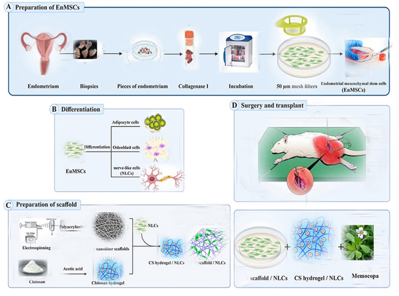
Objectives: This research aims to use neural-like cells (NLCs) derived from endometrial mesenchymal stem cells (EnMSCs) on a polylactic acid/chitosan scaffold (PLA/CS) along with the use of bacopa monnieri (Memocopa) in a rat sciatic nerve injury model for sciatic nerve regeneration. While previous studies have explored stem cell therapies and scaffold-based approaches for nerve regeneration, using EnMSCs in combination with a PLA/CS scaffold and Memocopa represents a novel, potentially synergistic approach.

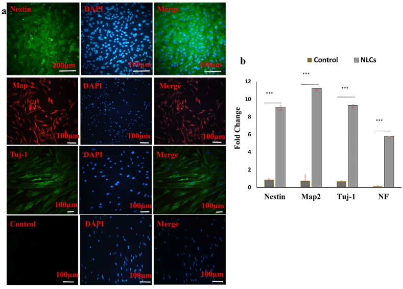
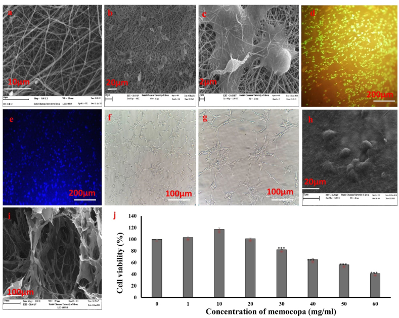
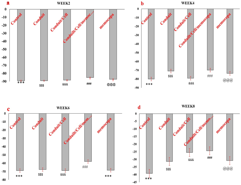
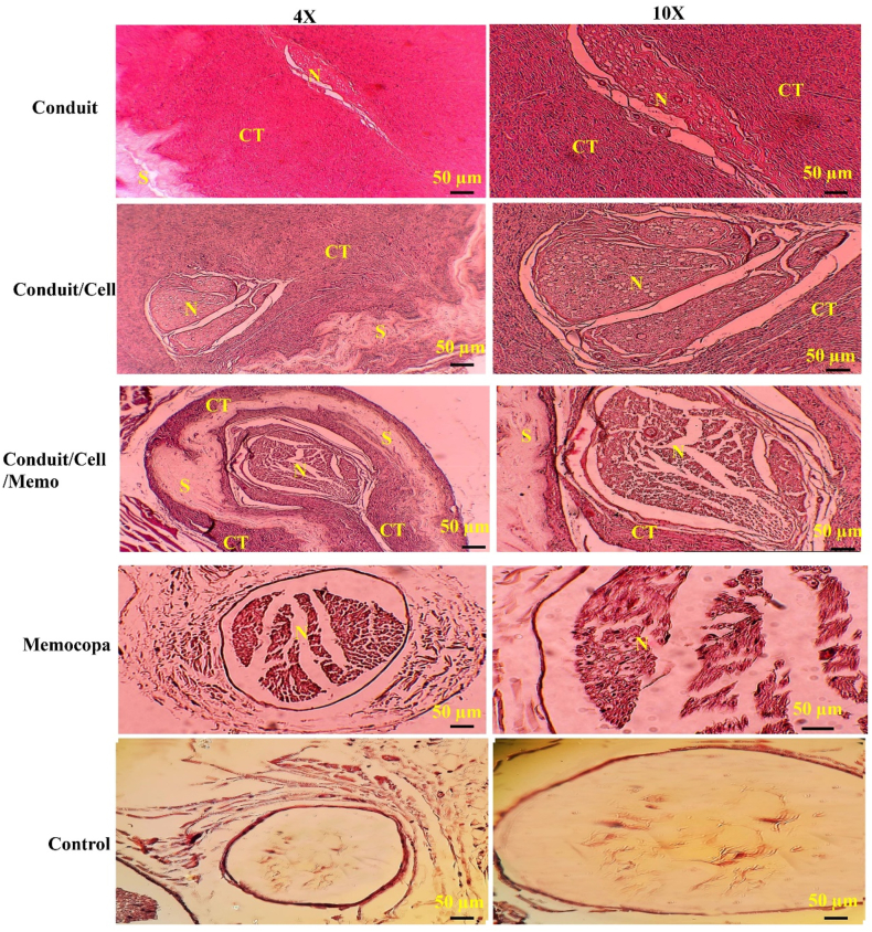
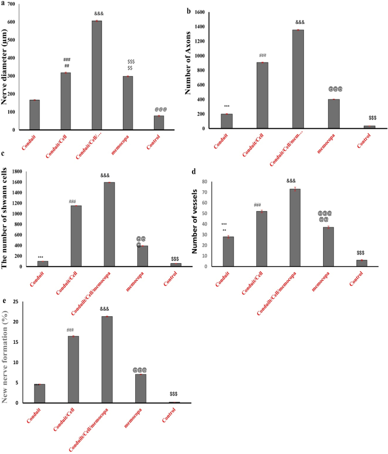
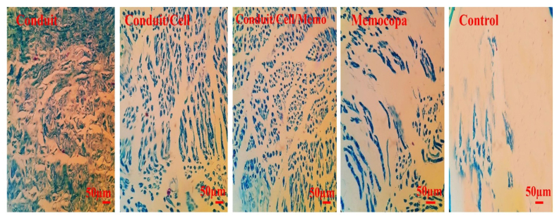
Method: EnMSCs were isolated, characterized, and differentiated in a study. The expression of specific genes in the differentiated cells was confirmed using RT-PCR and immunocytochemistry. PLA nanofiber and chitosan hydrogel scaffolds were created for neural tissue engineering. Memocopa was administered orally alongside scaffold and cell transplantation. The study involved 25 adult male Wistar rats with a 3 mm sciatic nerve gap, divided into five groups based on treatment. Animals were monitored for 8 weeks, during which SFI was measured. Tissue samples were then prepared for histological examination, including various staining techniques.

Results: The combination of scaffold, cells, and Memocopa showed significant improvements in sciatic nerve function, as indicated by the SFI results in the eighth week: conduit group -33.87, conduit/cells group -25.92, conduit/cells/Memocopa group -22.86, Memocopa group -30.93, and control group -38.87. Histological findings revealed improvements in various aspects, including percentages of new nerve formation across the different treatment groups: conduit group 4.62 %, conduit/cells group 16.45 %, conduit/cells/Memocopa group 21.32 %, Memocopa group 7.07 %, and control group 0.22 %.

Conclusions: The results of this study showed that efficient differentiation of EnMSCs into NLCs is possible, and with the help of PLA/CS scaffold and simultaneous use of Memocopa, it is possible to repair and improve sciatic nerve injury in a rat animal model.

Keywords: Bacopa monnieri; Conduit; Endometrial stem cells; Nerve regenerating; Neural differentiation.


Conflict of interest statement

The authors declare that they have no known competing financial interests or personal relationships that could have appeared to influence the work reported in this paper.

Figures

Image 1
Graphical abstract
Fig. 1
Fig. 1
Morphology examination and identification of mesenchymal stem cells derived from the endometrial uterus using an inverted microscope. (a) Isolated EnMSCs cells were cultured and found in the culture medium 24 h later. (b) Attached EnMSCs cells in the flask bottom in the third passage. (c) Differentiation of EnMSCs cells into adipocytes with Oil Red staining 21 days after differentiation, red areas indicate the formation of fat droplets. (d) Differentiation of EnMSCs cells into osteoblast cells with Alizarin Red staining 21 days after differentiation, red areas indicate the accumulation and deposition of calcium. (e) Flowcytometry analysis of EnMSCs cells in the third passage. Cells were labeled with specific monoclonal antibodies against specified molecules (colorful chart) and with isotype controls (colorless chart), positive markers CD73, CD90, and CD105, and negative markers CD34 and CD45 were examined.
Fig. 2
Fig. 2
(a) Immunocytochemistry analysis for the expression of Nestin, Map-2, and Tuj-1 markers as neuron-like specific markers. This image shows EnMSCs cells 14 days after neural differentiation. The cell nuclei are stained with DAPI. (b) RT-PCR analysis for the expression of specific genes Nestin, Map-2, Tuj-1, and NF-L in neuron-like cells and EnMSCs cells 14 days after treatment. Each experiment was repeated three times and the GAPDH gene was used as a housekeeping gene (∗ indicates a significant level with P ≤ 0.001). (SEM = 0.2).
Fig. 3
Fig. 3
Microscopic images of scaffolds and EnMSCs cell cultures on them. (a) SEM image of electrospun PLA scaffold without cells. (b–c) SEM image of EnMSCs cells cultured on the scaffold, demonstrating the biocompatibility of the scaffold with a large number of cells attached to it. (d) Acridine orange/ethidium bromide staining shows a large number of live cells appearing green. (e) DAPI staining showing cell nuclei in blue. (f–g) Inverted microscope image of EnMSCs cells differentiated into neural precursor cells in chitosan hydrogel, with the axonal network of neurons visible (10 ×) and (20 ×). (h) SEM image of chitosan hydrogel with pseudo-neuronal cells. (i) SEM image of chitosan hydrogel. (j) Evaluation of the viability of EnMSCs cells treated with different concentrations of Memocopa using the MTT test after 24 h (n = 3). ∗p < 0.001 compared to the control sample. (SEM = 2).
Fig. 4
Fig. 4
Evaluation of sciatic nerve function index. (a) Evaluation of sciatic nerve function index in different groups in the second week. (b) Evaluation of sciatic nerve function index in different groups in the fourth week. (c) Evaluation of sciatic nerve function index in different groups in the sixth week. (d) Evaluation of sciatic nerve function index in different groups in the eighth week. The graphs represent the average of 3 repetitions. Symbols that are not common between columns indicate that statistically, the difference in their averages is significant at a probability level of less than 0.05. ∗, ###, $$$, @@@, and $$$ indicate significant levels with p < 0.001 compared to other. (SEM = 2.5).
Fig. 5
Fig. 5
Histological examination of sciatic nerve samples in a rat model with H&E staining in different groups including the Candida group, Conduit/cell group, Conduit/cell/Memocopa group, Memocopa group, and control group under a light microscope 8 weeks after treatment. In the images, S indicates Schwann cells, N indicates neurons, and CT indicates connective tissue. The results of the H&E staining images showed significant improvement and regeneration in the nerve injury of the group with Conduit. The samples are shown in two magnifications, with details of the images on the left (10 ×) magnified further, and on the right (4 ×) visible.
Fig. 6
Fig. 6
Results related to histomorphometric evaluations. (a) The diameter of nerves formed in different groups 8 weeks after treatment (SEM = 0.5). The largest nerve diameter is observed in the Conduit/Cell/Memocopa group, and the smallest nerve diameter is observable in the control group. (b) The number of axons in the regenerated nerve in different groups 8 weeks after treatment. (SEM = 20). The highest number of axons is in the Conduit/Cell/Memocopa group, and the lowest number of axons is observable in the control group. (c) The number of Schwann cells in the regenerated nerve in different groups 8 weeks after treatment. (SEM = 20). The highest number of Schwann cells is in the Conduit/Cell/Memocopa group, and the lowest number of Schwann cells is observable in the control group. (d) The number of vessels formed in the regenerated nerve in different groups 8 weeks after treatment. (SEM = 2). The highest number of vessels is in the Conduit/Cell/Memocopa group, and the lowest number of vessels is observable in the control group. (e) Percentage of new nerve formation in different groups 8 weeks after treatment. (SEM = 1). The graphs represent the average of 3 repetitions. The non-common symbols between the columns indicate that statistically, the difference in their averages is significant at the 0.05 level. ∗, ###, $$$, @@@, and $$$ indicate significant levels with p < 0.001 compared to other groups. ## indicates significant levels with p < 0.01 compared to other mucosa.
Fig. 7
Fig. 7
Examination of nerve samples with LFB staining in different groups using light microscopy 8 weeks after treatment. In LFB staining, myelin appears blue. In this study, all groups showed significant improvement compared to the control. In the control group, we do not observe the formation of specific axons. In the Conduit/cell/Memocopa group, fibers and axons are denser, more regular, and have a higher intensity of staining. The scale indicates 50 mμ.
Fig. 8
Fig. 8
Evaluation of the expression of the NF-200 neural marker in sciatic nerve samples with immunohistochemical staining in different groups including the Conduit group, Conduit/cell group, Conduit/cell/Memocopa group, and Memocopa group using a fluorescent microscope 8 weeks after treatment. The higher the intensity of the green color, the more indicative of increased neurofilament expression. S: Indicates the presence of the scaffold. The nuclei were stained with DAPI color.

Similar articles

References

    1. Sulaiman W., Gordon T. Neurobiology of peripheral nerve injury, regeneration, and functional recovery: from bench top research to bedside application. Ochsner J. 2013;13(1):100–108. - PMC - PubMed
    1. Hussain G., Wang J., Rasul A., Anwar H., Qasim M., Zafar S., et al. Current status of therapeutic approaches against peripheral nerve injuries: a detailed story from injury to recovery. Int J Biol Sci. 2020;16(1):116. - PMC - PubMed
    1. Piñero G., Usach V., Soto P.A., Monje P.V., Setton-Avruj P. EGFP transgene: a useful tool to track transplanted bone marrow mononuclear cell contribution to peripheral remyelination. Transgenic Res. 2018;27:135–153. - PubMed
    1. Hoveizi E., Tavakol S. Therapeutic potential of human mesenchymal stem cells derived beta cell precursors on a nanofibrous scaffold: an approach to treat diabetes mellitus. J Cell Physiol. 2018;234(7):10196–10204. - PubMed
    1. Hoveizi E., Mohammadi T., Moazedi A.A., Zamani N., Eskandary A. Transplanted neural-like cells improve memory and Alzheimer-like pathology in a rat model. Cytotherapy. 2018;20(7):964–973. Epub 2018/07/22. - PubMed

LinkOut - more resources