Case Reports
. 2025 May 30:2025:8268690.
doi: 10.1155/crot/8268690. eCollection 2025.

Sternoclavicular Joint Tracheal Fistula: An Unusual Postradiation Complication in a Laryngectomee

Affiliations
Case Reports

Sternoclavicular Joint Tracheal Fistula: An Unusual Postradiation Complication in a Laryngectomee

Elena Dina et al. Case Rep Otolaryngol. .

Abstract

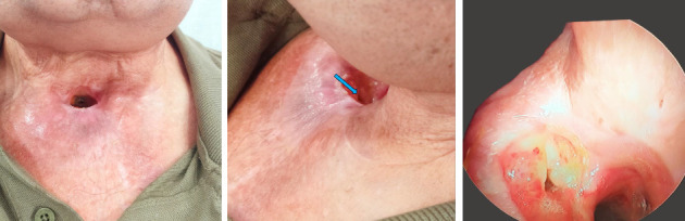
A 68-year-old man previously treated for a large laryngeal neoplasm (pT4 pN0 squamous cell carcinoma) developed osteomyelitis of the medial third of the right clavicle with the formation of a fistula between the sternoclavicular joint and tracheal wall near the tracheostomy border. The clinical course was tedious, required prolonged antibiotic trials, and extended surgical bone resection to control the infection. The final outcome was favorable with wound closure although the patient was left with permanent limitation of shoulder abduction (his shoulder mobility had been normal prior to this process). Histopathological examination of the resected bone suggested a diagnosis of both osteoradionecrosis and osteomyelitis. Indeed, differential diagnosis between these two entities can be challenging after radiotherapy. Here, we present a review of the relevant academic literature and discuss the therapeutic options.

Keywords: clavicular osteomyelitis; clavicular osteoradionecrosis; sternoclavicular joint fistula; sternoclavicular joint osteomyelitis; sternoclavicular joint osteoradionecrosis.


Conflict of interest statement

The authors declare no conflicts of interest.

Figures

Figure 1
Figure 1
(a) Initial case presentation: right SCJ and adjacent anterior chest wall cellulitis. First axial CT scan slides (b). The red arrow shows focal ulceration of the right side of the tracheostomy; the yellow arrows show soft tissue inflammatory changes (cellulitis). (c and d) Chest wall inflammatory changes (cellulitis) and right pectoral major myositis (asterisk) with gas formation (red circle).
Figure 2
Figure 2
Front and oblique view: better infection control, the blue arrow shows SCJ–tracheal fistula. Weeks later: a 30° endoscope view of the fistula with articular liquid and air bubbles.
Figure 3
Figure 3
CT scan at 3 weeks: (a) liquid collection containing gas (red circle) in the right pectoralis major. (b) Both clavicle and sternum bone erosions (red arrows).
Figure 4
Figure 4
CT scan at 6 weeks later after clinical worsening: (a and b) axial and (c and d) coronal slides. Signs of sternoclavicular joint arthritis with enlargement of the bone erosions, osteolysis, and soft tissue augmentation (red arrows).
Figure 5
Figure 5
Resection of the medial third of the clavicle, a fibrous capsule closed the fistula tract, and the space was filled with STIMULAN rapid cure beads soaked in vancomycin and gentamicin; blue star: lateral portion of the right clavicle; yellow star: right subclavicular region.
Figure 6
Figure 6
(a) Hematoxylin–eosin (HE) stained, ×10 magnification: ORN with dense fibrosis (), chronic inflammatory infiltrate (OSM), and empty bone trabeculae (arrows). (b) HE stained, ×20 magnification: the image of a residual bone trabecula with osteonecrosis and clusters of empty osteocytes without viable nuclei (arrow).
Figure 7
Figure 7
Postoperative at 1 month: local healing and limited arm abduction.
Figure 8
Figure 8
Postoperative at 6 months: clinical stability. Axial CT scan: fibrosis filling the clavicular resection space (blue arrows).

Similar articles

References

    1. Vu T., Yammine N. V., Al‐Hakami H., Hier M. P., Black M. J. Sternoclavicular Joint Osteomyelitis Following Head and Neck Surgery. The Laryngoscope . 2010;120(5):920–923. doi: 10.1002/lary.20849. - DOI - PubMed
    1. Piazza C., Magnoni L., Nicolai P. Clavicular Osteomyelitis: A Rare Complication After Surgery for Head and Neck Cancer. European Archives of Oto-Rhino-Laryngology . 2006;263(7):653–656. doi: 10.1007/s00405-006-0040-z. - DOI - PubMed
    1. Shodo R., Sato Y., Ota H., Horii A. Clavicle Fracture With Osteomyelitis After Neck Dissection and Post-Operative Radiotherapy: Case Report. Journal of Laryngology & Otology . 2017;131(11):1026–1029. doi: 10.1017/S0022215117001748. - DOI - PubMed
    1. Irizarry R., Shatzkes D. R., Teng S., Kohli N., Har‐El G. Osteoradionecrosis of the Sternoclavicular Joint After Laryngopharyngeal Radiation. The Laryngoscope . 2019;129(4):865–870. doi: 10.1002/lary.27324. - DOI - PubMed
    1. Stofman G. M., Lowry L. D., Cohn J. R., Jabourian Z. Osteoradionecrosis of the Head and Neck: A Case of a Clavicular-Tracheal Fistula Secondary to Osteoradionecrosis of the Sternoclavicular Joint. Annals of Otology, Rhinology & Laryngology . 1988;97(5):545–549. doi: 10.1177/000348948809700522. - DOI - PubMed

Publication types

LinkOut - more resources