. 2025 May 14:51:257-273.
doi: 10.1016/j.bioactmat.2025.05.007. eCollection 2025 Sep.

HydroWrap for T2DM-Related Fractures: A smart H2S-delivery controller modulating Macrophage senescence

Affiliations

HydroWrap for T2DM-Related Fractures: A smart H2S-delivery controller modulating Macrophage senescence

Siqi Zhang et al. Bioact Mater. .

Abstract

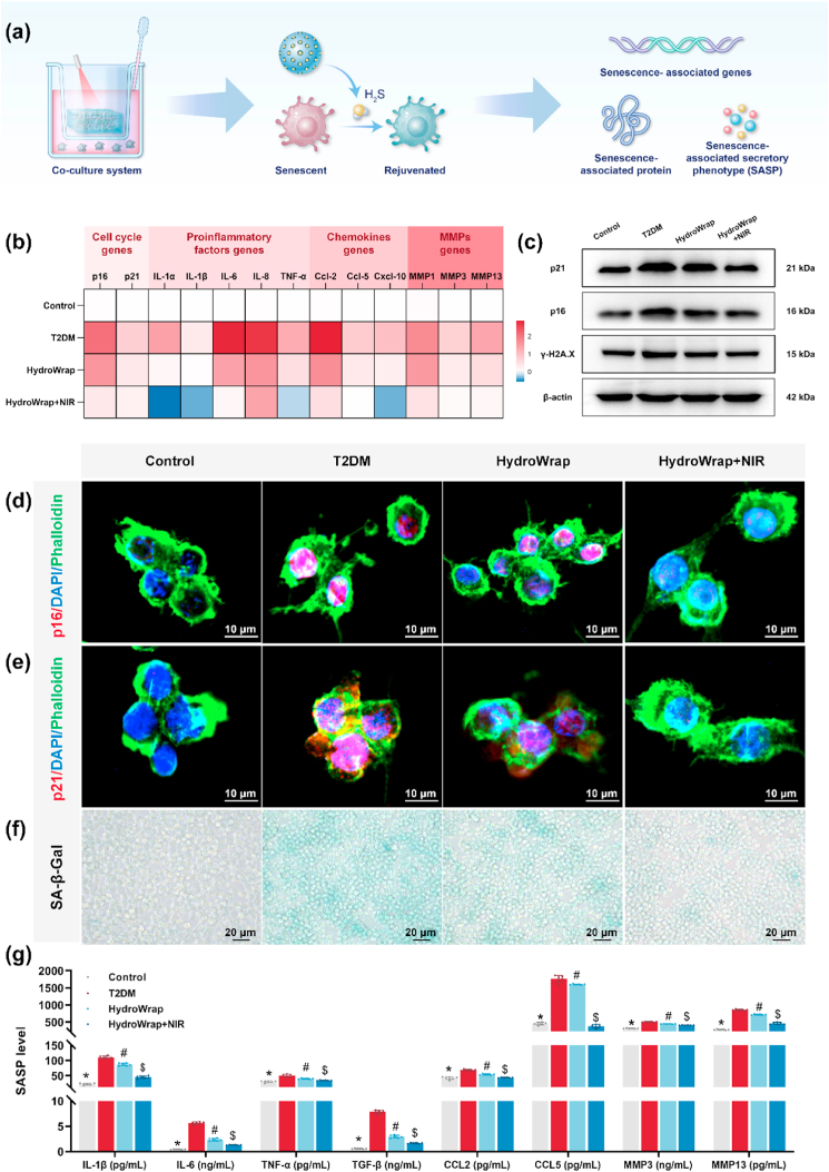
Impaired fracture healing in type 2 diabetes mellitus (T2DM) poses a significant clinical challenge, primarily due to a compromised bone microenvironment driven by senescent macrophages and their amplifying effects. Reduced hydrogen sulfide (H2S) levels are a critical contributor to this pathology. To address this, we developed HydroWrap, an advanced H2S-delivery controller designed to modulate distinct stages of macrophage senescence. Under near-infrared (NIR) irradiation, HydroWrap underwent an increase in temperature, causing the hydrogel network to contract and accelerate H2S generation. This rapid delivery restores H2S levels, alleviating mitochondrial dysfunction and suppressing senescence-associated secretory phenotypes (SASP), thereby interrupting the senescence cascade. In T2DM's hyperglycemic bone microenvironment, HydroWrap provides sustained, glucose-responsive H2S release, promoting mitophagy and preventing macrophage senescence progression. This dual mechanism addresses both acute and chronic dysfunctions associated with senescence. In vivo studies demonstrated that HydroWrap significantly improved fracture healing by reducing recovery time and enhancing bone quality. These findings underscore the therapeutic potential of modulating macrophage senescence in T2DM using a biocompatible drug delivery system. HydroWrap offers a promising strategy for improving fracture outcomes in diabetic patients and may hold broader applications in senescence-related diseases.

Keywords: Bone senescence; Diabetic fracture healing; Hydrogen Sulfide; Photothermal therapy; Responsive hydrogel.


Conflict of interest statement

The authors declare the following financial interests/personal relationships which may be considered as potential competing interests: Yafei Feng, Wei Lei reports financial support was provided by 10.13039/501100001809National Natural Science Foundation of China. If there are other authors, they declare that they have no known competing financial interests or personal relationships that could have appeared to influence the work reported in this paper.

Figures

Image 1
Graphical abstract
Scheme 1
Scheme 1
Schematic illustration of HydroWrap, a smart H2S-releasing controller, showcasing its dual mechanisms of rejuvenating senescent macrophages and disrupting the senescence cascade to accelerate fracture healing in T2DM.
Fig. 1
Fig. 1
Senescent Macrophages and Reduced H2S levels in T2DM patients. (a) Schematic diagram illustrating the methodology used to identify overlapping genes between DEGs in monocytes from T2DM patients and a cell senescence gene database. (b) Venn diagram and circular heatmap showing the common genes identified. (c) GO analysis of the shared DEGs. (d) Schematic representation of the process for obtaining femur head samples and the site selected for histological staining. (e) Immunofluorescence images showing co-localization of p16 and CD68 in femur head tissue. (f) Violin plots comparing H2S concentrations in plasma between healthy controls and T2DM patients (n = 20). (g, h) Logistic regression analysis exploring the association between plasma H2S concentration and BGL or plasma SASP concentrations. (i) Violin plots comparing H2S concentrations in monocyte culture supernatants between healthy controls and T2DM patients (n = 20). (j) mRNA expression levels of key H2S-synthesizing enzymes (CBS, CSE, and 3-MST) in monocytes. (k) Summary diagram highlighting the key findings: increased senescent macrophages and elevated SASP levels, accompanied by reduced H2S secretion in T2DM patients compared to controls.
Fig. 2
Fig. 2
Synthesis and Characterization of HydroWrap. (a) Schematic diagram illustrating the fabrication process of mPDA-H2S@ABA NPs (yellow dots referring to ADT). (b) TEM image with EDS mapping showing the elemental composition of mPDA-H2S@ABA. (c) Zeta potential analysis of mPDA NPs and mPDA-H2S NPs. (d) Particle size distribution of mPDA NPs and mPDA-H2S@ABA NPs. (e) Schematic representation of the fabrication process for HydroWrap. (f) SEM images showing the morphologies of SilMA/pNIPAM hydrogel and the final HydroWrap. (g) Frequency-dependent storage and loss moduli from oscillatory frequency sweep tests performed on composites containing different concentrations of mPDA-H2S@ABA NPs. (h) Photothermal stability of HydroWrap demonstrated over five NIR laser on/off cycles. (i) Comparative cumulative H2S release profiles from HydroWrap with (42 °C) and without (25 °C) NIR laser exposure. (j) Viscosity variations of HydroWrap in glucose solutions at different concentrations. (k) Cumulative H2S release profiles from HydroWrap in glucose solutions of varying concentrations over 72 h. (l) Photographs showing shape changes of HydroWrap under NIR laser irradiation or in glucose solutions. (m) Schematic diagram illustrating the therapeutic mechanism of HydroWrap.
Fig. 3
Fig. 3
In Vitro Anti-senescence Effects on Macrophages. (a) Schematic diagram illustrating the co-culture system and proposed therapeutic mechanism. (b) Heatmap showing relative mRNA expression levels of senescence-associated genes. (c) WB analysis of p16, p21, and γ-H2A.X protein expression levels. (d, e) Confocal immunofluorescence images showing p16 and p21 localization (red: p16/p21; green: phalloidin; blue: DAPI). (f) SA-β-gal staining of macrophages to assess senescence. (g) SASP levels in co-culture supernatants measured by ELISA (n = 6 per group). Statistical significance is indicated as follows: ∗, #, and $ denote p < 0.05 when comparing the Control, HydroWrap, and HydroWrap + NIR groups to the T2DM group, respectively.
Fig. 4
Fig. 4
Macrophage Rejuvenation Enhances Angiogenesis and Osteogenesis. (a) Schematic illustration of the experimental approach to evaluate the impact of conditioned medium from macrophages on angiogenesis and osteogenesis. (b) Crystal violet staining of HUVECs in the wound healing assay, and (c) quantitative analysis of wound closure. (d) Representative images from the tube formation assay and (e) quantitative analysis of vessel area percentage. (f) Representative images of ALP and ARS staining in MC3T3-E1 cells, accompanied by (g) quantitative analysis of ALP activity and (h) mineralization levels. (i) Relative mRNA expression levels of angiogenesis-related genes in HUVECs and (j) osteogenesis-related genes in MC3T3-E1 cells (n = 6 per group). Statistical significance is indicated as follows: ∗, #, and $ denote p < 0.05 when comparing the Control, HydroWrap, and HydroWrap + NIR groups to the T2DM group, respectively.
Fig. 5
Fig. 5
In Vivo Assessment of Diabetic Fracture Healing. (a) Schematic representation of the experimental design for animal studies. (b) In vivo photothermal performance of the HydroWrap + NIR group. (c) X-ray radiographs showing fracture sites at 1 and 2 weeks post-surgery, with yellow arrows indicating callus formation. (d) Micro-CT images and 3D reconstructions of fracture sites at 4 and 8 weeks post-surgery. (e) Quantitative assessment of fracture healing scores at 1 and 2 weeks. (f–g) Measurements of BMD, BV, and BV/TV in callus regions (n = 6 per group). Statistical significance is indicated as follows: ∗, #, and $ denote p < 0.05 when comparing the Control, HydroWrap, and HydroWrap + NIR groups to the T2DM group, respectively.
Fig. 6
Fig. 6
Histological Evaluation of HydroWrap's Efficacy in Promoting Diabetic Fracture Healing. (a, b) Histological analysis of fractured femurs at 4 and 8 weeks post-surgery using H&E and Masson staining. (c) Immunohistochemical (IHC) staining at 4 and 8 weeks post-surgery for OCN, an osteogenesis marker, and VEGF, an angiogenesis marker, at the fracture site. (d–f) Quantitative evaluation of callus width, callus area, and collagen fiber area at 4 and 8 weeks post-surgery. (g, h) Quantification of OCN/VEGF positive cell areas. (Data are presented as mean ± standard deviation; n = 6 per group. Statistical significance is indicated as follows: ∗, #, and $ denote p < 0.05 when comparing the Control, HydroWrap, and HydroWrap + NIR groups to the T2DM group, respectively.).
Fig. 7
Fig. 7
Transcriptome sequencing analysis of DEGs. (a, b) Venn diagram and volcano plots showing DEGs across groups. (c) Heatmap of DEGs associated with cellular senescence. (d) GO enrichment circle plots highlighting the top 10 enriched terms. (e) GSEA of DNA replication and cell cycle pathways. (f) Reactome analysis categorizing DEGs into five key terms. (g) Network diagram illustrating protein interactions among three key pathways.
Fig. 8
Fig. 8
Mechanisms by which HydroWrap Reduces Macrophage Senescence. (a) WB analysis of proteins involved in the NF-κB pathway. (b) qRT-PCR quantification of expression levels of three key H2S-synthesizing enzymes (CBS, CSE, and 3-MST). (c) WB analysis of proteins in the Pink1/Parkin signaling pathway. (d) JC-1 staining of macrophages to assess mitochondrial membrane potential (red: JC-1 aggregates; green: JC-1 monomers). (e) Fluorescence microscopy of macrophages transfected with Ad-EGFP-LC3B and stained with Mitotracker-Red to visualize mitophgy and mitochondrial dynamics.

References

    1. Ahmad E., Lim S., Lamptey R., Webb D.R., Davies M.J. Type 2 diabetes. Lancet. 2022;400(10365):1803–1820. doi: 10.1016/S0140-6736(22)01655-5. - DOI - PubMed
    1. Khosla S., Samakkarnthai P., Monroe D.G., Farr J.N. Update on the pathogenesis and treatment of skeletal fragility in type 2 diabetes mellitus. Nat. Rev. Endocrinol. 2021;17(11):685–697. doi: 10.1038/s41574-021-00555-5. - DOI - PMC - PubMed
    1. Vilaca T., Schini M., Harnan S., Sutton A., Poku E., Allen I.E., Cummings S.R., Eastell R. The risk of hip and non-vertebral fractures in type 1 and type 2 diabetes: a systematic review and meta-analysis update. Bone. 2020;137 doi: 10.1016/j.bone.2020.115457. - DOI - PubMed
    1. Hofbauer L.C., Busse B., Eastell R., Ferrari S., Frost M., Muller R., Burden A.M., Rivadeneira F., Napoli N., Rauner M. Bone fragility in diabetes: novel concepts and clinical implications. Lancet Diabetes Endocrinol. 2022;10(3):207–220. doi: 10.1016/S2213-8587(21)00347-8. - DOI - PubMed
    1. Leungsuwan D.S., Chandran M. Bone fragility in diabetes and its Management: a narrative review. Drugs. 2024;84(9):1111–1134. doi: 10.1007/s40265-024-02078-5. - DOI - PubMed

LinkOut - more resources