. 2025 May 8;9(2):631-660.
doi: 10.1162/netn_a_00420. eCollection 2025.

Whole-brain modular dynamics at rest predict sensorimotor learning performance

Affiliations

Whole-brain modular dynamics at rest predict sensorimotor learning performance

Dominic I Standage et al. Netw Neurosci. .

Abstract

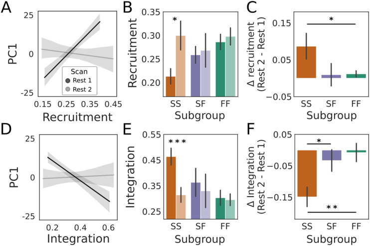
Neural measures that predict cognitive performance are informative about the mechanisms underlying cognitive phenomena, with diagnostic potential for neuropathologies with cognitive symptoms. Among such markers, the modularity (subnetwork composition) of whole-brain functional networks is especially promising due to its longstanding theoretical foundations and recent success in predicting clinical outcomes. We used functional magnetic resonance imaging to identify whole-brain modules at rest, calculating metrics of their spatiotemporal dynamics before and after a sensorimotor learning task on which fast learning is widely believed to be supported by a cognitive strategy. We found that participants' learning performance was predicted by the degree of coordination of modular reconfiguration and the strength of recruitment and integration of networks derived during the task itself. Our findings identify these whole-brain metrics as promising network-based markers of cognition, with relevance to basic neuroscience and the potential for clinical application.

Keywords: Cognition; Dynamic modularity; Predictive markers; Resting-state fMRI; Sensorimotor learning.

Plain language summary

The prediction of cognitive performance by measures derived from neuroimaging signals is informative about the neural processes underlying those signals, with the potential for use in the diagnosis of cognitive abnormalities, such as those associated with aging and Alzheimer’s disease. These neural markers include statistics of functional networks, characterizing the cooperation of distributed brain regions in large-scale networks. We investigated dynamics (changes over time) of functional networks prior to a task on which participants differ in their use of strategies. We found that performance on the upcoming task was predicted by several measures of whole-brain network dynamics.


Conflict of interest statement

Competing Interests: The authors have declared that no competing interests exist.

Figures

<b>Figure 1.</b>
Figure 1.
Overview of the task and neural analyses. (A) Participants underwent a resting scan before and after a VMR task, during which the viewed cursor, controlled by the hand, was rotated about the movement start location. (B) For each participant, the cerebral cortex, striatum, and cerebellum were parcellated into discrete regions and the average %BOLD time-series was extracted from each region during resting and task scans (three example cortical regions are shown). (C) The coherence of (Haar family) wavelet coefficients was calculated for each pair of regions in sliding time windows to construct functional connectivity matrices for each window (w1 – wN, see the Methods section). Time-resolved clustering methods were applied to the resulting multislice networks, identifying dynamic modules across time slices (four modules in the schematic). (D) Module allegiance matrix (left) showing the probability that each pair of brain regions was in the same module during early learning, calculated over all participants, modular partitions, and time windows (left). The arrow depicts the clustering of this matrix, identifying networks (right) that summarize the modular dynamics (see the Methods section). Brain plots of these networks are shown in Supporting Information Figure S1.
<b>Figure 2.</b>
Figure 2.
Participants were assigned to one of three subgroups by a clustering of their behavioral data. (A) Mean (across participants) of bin median errors during baseline (nonrotation), learning (45° rotation of the cursor), and “washout” (nonrotation, to unlearn the mapping prior to the second day) on Day 1 (pink) and Day 2 (cyan). Bins consisted of eight consecutive trials, during which the target was chosen at random without replacement from eight equidistant locations around an invisible circle. Early error and late error were defined as the first and last three learning bins, respectively (gray shading). Ribbons show standard error (±1 SE), and the dashed vertical lines demarcate the three task blocks. Savings (inset, early error on Day 1 minus early error on Day 2) was significant at the group level (paired t test; t[31] = 6.122, P = 8.666e-7). (B) The group-averaged approach in panel A obscures individual differences in learning. The trajectory of errors by three example participants shows a participant who learned quickly on both days (top, error is reduced quickly on Day 1 [magenta] and on Day 2 [cyan]), a participant who learned slowly on both days (middle, error is reduced slowly on both days) and a participant who learned slowly on Day 1 and quickly on Day 2 (bottom). (C) A clustering of participants’ early error on each day, late error on each day, and savings (five variables in total) identified three subgroups of participants whose mean bin median errors resemble the example participants in panel B on Day 1 (left) and Day 2 (right). We refer to these subgroups as FF (fast learning on both days), SS (slow learning on both days), and SF (slow learning on Day 1 and fast learning on Day 2). (D) Top two PCA components for the five learning measures across participants. Data points correspond to participants, color-coded by their cluster-assigned subgroup (legend in panel C). The horizontal axis shows that PC1 accurately classifies 31 of 32 participants (97% accuracy). Scatter plots on the right show that PC1 and PC2 closely correspond to mean early error across days and savings, respectively.
<b>Figure 3.</b>
Figure 3.
Prior to learning, greater dynamic modularity scores did not predict better task performance. The scatter plot shows PC1 for each subject, our proxy for behavioral subgroup membership, as a function of the mean quality function score Q (quantifying modularity, see text). Filled circles correspond to the FF, SS, and SF behavioral subgroups (see legend). The fitted line shows a linear regression model (least squares fit) where the shaded area corresponds to ±1 SE. Subgroup means ±1 SE are shown as bar graphs below the scatter plots. Contrary to our hypothesis, the fit was nonsignificant (R2 = 0.070, F[1, 30] = 2.246, p = 0.144).
<b>Figure 4.</b>
Figure 4.
Coordinated (A) and uncoordinated (B) modular reconfiguration at rest predicted fast and slow learning profiles on the upcoming task, respectively. (A) PC1 as a function of mean cohesive flexibility (cohesion strength, see text). The fit of a linear regression model (black line; gray shading shows ±1 SE) showed a positive predictive relationship between cohesion strength (the predictor) and PC1 (R2 = 0.198, F[1, 30] = 7.401, p = 0.011), where FF was more cohesive than SS (two-sample t test: t[23] = 2.518, p = 0.019), but SF did not differ significantly from FF (t[20] = 1.233, p = 0.232) or SS (t[15] = 1.176, p = 0.258). (B) PC1 as a function of mean disjointed flexibility (disjointedness, see text). The fit of a linear regression model (black line) showed a negative predictive relationship between disjointedness (the predictor) and PC1 (R2 = 0.186, F[1, 30] = 6.852, p = 0.014), where SS was more disjointed than FF (t[23] = −2.089, p = 0.048), but SF did not differ significantly from SS (t[15] = −1.115, p = 0.282) or FF (t[20] = −0.596, p = 0.558). (C) Brain plots show Pearson’s correlation coefficient (r=R2) for cohesion strength and PC1 (upper plots) and disjointedness and PC1 (lower plots) for individual brain regions. Correlation coefficients associated with cohesion strength (mostly positive) and disjointedness (mostly negative) are shown with a divergent color scheme, ranging from strongly negative (dark blue) to strongly positive (dark red). In panels A and B, subgroup means ±1 SE are shown as bar graphs below the scatter plots, where stars indicate significant differences (p < 0.05).
<b>Figure 5.</b>
Figure 5.
Prior to learning, stronger recruitment of the relearning network, and weaker integration between the learning and relearning networks, predicted faster learning and thereby a more strategic (cognitive) approach to the task. (A) The relearning network (left) consisted of regions spanning contralateral motor cortex, bilateral cerebellum, medial prefrontal cortex, and several subcortical structures (bilateral hippocampus, pallidum, amygdala, and accumbens). Recruitment of this network at rest was a significant predictor of PC1, as determined by the fit of a linear regression model (black line; R2 = 0.253, F[1, 30] = 10.15, p = 0.003, FDR-adjusted p = 0.017). Gray shading shows ±1 SE. Recruitment by FF was stronger than SS (t[23] = 3.028, p = 0.006), but recruitment by SF did not differ statistically from that of FF (t[20] = 1.013, p = 0.323) or SS (t[15] = 1.858, p = 0.083). (B) The learning network (left) consisted of regions spanning the anterior temporal pole, inferior and superior parietal, dorsolateral prefrontal cortex, and the bilateral caudate. Its pre-task integration with the relearning network was (negatively) predictive of PC1 (R2 = 0.199, F[1, 30] = 7.447, p = 0.010), where integration was stronger by the SS subgroup than FF (t[23] = 3.574, p = 0.002) but did not differ statistically between SF and FF (t[20] = 1.061, p = 0.301) or SS (t[15] = −1.673, p = 0.115). Error bars show ±1 SE. In bar plots, two stars indicate significant differences (p < 0.01).
<b>Figure 6.</b>
Figure 6.
Prior to learning, stronger RSFC predicted better task performance. The scatter plot shows PC1 as a function of node strength (mean strength of connectivity over all brain regions over all temporal windows for each subject), where the black line shows the significant fit of linear regression model +1 SE (R2 = 0.188, F[1, 30] = 6.968, p = 0.013). Node strength was stronger among the FF subgroup than the SS subgroup (two-sample t test: t[23] = 2.368, p = 0.027), but did not differ statistically between FF and SF (t[20] = 0.488, p = 0.631) or SS and SF (t[15] = 1.556, p = 0.141). The star in the bar plot indicates statistical significance (p < 0.05).
<b>Figure 7.</b>
Figure 7.
(A) The Pearson correlation between recruitment of the relearning network and PC1 was stronger (Steiger’s test for dependent correlations: t[30 ] = 3.636 p = 0.001, corrected p = 0.005) during the first (pre-task) resting scan (Rest 1, black: r = 0.501, p = 0.003; r = R2 per Figure 5A) than the second (post-task) resting scan (Rest 2, gray: r = −0.087, p = 0.635). (B) The change in recruitment was driven by the SS subgroup, whose recruitment was significantly greater (paired t test: t[9] = 2.475, p = 0.035) during the second resting scan (lighter shade) than the first (darker shade). Neither of the FF (t[14] = 1.386, p = 0.187) or SF (t[6] = 0.311, p = 0.766) subgroups differed significantly between resting scans. (C) The change in recruitment by SS was significantly greater than that by FF (two-sample t test: t[23] = −2.487, p = 0.021) but did not differ significantly between SS and SF (t[15] = 0.096, p = 0.924) or between SF and FF (t[20] = −1.573, p = 0.137). (D) The correlation between integration (of the relearning network with the regions of the learning network) and PC1 was weaker (Steiger’s test for dependent correlations: t[30 ] = −3.136, p = 0.004, corrected p = 0.010) during the first resting scan (Rest 1, black, r = −0.446, p = 0.010; ∣r∣ = R2 per Figure 5B) than the second resting scan (Rest 2, gray, r = 0.025, p = 0.890). (E) The change was again driven by the SS subgroup, whose integration was significantly weaker (t[9] = −4.798, p = 9.758e-4) during the second resting (lighter shade) scan than during the first (darker shade). The FF (t[14] = −0.266, p = 0.794) and SF (t[6] = −0.964, p = 0.372) subgroups did not differ significantly between resting scans. (F) The change in integration by SS was significantly greater than that by SF (two-sample t test: t[15] = −2.468, p = 0.027) and FF (t[23] = 3.230, p = 0.004), but FF and SF were not significantly different (t[20] = 0.520, p = 0.609). In all panels, stars indicate statistical significance (one star: p < 0.05; two stars: p < 0.01; three stars: p < 1e-3).
<b>Figure 8.</b>
Figure 8.
The organization of dynamic, resting-state functional networks was more similar during pre-task (Rest 1) and post-task (Rest 2) rest than when either resting scan was compared with learning. (A) Linear fit to whole-group module allegiance probabilities (P, the probability that each of the 10,011 pairs of brain regions were in the same module; see text) for Rest 1 and Rest 2 (black, r = 0.838), Rest 1 and early learning (gray, r = 0.765), and Rest 2 and early learning (gray, r = 0.761). The gray lines are nearly indiscernible, as is the standard error in all fits. Fitted data points are omitted for clarity. (B) Mean similarity (Pearson’s r) of module allegiance matrices across all participants for each pair of scans (R1: Rest 1; R2: Rest 2: EL: early learning). Rest 1 and Rest 2 were significantly more similar than Rest 1 and early learning (paired one-tailed t test: t[31] = 2.174, p = 0.019) and Rest 2 and early learning (R2-EL; t[31] = 1.918, p = 0.032), whereas the correlation between Rest 1 and early learning was not statistically different from Rest 2 and early learning (paired two-tailed t test: t[31] = −0.269, p = 0.790). Note that our use of one-tailed t tests reflects our testing of predictions in panel “A.” (C) Mean similarity of each pair of scans for each behavioral subgroup (left, SS; middle, SF; right, FF). Among FF participants, module allegiance matrices from the resting scans were more similar than Rest 1 and early learning (paired one-tailed t test: t[14 ) = −2.112, p = 0.027) or Rest 2 and early learning (t[14] = −2.574, p = 0.011), whereas the resting scans’ correlations with learning were not significantly different (paired two-tailed t test: t[14] = 0.108, p = 0.542). Among SF and SS participants, Rest 1 and Rest 2 were not significantly more similar than Rest 1 and early learning (paired one-tailed t test, SF: t[6] = −0.069, p = 0.474; SS: t[9] = −1.648, p = 0.067) or Rest 2 and early learning (SF: t[6] = −0.015, p = 0.494; SS: t[9] = −0.489, p = 0.318) and the correlation between Rest 1 and early learning was not statistically different from Rest 2 and early learning (paired two-tailed t test, SF: t[6] = 0.102, p = 0.539; SS: t[9] = 0.241, p = 0.592). Single stars in panels B and C indicate statistical significance (p < 0.05).

References

    1. Abraham, A., Pedregosa, F., Eickenberg, M., Gervais, P., Mueller, A., Kossaifi, J., … Varoquaux, G. (2014). Machine learning for neuroimaging with scikit-learn. Frontiers in Neuroinformatics, 8, 14. 10.3389/fninf.2014.00014, - DOI - PMC - PubMed
    1. Albert, N. B., Robertson, E. M., & Miall, R. C. (2009). The resting human brain and motor learning. Current Biology, 19(12), 1023–1027. 10.1016/j.cub.2009.04.028, - DOI - PMC - PubMed
    1. Andrews-Hanna, J. R., Snyder, A. Z., Vincent, J. L., Lustig, C., Head, D., Raichle, M. E., & Buckner, R. L. (2007). Disruption of large-scale brain systems in advanced aging. Neuron, 56(5), 924–935. 10.1016/j.neuron.2007.10.038, - DOI - PMC - PubMed
    1. Arnemann, K. L., Chen, A. J.-W., Novakovic-Agopian, T., Gratton, C., Nomura, E. M., & D’Esposito, M. (2015). Functional brain network modularity predicts response to cognitive training after brain injury. Neurology, 84(15), 1568–1574. 10.1212/WNL.0000000000001476, - DOI - PMC - PubMed
    1. Avants, B. B., Epstein, C. L., Grossman, M., & Gee, J. C. (2008). Symmetric diffeomorphic image registration with cross-correlation: Evaluating automated labeling of elderly and neurodegenerative brain. Medical Image Analysis, 12(1), 26–41. 10.1016/j.media.2007.06.004, - DOI - PMC - PubMed

LinkOut - more resources