. 2025 May 23:16:1600960.
doi: 10.3389/fphar.2025.1600960. eCollection 2025.

Exploring the anti-gastric cancer mechanisms of Diosgenin through integrated network analysis, bioinformatics, single-cell sequencing, and cell experiments

Affiliations

Exploring the anti-gastric cancer mechanisms of Diosgenin through integrated network analysis, bioinformatics, single-cell sequencing, and cell experiments

Zhangjun Yun et al. Front Pharmacol. .

Abstract

Background: To comprehensively investigate the mechanism of action of Diosgenin elements against gastric cancer (GC).

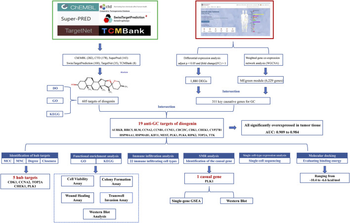
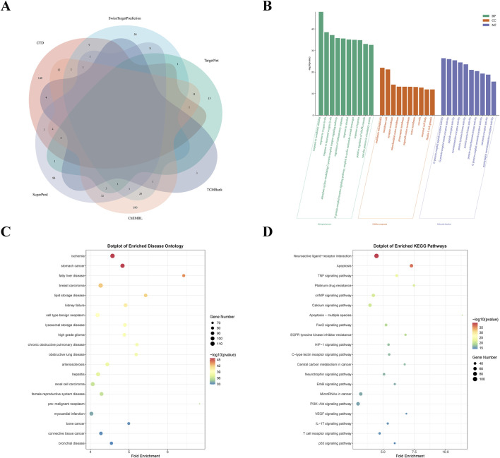
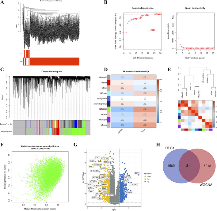
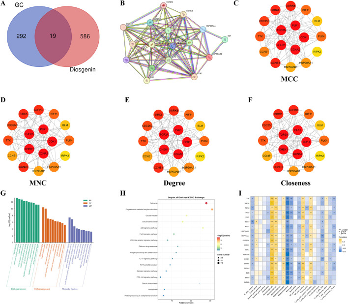
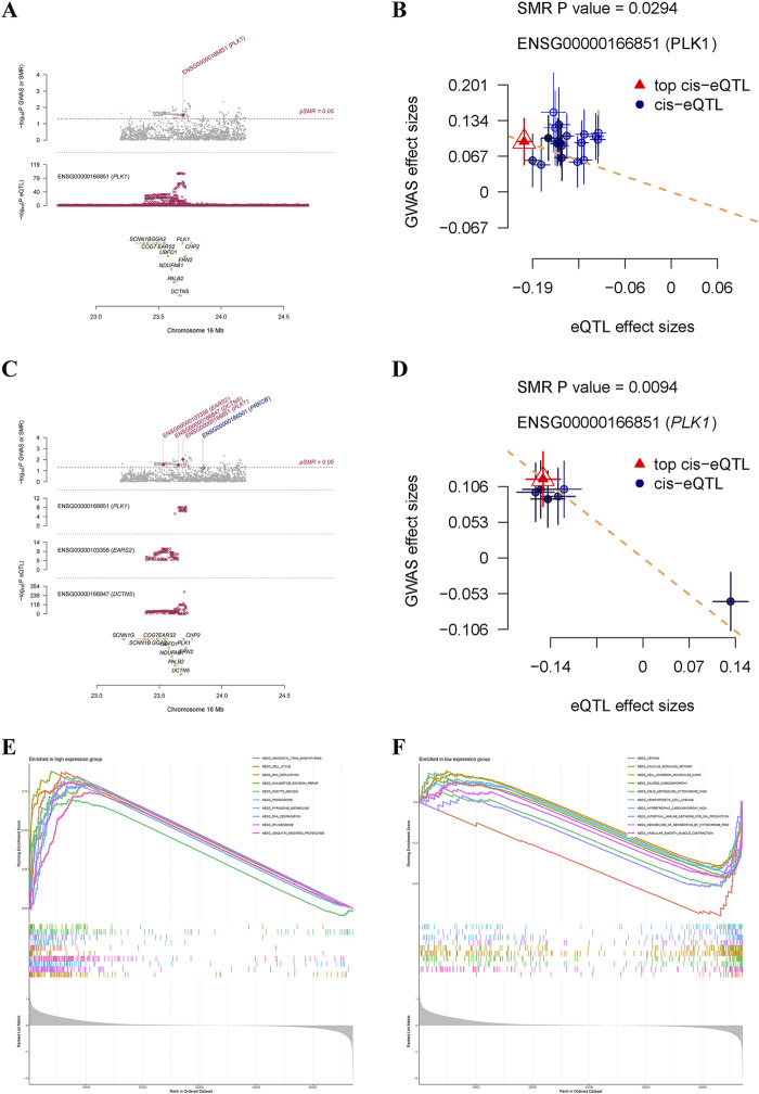
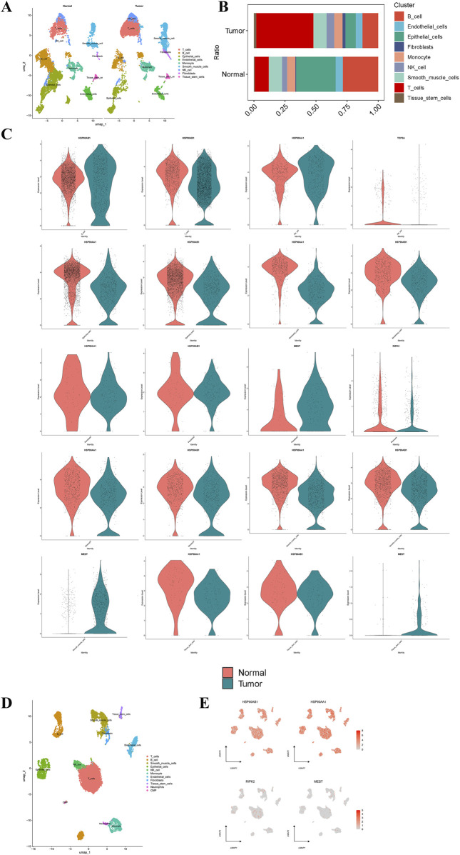
Methods: Targets of Diosgenin were collected from six databases, and enrichment analysis was used to identify its associated diseases and biological pathways. GC-related genes were identified using weighted gene co-expression network analysis. A multi-approach strategy, including network analysis, bioinformatics, single-cell RNA sequencing, Mendelian randomization, and cell experiments, was used to explore the anti-GC mechanisms of Diosgenin.

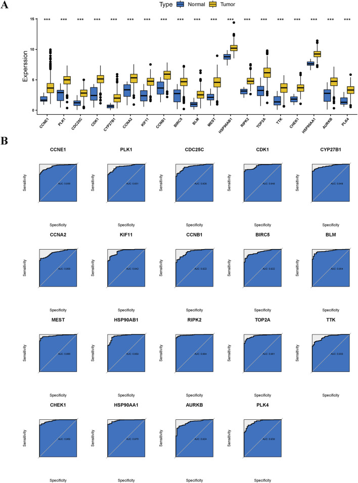
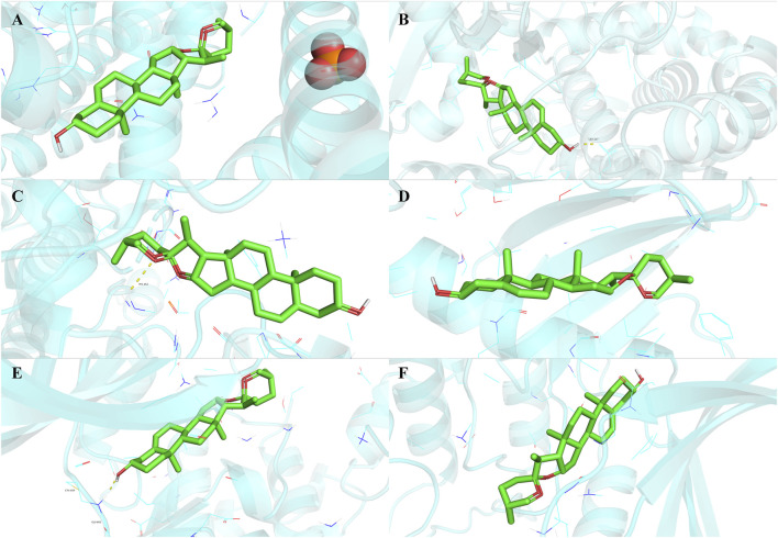
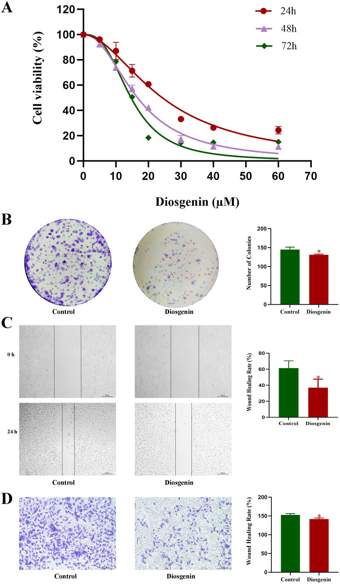
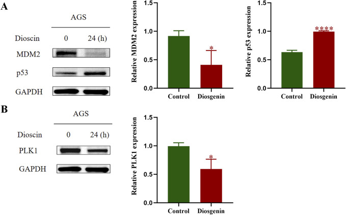
Results: In this study, 605 Diosgenin targets were identified, with key involvement in cell apoptosis, TNF signaling, and platinum resistance pathways, demonstrating significant enrichment in GC. Diosgenin may exert its anti-GC effects through 311 targets, involving regulation of the cell cycle, p53, and FoxO signaling pathway. Key effectors, including CDK1, CCNA2, TOP2A, CHEK1, and PLK1, were identified. Single-cell sequencing indicated that TOP2A, HSP90AA1, and HSP90AB1 might be crucial immune regulatory targets of Diosgenin. Diosgenin significantly inhibited GC cell proliferation, colony formation, migration, and invasion. Evidence from western blot analysis indicates that Diosgenin exerts anti-GC effects by suppressing the expression of PLK1 and MDM2 proteins while upregulating p53 protein levels.

Conclusion: These findings highlight Diosgenin's potential as a promising therapeutic agent for GC, offering a foundation for future research and clinical applications.

Keywords: Diosgenin; MDM2; gastric cancer; mechanism; network analysis; p53; plk1.


Conflict of interest statement

The authors declare that the research was conducted in the absence of any commercial or financial relationships that could be construed as a potential conflict of interest.

Figures

FIGURE 1
FIGURE 1
Elucidating the anti-GC mechanisms of diosgenin: a comprehensive flowchart approach.
FIGURE 2
FIGURE 2
Screening analysis of Diosgenin targets. (A) Venn diagram of Diosgenin in the six databases. (B) Gene Ontology (GO) enrichment analysis (BP, biological process; CC, cellular component; MF, molecular function) of Diosgenin targets. (C) Disease Ontology (DO) enrichment analysis of Diosgenin targets. (D) Kyoto Encyclopedia of Genes and Genomes (KEGG) enrichment analysis of Diosgenin targets.
FIGURE 3
FIGURE 3
Identification of critical causative genes in gastric cancer through gene co‐expression network analysis (WGCNA). (A) Sample dendrogram and trait heatmap. (B) Selection of soft thresholds. (C) Cluster dendrogram of WGCNA. (D) Correlations between gene modules and melanoma status. (E) Correlation between modules. (F) Correlation between brown module memberships and gene significance. (G) A volcano plot presented the differentially expressed genes (DEGs) in gastric cancer. (H) Venn diagram showed the intersection genes of genes identified by WGCNA and DEGs.
FIGURE 4
FIGURE 4
Functional enrichment and immune infiltration analysis for anti-gastric cancer targets. (A) Venn diagram illustrated the intersection genes of Critical causative genes in gastric cancer and targets of Diosgenin. GC: gastric cancer. (B) Construct a protein-protein interaction network of Diosgenin in the treatment of gastric cancer using the STRING database. (C–F) The network plot displayed the importance of each gene evaluated using four algorithms (Matthews Correlation Coefficient (MCC), Maximum Neighborhood Component (MNC), Degree, and Closeness) in Cytoscape 3.10.1. Darker colors of nodes indicate greater importance of the gene. (G) Gene Ontology (GO) enrichment analysis (BP, biological process; CC, cellular component; MF, molecular function) of anti-gastric cancer targets. (H) Kyoto Encyclopedia of Genes and Genomes (KEGG) enrichment analysis of anti-gastric cancer targets. (I) Heatmap illustrated the correlation between anti-gastric cancer targets and 22 immune cells.
FIGURE 5
FIGURE 5
Differential expression and diagnostic efficacy for anti-gastric cancer targets. (A) Box plots showed the differential expression of anti-gastric cancer targets in tumor tissues and normal tissues. P value < 0.001***. (B) Receiver operating characteristic illustrated the diagnostic efficacy of anti-gastric cancer targets.
FIGURE 6
FIGURE 6
Summary-data-based Mendelian randomization analysis and single-gene gene set enrichment analysis. Locus zoom plot (A) and scatter plot (B) displayed causal effects between PLK1 and gastric cancer based on cis eQTLs from the eQTLGen consortium. Locus zoom plot (C) and scatter plot (D) displayed causal effects between PLK1 and gastric cancer based on cis eQTLs from the GTEx Consortium V8. Top 10 pathways significantly enriched in PLK1 high (E) and low (F) expression groups.
FIGURE 7
FIGURE 7
Single‐cell type expression in gastric tumor tissue for the anti-gastric cancer targets. (A) A total of 9 cell types were identified in normal and gastric cancer tissues. (B) Proportion of 9 cell types in normal and gastric cancer tissues. (C) Differential expression of the anti-gastric cancer targets of Diosgenin at the cell level in normal and gastric cancer tissues. (D) A total of 11 cell types were identified in gastric cancer tissues. (E) Specific expression of the anti-gastric cancer target of Diosgenin on cell types in gastric cancer tissues.
FIGURE 8
FIGURE 8
Molecular docking results of Diosgenin with anti-gastric cancer targets. (A) BLM. (B) CYP27B1. (C) CCNE1. (D) PLK4. (E) TTK. (F) CHEK1.
FIGURE 9
FIGURE 9
Diosgenin modulates the proliferation, migration, and invasive capabilities of GC cells. (A) Cell viability was measured using the CCK-8 assay. (B) Colony formation assays were performed to assess clonogenic potential. (C) Wound healing assays were used to evaluate cell migration. (D) Transwell assays were conducted to determine cell invasiveness. P value < 0.05*
FIGURE 10
FIGURE 10
Western blot was performed to detect the protein expression levels of MDM2, p53, and PLK1 in GC cells. (A) Western blot results of MDM2, p53. (B) Western blot results of PLK1. P value < 0.05*, P value < 0.001***.

Similar articles

References

    1. Aran D., Looney A. P., Liu L., Wu E., Fong V., Hsu A., et al. (2019). Reference-based analysis of lung single-cell sequencing reveals a transitional profibrotic macrophage. Nat. Immunol. 20 (2), 163–172. 10.1038/s41590-018-0276-y - DOI - PMC - PubMed
    1. Bendris N., Arsic N., Lemmers B., Blanchard J. M., Cyclin A. (2012). Cyclin A2, Rho GTPases and EMT. Small GTPases 3 (4), 225–228. 10.4161/sgtp.20791 - DOI - PMC - PubMed
    1. Bray F., Laversanne M., Sung H., Ferlay J., Siegel R. L., Soerjomataram I., et al. (2024). Global cancer statistics 2022: GLOBOCAN estimates of incidence and mortality worldwide for 36 cancers in 185 countries. CA Cancer J. Clin. 74 (3), 229–263. 10.3322/caac.21834 - DOI - PubMed
    1. Butler A., Hoffman P., Smibert P., Papalexi E., Satija R. (2018). Integrating single-cell transcriptomic data across different conditions, technologies, and species. Nat. Biotechnol. 36 (5), 411–420. 10.1038/nbt.4096 - DOI - PMC - PubMed
    1. Cai X. P., Chen L. D., Song H. B., Zhang C. X., Yuan Z. W., Xiang Z. X. (2016). PLK1 promotes epithelial-mesenchymal transition and metastasis of gastric carcinoma cells. Am. J. Transl. Res. 8 (10), 4172–4183. - PMC - PubMed

LinkOut - more resources