. 2025 Apr 29;28(6):112552.
doi: 10.1016/j.isci.2025.112552. eCollection 2025 Jun 20.

Young rat vascular endothelial cells promote neurological recovery of stroke aged rat via HIF-1α

Affiliations

Young rat vascular endothelial cells promote neurological recovery of stroke aged rat via HIF-1α

Xianglong Li et al. iScience. .

Abstract

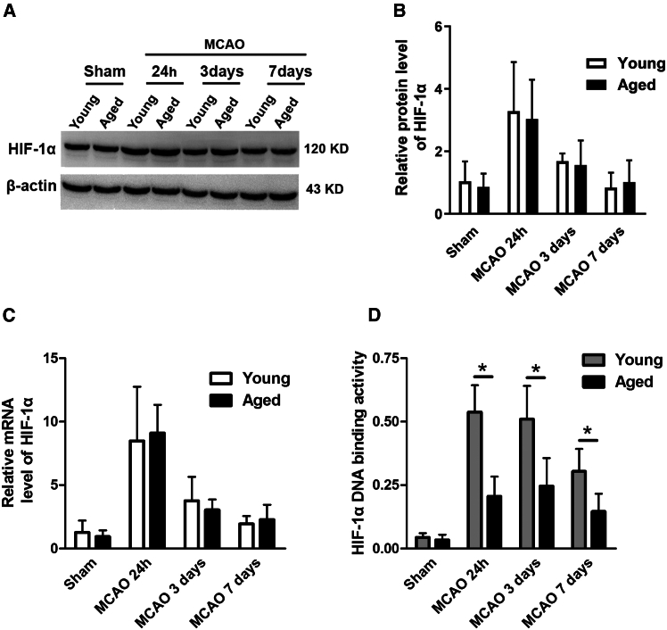
This study aims to explore whether vascular endothelial cells (VECs) derived from young rats enhance neurological recovery following ischemic stroke in aged rats. Middle cerebral artery occlusion models were established, and the rats received lateral ventricle injections of VECs isolated from the cerebral cortex of either young or aged rats. Young VECs treatment facilitated neurorestoration in aged rats on days 3, 7, and 21 following cerebral ischemia. Aged subjects treated with young VECs exhibited reduced neuronal apoptosis, smaller infarct volumes, increased microvessel density, and elevated nerve growth factor expression relative to those receiving aged VECs. Protein levels did not differ between age groups on days 1, 3, or 7 post-stroke (p > 0.05), while DNA binding activity of hypoxia-inducible factor 1 alpha (HIF-1α) was significantly greater in young than in aged rats (p < 0.05). Young-derived VECs promote neurological recovery after cerebral ischemic stroke in aged rats through HIF-1α.

Keywords: Molecular biology; Neuroscience.


Conflict of interest statement

The authors declare that they have no competing interests.

Figures

None
Graphical abstract
Figure 1
Figure 1
The recovery of neurologic function in aged rats following ischemic stroke was impaired compared with young rats The MCAO model was established with rats. Neurological function was assessed using the corner test (A) and MNSS (B). After ischemic brain injury, the recovery of neurological function was impaired in aged rats compared with young rats. Data were presented as mean ± SD, ∗p < 0.05 vs. young MCAO (unpaired t-test). MNSS, modified neurology severity score; MCAO, middle cerebral artery occlusion. R, right; L, left; R + L = 10.
Figure 2
Figure 2
The protein level, mRNA expression level, and DNA binding activity of HIF-1α in the ischemic penumbra at different time points There was no significant difference in protein (A and B) or mRNA levels (C) between young and aged rats after cerebral ischemic injury. The DNA binding activity of HIF-1α was significantly reduced in aged rats on days 1, 3, and 7 after MCAO compared with young rats (D). Data were presented as mean ± SD (n = 5 rats per group), ∗p < 0.05 (unpaired t-test).
Figure 3
Figure 3
The protein expression and DNA binding activity of HIF-1α in aged VECs and young VECs (A) The VECs were characterized by immunofluorescence technique. (B) The DNA binding activity of HIF-1α was reduced in aged VECs compared with young VECs. (C and D) No significant difference was observed in HIF-1α protein level between aged and young VECs, and the HIF-1α protein expression could be significantly inhibited by siRNA in the young VECs. Data were presented as mean ± SD (n = 5 rats per group), ∗p < 0.05 (unpaired t-test).
Figure 4
Figure 4
Young VECs promoted neurologic function recovery of aged rats by lateral ventricle transplantation Aged rats with MCAO were treated with vehicle, aged VECs, young VECs or Young VEC with HIF-1α siRNA. (A) Compared with the other three groups, the MCAO + young VEC group showed significant greater neurological recovery. ∗p < 0.05 vs. MCAO + young VEC (two-way repeated measures ANOVA). (B and C) At day 3 after MCAO, TTC staining was used to measure infarct volume. The infarct volumes of the MCAO + young VEC group were significantly decreased compared with that of the other four groups. n = 10 rats per group, ∗p < 0.05 (two-way repeated measures ANOVA). Data were presented as mean ± SD.
Figure 5
Figure 5
Young VECs decreased nerve cells apoptosis in the aged rats after ischemic stroke TUNEL staining revealed that young VECs treatment significantly suppressed apoptosis in the ischemic area of aged rats 24 h after MCAO compared with aged VECs treatment. Data were presented as mean ± SD (n = 10 rats per group), ∗p < 0.05 (two-way repeated measures ANOVA).
Figure 6
Figure 6
Young VECs treatment increased microvessels density and NGF expression in ischemic penumbra cortex of the aged rats (A) On the 7th day after VECs treatment, the MCAO + young VEC group present significantly higher microvessels density than other four groups. (B) At 24 h after VECs treatment, NGF protein level was significantly increased in the MCAO + young VEC group compared with the other four groups. Data were presented as mean ± SD (n = 10 rats per group), ∗p < 0.05 (two-way repeated measures ANOVA).

Similar articles

Cited by

References

    1. Feigin V.L., Lawes C.M.M., Bennett D.A., Anderson C.S. Stroke epidemiology: a review of population-based studies of incidence, prevalence, and case-fatality in the late 20th century. Lancet Neurol. 2003;2:43–53. - PubMed
    1. Yang X., Zhang Y., Geng K., Yang K., Shao J., Xia W. Sirt3 Protects Against Ischemic Stroke Injury by Regulating HIF-1α/VEGF Signaling and Blood-Brain Barrier Integrity. Cell. Mol. Neurobiol. 2021;41:1203–1215. - PMC - PubMed
    1. Barteczek P., Li L., Ernst A.S., Böhler L.I., Marti H.H., Kunze R. Neuronal HIF-1α and HIF-2α deficiency improves neuronal survival and sensorimotor function in the early acute phase after ischemic stroke. J. Cereb. Blood Flow Metab. 2017;37:291–306. - PMC - PubMed
    1. Chakrabarti S., Rizvi M., Morin K., Garg R., Freedman J.E. The role of CD40L and VEGF in the modulation of angiogenesis and inflammation. Vascul. Pharmacol. 2010;53:130–137. - PubMed
    1. Ashina K., Tsubosaka Y., Kobayashi K., Omori K., Murata T. VEGF-induced blood flow increase causes vascular hyper-permeability in vivo. Biochem. Biophys. Res. Commun. 2015;464:590–595. - PubMed

LinkOut - more resources