. 2025 May 15;16(5):101447.
doi: 10.4239/wjd.v16.i5.101447.

Exercise training benefits pancreatic islet by modulating the insulin-like growth factor 1/phosphatidylinositol 3-kinase/protein kinase B pathway

Affiliations

Exercise training benefits pancreatic islet by modulating the insulin-like growth factor 1/phosphatidylinositol 3-kinase/protein kinase B pathway

Ya-Wen Wu et al. World J Diabetes. .

Abstract

Background: Diabetes is characterized by insulin resistance as well as impaired insulin production, with β-cell dysfunction playing a critical role in disease progression. Exercise is known to improve insulin sensitivity, but its effects on pancreatic islet quality and function remain poorly understood. This work hypothesized that swimming training enhances glycemic control and insulin secretion by upregulating the insulin-like growth factor 1 (IGF-1)/phosphatidylinositol 3-kinase/protein kinase B (PI3K/AKT) pathway in streptozotocin (STZ)-induced diabetic rats.

Aim: To investigate the effects of swimming on pancreatic islet quality and function in STZ-induced diabetic rats via the IGF-1/PI3K/AKT pathway.

Methods: Twenty-six Sprague-Dawley rats were grouped into diabetic and control groups, with each group further split into exercise and sedentary subgroups. Diabetic rats were induced with STZ. The exercise groups underwent swimming training for 60 minutes/day, 5 days/week, for 8 weeks. Body weight, food intake, blood glucose, insulin, lipids, and muscle glycogen were measured. Pancreatic islet morphology and the protein expression levels of IGF-1, PI3K, and AKT were analyzed. Data were analyzed using two-way repeated-measure ANOVA, followed by Tukey's post-hoc test.

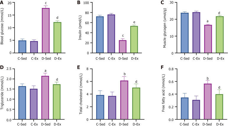
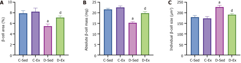
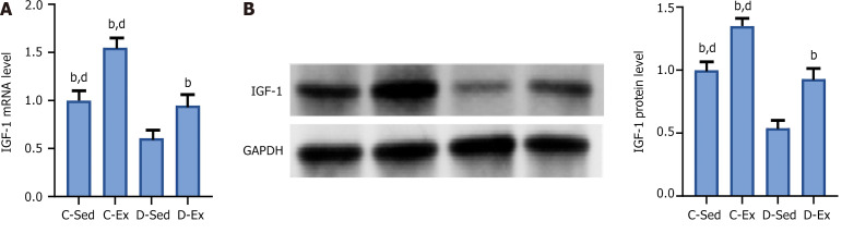
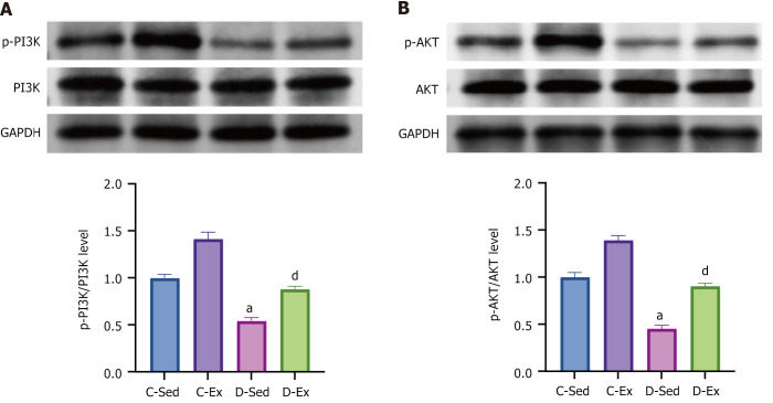
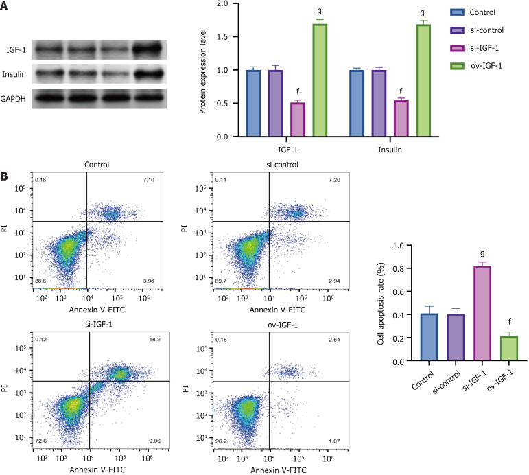
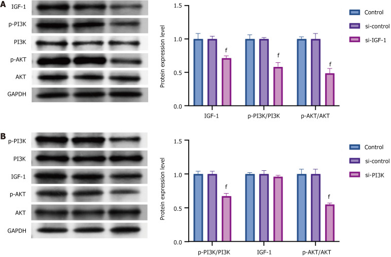
Results: Exercise training significantly improved body weight [diabetic exercise group (D-Ex): 390.66 ± 50.14 g vs diabetic sedentary group (D-Sed): 315.89 ± 50.12 g, P < 0.05], reduced blood glucose (D-Ex: 12.21 ± 4.43 mmol/L vs D-Sed: 17.79 ± 2.05 mmol/L, P < 0.05), and increased insulin levels (D-Ex: 53.50 ± 15.31 pmol/L vs D-Sed: 25.31 ± 10.23 pmol/L, P < 0.05) in diabetic rats. It also enhanced islet morphology, increased IGF-1 expression, and activated the PI3K/AKT pathway (P < 0.05). In-vitro experiments confirmed that IGF-1 positively regulated insulin expression and inhibited β-cell apoptosis via the PI3K/AKT pathway.

Conclusion: Exercise training improves pancreatic islet quality and function in diabetic rats by modulating the IGF-1/PI3K/AKT pathway, highlighting its therapeutic potential for diabetes management.

Keywords: Diabetes; Exercise training; Insulin-like growth factor 1; Islet; Phosphatidylinositol 3-kinase/protein kinase B.


Conflict of interest statement

Conflict-of-interest statement: All the authors report no relevant conflicts of interest for this article.

Figures

Figure 1
Figure 1
Features of the diabetic rat model and the impact of physical activity. A: Measurement of rat body weight post-experiment; B: Recording of daily average food intake for rats during the experiment; C: Pancreatic morphology of rats from the four groups (× 400 magnification); D: Measurement of the islet longest diameter through observation of pancreatic sections; E: Measurement of islet area through observation of pancreatic sections; F: Measurement of islet number through observation of pancreatic sections. aP < 0.05, bP < 0.01, cP < 0.001 vs the control sedentary group; dP < 0.05, eP < 0.01 vs the diabetic sedentary group. D-Ex: The diabetic exercise group; D-Sed: The diabetic sedentary group; C-Ex: The control exercise group; C-Sed: The control sedentary group.
Figure 2
Figure 2
Serum biomarkers of rat model of diabetes and effect of exercise. A: Measurement of blood glucose levels from blood collected via tail vein; B: Detection of insulin levels from blood collected via tail vein; C: Measurement of glycogen content from the gastrocnemius muscle; D: Detection of triglyceride concentration from blood collected via tail vein; E: Detection of total cholesterol concentration from blood collected via tail vein; F: Detection of free fatty acid concentration from blood collected via tail vein. aP < 0.05, bP < 0.01, cP < 0.001 vs the control sedentary group; dP < 0.05, eP < 0.01 vs the diabetic sedentary group. D-Ex: The diabetic exercise group; D-Sed: The diabetic sedentary group; C-Ex: The control exercise group; C-Sed: The control sedentary group.
Figure 3
Figure 3
Impact of diabetes and exercise on islet β-cell. A: Microscopic measurement of islet β-cell area; B: Calculation of islet β-cell mass through the percentage of insulin-positive area multiplied by pancreatic weight; C: Determination of the size of individual islet β-cells by dividing the insulin-positive area by the number of nuclei counted in the corresponding insulin-positive structures. aP < 0.05 vs the control sedentary group; dP < 0.05 vs the diabetic sedentary group. D-Ex: The diabetic exercise group; D-Sed: The diabetic sedentary group; C-Ex: The control exercise group; C-Sed: The control sedentary group.
Figure 4
Figure 4
Effect of diabetes and exercise on the expression of insulin-like growth factor 1. A: Measurement of insulin-like growth factor 1 (IGF-1) mRNA levels through real-time quantitative polymerase chain reaction; B: Determination of IGF-1 protein levels through Western blot. bP < 0.01 vs the control sedentary group; dP < 0.05 vs the diabetic sedentary group. IGF-1: Insulin-like growth factor 1; RT-qPCR: Real-time quantitative polymerase chain reaction; GADPH: Glyceraldehyde-3-phosphate dehydrogenase; D-Ex: The diabetic exercise group; D-Sed: The diabetic sedentary group; C-Ex: The control exercise group; C-Sed: The control sedentary group.
Figure 5
Figure 5
Impact of diabetes and swimming training on the expression of phosphatidylinositol 3-kinase/protein kinase B. A: Determination of p-phosphatidylinositol 3-kinase (PI3K)/PI3K protein expression levels through Western blot (WB); B: Assessment of p-protein kinase B (AKT)/AKT protein expression levels through WB. aP < 0.05 vs the control sedentary group; dP < 0.05 vs the diabetic sedentary group. PI3K: Phosphatidylinositol 3-kinase; AKT: Protein kinase B; GADPH: Glyceraldehyde-3-phosphate dehydrogenase; D-Ex: The diabetic exercise group; D-Sed: The diabetic sedentary group; C-Ex: The control exercise group; C-Sed: The control sedentary group.
Figure 6
Figure 6
Effects of insulin-like growth factor 1 on islet β-cell function in vitro. A: Determination of insulin-like growth factor 1 and insulin protein levels through Western blot; B: Cell apoptosis rate by flow cytometry. fP < 0.05, gP < 0.01 vs si-control group. IGF-1: Insulin-like growth factor 1; GADPH: Glyceraldehyde-3-phosphate dehydrogenase.
Figure 7
Figure 7
Relationship between insulin-like growth factor 1 and phosphatidylinositol 3-kinase/protein kinase B in vitro. A: Detection of protein expression levels of insulin-like growth factor 1 (IGF-1), phosphatidylinositol 3-kinase (PI3K), and Protein kinase B (AKT) through Western blot (WB) after knocking down IGF-1; B: Assessment of protein expression levels of PI3K, IGF-1, and AKT through WB after knocking down PI3K. fP < 0.05 vs si-control group; IGF-1: Insulin-like growth factor 1; PI3K: Phosphatidylinositol 3-kinase; AKT: Protein kinase B; GADPH: Glyceraldehyde-3-phosphate dehydrogenase.

Similar articles

Cited by

References

    1. Wajchenberg BL. beta-cell failure in diabetes and preservation by clinical treatment. Endocr Rev. 2007;28:187–218. - PubMed
    1. Qian L, Xu L, Wang X, Fu X, Gu Y, Lin F, Peng Y, Li G, Luo M. Early insulin secretion failure leads to diabetes in Chinese subjects with impaired glucose regulation. Diabetes Metab Res Rev. 2009;25:144–149. - PubMed
    1. Park S, Hong SM, Lee JE, Sung SR. Exercise improves glucose homeostasis that has been impaired by a high-fat diet by potentiating pancreatic beta-cell function and mass through IRS2 in diabetic rats. J Appl Physiol (1985) 2007;103:1764–1771. - PubMed
    1. Min HK. Non-insulin-dependent diabetes mellitus (NIDDM) in Korea. Diabet Med. 1996;13:S13–S15. - PubMed
    1. Weir GC, Bonner-Weir S. Five stages of evolving beta-cell dysfunction during progression to diabetes. Diabetes. 2004;53 Suppl 3:S16–S21. - PubMed

LinkOut - more resources