. 2025 May 23:16:1606326.
doi: 10.3389/fendo.2025.1606326. eCollection 2025.

Baoshentongluo Formula relieves podocyte injury in diabetic kidney disease through regulating mitophagy via PINK1/Parkin signaling pathway

Affiliations

Baoshentongluo Formula relieves podocyte injury in diabetic kidney disease through regulating mitophagy via PINK1/Parkin signaling pathway

Yanyu Pang et al. Front Endocrinol (Lausanne). .

Abstract

Introduction: Diabetic kidney disease (DKD) progression is strongly associated with podocyte mitochondrial dysfunction. The clinically effective Chinese herbal Baoshentongluo formula (BSTL) has demonstrated significant proteinuria reduction in DKD patients. HPLC-ESI-MS analysis identified characteristic bioactive components in BSTL including astragalosides, rehmanniosides, and tanshinones. However, the molecular mechanisms through which BSTL maintains podocyte homeostasis remain incompletely understood.

Methods: Mouse podocyte clone-5 (MPC-5) cells and db/db mice were used. Db/db mice were randomized into db/db and db/db + BSTL (16.5 g/kg/d, intragastric administration for 12 weeks). A group of m/m mice served as the control. Renal function, urinary albumin-to-creatinine ratio (UACR), histopathological analysis, apoptotic, and mitophagy-related protein levels were evaluated. MPC-5 cells were exposed to high glucose (HG, 30 mM) and BSTL drug-containing serum (8%) for 24 h grouping as control, HG, HG + BSTL, and HG + siPINK1. Podocyte apoptosis, mitophagy levels, and expression of PTEN-induced putative kinase 1 (PINK1) and E3 ubiquitin ligase (Parkin) were assessed.

Results: In db/db diabetic mice, oral administration of BSTL significantly lowered urinary albumin-to-creatinine ratio (P<0.05), improved glomerular filtration rate, and ameliorated renal histopathological changes, decreased LC3-II/LC3-I ratio, and downregulated expression of mitophagy-related proteins PINK1, Parkin, ATG5 and Beclin-1. Treatment with 8% BSTL-containing serum significantly attenuated HG-induced podocyte apoptosis (P<0.01) and suppressed excessive mitophagy, as evidenced by reduced TOM20/LC3 co-localization (P<0.01). Notably, BSTL treatment markedly reduced protein levels of both PINK1 and Parkin (P<0.01), key regulators of mitophagy initiation. Genetic silencing of PINK1 in podocytes phenocopied BSTL's protective effects, confirming the pathway specificity.

Discussion: Our integrated in vitro and in vivo findings establish that BSTL protects against DKD progression by selectively inhibiting PINK1/Parkin-dependent mitophagy in podocytes to inhibit podocyte injury, which provides both mechanistic insights and therapeutic potential for clinical DKD management.

Keywords: apoptosis; diabetic kidney disease; mitochondria; podocyte; traditional Chinese medicine.


Conflict of interest statement

The authors declare that the research was conducted in the absence of any commercial or financial relationships that could be construed as a potential conflict of interest.

Figures
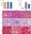
Figure 1
Figure 1
BSTL treatment improved renal function and attenuated renal histological damage in db/db mice. (A) Quantitative assessment of Scr in mice. (B) Quantitative assessment of UACR in mice. (C) Representative micrographs of HE-stained kidney sections (×200), PAS-stained kidney sections (×400), and Masson’s trichrome-stained kidney sections (×400) from different groups. Red arrows: glomerular basement membrane; green arrows: endothelial cells; yellow arrows: mesangial cells; black arrows: mesangial matrix. *P<0.05 vs. con; #P<0.05 vs. DKD. con, control mice; DKD, db/db mice; BSTL, db/db mice treated with BSTL.
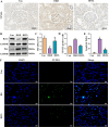
Figure 2
Figure 2
BSTL treatment inhibited podocyte injury and apoptosis in db/db mice and HG-treated podocytes. (A) TUNEL staining in renal sections from different groups (×400). (B) Representative Western blot of Bcl-2 and Bax in the kidneys of mice. (C) Quantitative assessment of Bcl-2 and Bax in the kidneys of mice. (D) Quantitative assessment of TUNEL staining in renal sections of different groups. *P<0.05 vs. con; **P<0.01 vs. Con; #P<0.05 vs. DKD; ##P<0.01 vs. DKD. con, control mice; DKD, db/db mice; BSTL, db/db mice treated with BSTL. (E) Quantitative assessment of TUNEL staining in podocytes from different groups. (F) TUNEL staining in podocytes of different groups (×200). *P<0.05 vs. con; #P<0.05 vs. HG. con, normal glucose; HG, high glucose; BSTL, high glucose combined with BSTL drug-containing serum.
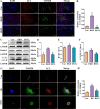
Figure 3
Figure 3
BSTL treatment reduced the expression of mitophagy-related proteins in the renal tissues of db/db mice and HG-treated podocytes. (A) Representative images of immunofluorescent co-localization staining of glomerular LC3 and TOM20 in different mice (×200). (B) Quantitative assessment of co-localization staining of glomerular LC3 and TOM20 in the kidneys of mice. (C) Representative western blot of ATG5, Beclin-1 and LC3 in the kidneys of mice. (D-F) Quantitative assessment of ATG5, Beclin-1 and LC3 in the kidneys of mice. *P<0.05 vs. con; **P<0.01 vs. Con; #P<0.05 vs. DKD; ##P<0.01 vs. DKD. con, control mice; DKD, db/db mice; BSTL, db/db mice treated with BSTL. (G) Representative images of immunofluorescent co-localization staining of LC3 and TOM20 in different cells (×200). (H) Quantitative assessment of co-localization staining of LC3 and TOM20 in different cells. *P<0.05 vs. con; #P<0.05 vs. HG. con, normal glucose; HG, high glucose; BSTL, high glucose combined with BSTL drug-containing serum.
Figure 4
Figure 4
BSTL restored mitophagy in db/db mice and HG-cultured podocytes via the PINK1/Parkin signaling pathway. (A) Representative Western blots of PINK1 and the Parkin protein in the kidneys of mice. (B, C) Quantitative assessment of PINK1 and Parkin protein in the kidneys of mice. (D, E) Quantitative assessment of IHC staining of renal sections for the PINK1 and Parkin protein in different groups. (F) IHC staining of renal sections for PINK1 and Parkin protein in different groups (×400). *P<0.05 vs. con; **P<0.01 vs. Con; #P<0.05 vs. DKD; ##P<0.01 vs. DKD. con, control mice; DKD, db/db mice; BSTL, db/db mice treated with BSTL. (G) Representative Western blot and quantitative assessment of PINK1 and Parkin in podocytes. (H, I) Quantitative assessment of PINK1 and Parkin in podocytes. *P<0.05 vs. con; #P<0.05 vs. HG. con, normal glucose; HG, high glucose; BSTL, high glucose combined with BSTL drug-containing serum.
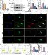
Figure 5
Figure 5
PINK1 deficiency in MPC-5 cells alleviates podocyte injury under the HG environment. (A) Quantitative assessment of the PINK1 gene in podocytes. (B-D) Representative Western blots of the PINK1 and Parkin protein in podocytes. (E) Representative images of immunofluorescent co-localization staining of LC3 and TOM20 in different cells (×200). (F) Representative flow cytometry analysis depicting the apoptosis detection in podocytes with different treatments (Q2 represented the ratio of late apoptotic cells, Q3 represented the ratio of early apoptotic cells, Q2+Q3 represented the total ratio of apoptotic cells). (G) Quantitative data expressing the overall percentage of podocyte apoptosis. (H) Quantitative assessment of co-localization staining of LC3 and TOM20 in different cells. *P<0.05 vs. con; #P<0.05 vs. HG. con, normal glucose; HG, high glucose; BSTL, high glucose combined with BSTL drug-containing serum; PINK1 siRNA, silenced PINK1 in HG condition.

Similar articles

References

    1. Alicic RZ, Rooney MT, Tuttle KR. Diabetic kidney disease: challenges, progress, and possibilities. Clin J Am Soc Nephrol: CJASN. (2017) 12:2032–45. doi: 10.2215/cjn.11491116 - DOI - PMC - PubMed
    1. Ma RCW. Epidemiology of diabetes and diabetic complications in China. Diabetologia. (2018) 61:1249–60. doi: 10.1007/s00125-018-4557-7 - DOI - PubMed
    1. Huang R, Fu P, Ma L. Kidney fibrosis: from mechanisms to therapeutic medicines. Signal Transduct Target Ther. (2023) 8:129. doi: 10.1038/s41392-023-01379-7 - DOI - PMC - PubMed
    1. Song C, Zhu Z, Liu L, Liu S, Li Y, Xiao Y, et al. . The efficacy and safety of niaoduqing granules in the treatment of diabetic kidney disease: A systematic review and meta-analysis. Front Pharmacol. (2023) 14:1180751. doi: 10.3389/fphar.2023.1180751 - DOI - PMC - PubMed
    1. Kopp JB, Anders HJ, Susztak K, Podestà MA, Remuzzi G, Hildebrandt F, et al. . Podocytopathies. Nat Rev Dis Primers. (2020) 6:68. doi: 10.1038/s41572-020-0196-7 - DOI - PMC - PubMed

MeSH terms