. 2025 Mar 29;5(4):100496.
doi: 10.1016/j.bpsgos.2025.100496. eCollection 2025 Jul.

Astrogliosis Occurs Selectively in Amygdala of Adolescent Primate and Rodent Following Daily Δ9-Tetrahydrocannabinol, Prevented by Cannabidiol Co-Treatment

Affiliations

Astrogliosis Occurs Selectively in Amygdala of Adolescent Primate and Rodent Following Daily Δ9-Tetrahydrocannabinol, Prevented by Cannabidiol Co-Treatment

Yalin Sun et al. Biol Psychiatry Glob Open Sci. .

Abstract

Background: Adolescent-onset cannabis use confers higher risk for neuropsychiatric disorders, implicating amygdala dysfunction. However, the mechanisms that mediate Δ9-tetrahydrocannabinol (THC)-triggered neuroadaptive changes in the maturing amygdala remain unclear.

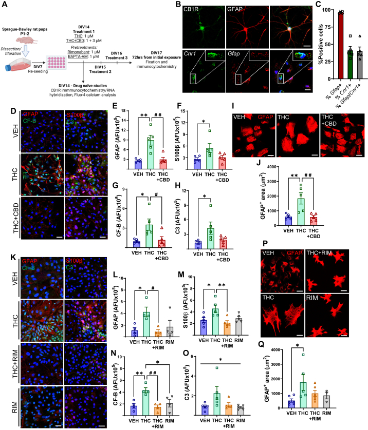
Methods: Proteomic analysis of amygdala tissue from male adolescent Saimiri boliviensis nonhuman primates chronically treated with THC provided leads for targeted analyses of glial fibrillary acidic protein (GFAP), stathmin-1, and neuronal cell adhesion molecule (NrCAM) in a second species of male adolescent (postnatal day [P]35) and adult (P70) Sprague-Dawley rats. Primate activity monitoring and rat behavioral testing revealed THC-disrupted sleep architecture and anxiety-related behavior, respectively. Primary rat astrocyte cultures provided mechanistic insight into THC activation of astrocyte inflammatory function.

Results: THC-induced upregulation of GFAP and complement factor-B (CF-B) signified proinflammatory glial activation exclusively in the adolescent amygdala, an effect absent in other brain regions and in adults. THC attenuated synaptic plasticity enhancers, stathmin-1 and NrCAM, effects not recapitulated in adults. Co-administered cannabidiol (CBD) prevented astrogliosis but did not restore synaptic plasticity marker levels. Astrogliosis was correlated with fragmented sleep, and attenuated plasticity markers were correlated with anxiety. THC-induced GFAP and CF-B upregulation with attenuation by CBD were replicated in cultured astrocytes, requiring cannabinoid type 1 receptor (CB1R)-activated calcium signaling. Elevated CB1R expression in the maturing brain was astrocyte-localized in the amygdala, but neuronal in the cortex and striatum.

Conclusions: Brain region- and age-specific regulation of CB1R in astrocytes critically links THC and unique adolescent amygdala vulnerability to inflammatory gliosis, impairing behaviors implicated in neuropsychiatric disorders. Mitigation of specific THC-induced changes by CBD offers leads for attenuating some adverse effects of THC.

Keywords: Adolescent neurodevelopment; Amygdala; Anxiety; Astrocytes; Cannabinoids; Neuroinflammation; Sleep.

Plain language summary

Brain maturation is still incomplete during adolescence, which makes it a period during which the brain is highly sensitive to exposure to drugs, such as cannabis. Early cannabis use may alter the brain in ways that increase a person’s risk of encountering psychiatric disturbances, which involve malfunction of the anxiety hub of the brain, the amygdala. In the current study, inflammation and reduced neuron communication machinery occurred in the amygdala after early exposure to the active component of cannabis (Δ9-tetrahydrocannabinol). This did not occur if exposure occurred during adulthood, highlighting the sensitivity of the adolescent brain to external influences. A different component of cannabis with anti-inflammatory benefits, cannabidiol, effectively blocked the inflammation caused by THC, potentially demonstrating a medicinal benefit in reversing certain harmful THC effects.


Figures

Figure 1
Figure 1
Exposure to THC in adolescence induces proinflammatory astrogliosis in the primate amygdala. (A) Drug administration regimen in male adolescent Saimiriboliviensis (squirrel monkey) primates. (B) Proteomic analysis of differentially expressed proteins in adolescent primate amygdala (n = 3 per group) for GFAP (THC vs. VEH q = .7884, THC+CBD vs. VEH q = 1.000, THC+CBD vs. THC q = .0463), stathmin-1 (THC vs. VEH q = 2.487 × 10⁻⁵, THC+CBD vs. VEH q = .0446, THC+CBD vs. THC q = .0197), and NrCAM (THC vs. VEH q = .0093, THC+CBD vs. VEH q = 1.000, THC+CBD vs. THC q = .0197). (C) GFAP and CF-B IHC from adolescent primate left hemisphere amygdala, scale bar = 10 μm. (D, E) GFAP and CF-B fluorescence (AFU), n = 4 per group (GFAP: p = .004, F2,9 = 10.83, post hoc: ∗∗p = .0046, ∗p = .0147. CF-B: p = .0031, F2,9 = 11.73, post hoc: ∗∗p = .0069, ##p = .0051). (F) GFAP+ fraction of total cell density, n = 4 (p = .0057, F2,9 = 9.696; post hoc: ∗∗p = .0067, ∗p = .0187). (G) Pearson’s 2-tailed correlation, linear regression and 95% CI between percentage (%) GFAP-positive astrocytes and CF-B (top), GFAP (bottom), p and R2 values above plots. (H) GFAP IHC from adolescent primate right hemisphere CeA and BLA, scale bar = 10 μm. (I) S100β, CB1R, GFAP IHC from adolescent primate right hemisphere BA (n = 3 per group). Scale bars = 40 μm for nonmagnified images, 5 μm for magnified images (last column). (J) Amygdala S100β-positive cells/mm3 (p = .0471, F2,6 = 5.308; post hoc: ∗p = .0396). (K, L) Amygdala CB1R and GFAP fluorescence intensity as % of vehicle (CB1R: p = .0371, F2,6 = 5.998, post hoc: ∗p = .0346. GFAP: p = .0005, F2,6 = 35.58, post hoc: ∗∗∗p = .0007, ∗∗p = .0011). (M) Pearson’s 2-tailed correlation, linear regression, and 95% CI between S100β-positive astrocytes and GFAP, p and R2 value above plot. (N, O) Stathmin-1 IHC and quantification from adolescent primate left hemisphere amygdala, n = 4 (p = .1269, F2,9 = 2.619). (P–R) GFAP IHC and quantification from adolescent PFC and CN, n = 4 (PFC: p = .5976, F2,9 = 0.5455; CN: p = .2337, F2,9 = 1.716). Scale bar = 10 μm. (S, T) GFAP IHC with quantification from adult Rhesus macaque primate amygdala, n = 2–3 per group (p = .5879, F2,9 = 0.5919). Scale bar = 50 μm. All bar graphs show mean ± SEM and were analyzed by 1-way analysis of variance with Tukey’s post hoc tests. All significance symbols displayed in graphs for comparisons are derived from post hoc analysis. AFU, arbitrary fluorescence units; AU, arbitrary units; BA, basal amygdala; BLA, basolateral amygdala; CB1R, CB1 receptor; CBD, cannabidiol; CeA, central amygdala; CN, caudate nucleus; CF-B, complement factor-B; GFAP, glial fibrillary acid protein; IHC, immunohistochemistry; NrCAM, neuronal cell adhesion molecule; ns, nonsignificant; PFC, prefrontal cortex; THC, Δ9-tetrahydrocannabinol; VEH, vehicle.
Figure 2
Figure 2
Adolescent but not adult exposure to THC induces proinflammatory astrogliosis with reduced stathmin-1 and NrCAM expression in rat amygdala. (A) Schematic of chronic drug administration regimen in male adolescent (P35) and adult (P70) Sprague-Dawley rats. (B) Rat amygdala GFAP and CF-B fluorescent IHC from vehicle (adolescent n = 5, adult n = 5), THC (adolescent n = 7, adult n = 4), and THC+CBD (adolescent n = 5, adult n = 5), scale bar = 15 μm. (C–F) Amygdala GFAP fluorescence values (AFUs) for adolescent rat (GFAP: Brown–Forsythe ANOVA, p = .0306, W2.000,8.715 = 3.013; Welch’s post hoc test: ∗p = .0456, #p = .0422. CF-B: p = .0141, F2,15 = 5.744; post hoc: ∗p = .0229, #p = .0387) and adult rat (GFAP: p = .1400, F2,10 = 2.408; CF-B: p = .4036, F2,10 = 0.9950). (G) Amygdala stathmin-1 IHC from vehicle- (adolescent n = 5, adult n = 6) and THC (adolescent n = 5, adult n = 6)-treated rats, scale bar = 15 μm. (H, I) Amygdala stathmin-1 fluorescence values for adolescent rat (2-tailed unpaired parametric t test, ∗p = .0498, t8 = 2.309) and adult rat (2-tailed unpaired parametric t test, p = .1123, t10 = 1.741). (J) Amygdala NrCAM IHC from vehicle-, THC-, and THC+CBD-treated rats (n = 6 per age and treatment), scale bar = 15 μm. (K,L) Amygdala NrCAM levels (AFU) for adolescent rats (K) (p = .0035, F2,15 = 8.464; post hoc: ∗∗p = .0041, ∗p = .0171) and adult rats (L) (p = .0549, F2,15 = 3.543). All bar graphs show mean ± SEM and significance derived from post hoc analysis. Unless otherwise indicated, data were analyzed by 1-way ANOVA with Tukey’s post hoc tests. AFU, arbitrary fluorescent unit; ANOVA, analysis of variance; CBD, cannabidiol; CF-B, complement factor-B; GFAP, glial fibrillary acid protein; IHC, immunohistochemistry; P, postnatal day; THC, Δ9-tetrahydrocannabinol; NrCAM, neuronal cell adhesion molecule; ns, nonsignificant; VEH, vehicle.
Figure 3
Figure 3
THC-induced sleep disruption is correlated with astrogliosis levels in nonhuman primate amygdala. (A) Trajectory of adolescent primate sleep fragmentation frequency over time represented as mean of 3-week epochs for vehicle (n = 4), THC (n = 4), and THC+CBD (n = 4). (B) Individual values with mean ± SEM for difference in sleep fragmentation frequency between averaged last 3 weeks of drug administration and first 3 weeks of drug administration in treated adolescent primates (1-way analysis of variance, p = .0457, F2,9 = 4.432; Tukey’s post hoc test, ∗p = .0471). (C–K) Pearson’s 2-tailed correlation analysis between sleep fragmentation frequency detected during weeks 1 to 3, 10 to 12, and 14 to 16 of drug administration with amygdala CF-B (C–E), GFAP (F–H), and proportion of GFAP-positive cells (I–K) for vehicle-, THC-, and THC+CBD-treated adolescent primate (n = 4 per group). CF-B and GFAP expression levels are expressed as AFUs, quantified from treated adolescent primate amygdala immunohistochemistry. Pearson’s 2-tailed analysis with linear regression was performed for all correlation data; 95% CI, p, and R2 values are indicated within all graphs. Individual subjects are plotted for all correlations. AFU, arbitrary fluorescence unit; CBD, cannabidiol; CF-B, complement factor-B; GFAP, glial fibrillary acid protein; NrCAM, neuronal cell adhesion molecule; THC, Δ9-tetrahydrocannabinol; VEH, vehicle.
Figure 4
Figure 4
Attenuation of amygdala stathmin-1 and NrCAM expression by THC and THC+CBD are correlated with anxiety-like behaviors in adolescent but not adult rats. Correlation between the volume of milk consumed during the novelty-induced hypophagia test and amygdala stathmin-1 levels (AFU) in adolescent (A) and adult (D) vehicle-treated (adolescent n = 5, adult n = 4) and THC-treated (adolescent n = 5, adult n = 4) rats. Correlation between light-dark box percentage time in light compartment (B, E) or elevated plus maze percentage time in open arms (C, F) with amygdala NrCAM levels (AFU) for rats treated with vehicle (adolescent n = 6, adult n = 6), THC (adolescent n = 6, adult n = 6), and THC+CBD (adolescent n = 5, adult n = 5). Pearson’s 2-tailed correlation analysis with linear regression was performed for all data; 95% CI, p, and R2 values are indicated within all graphs. Individual subjects are plotted for all correlations. AFU, arbitrary fluorescence unit, CBD, cannabidiol; NrCAM, neuronal cell adhesion molecule; THC, Δ9-tetrahydrocannabinol; VEH, vehicle.
Figure 5
Figure 5
THC induces CB1R-dependent transition to reactive astrocyte phenotype in primary astrocyte cultures. (A) Experimental design for in vitro primary astrocyte culture experiments. (B) GFAP-positive primary rat astrocytes showing CB1R protein (top, CB1R) and mRNA (bottom, Cnr1) levels, scale bars = 10 μm. (C) In situ mRNA % positivity quantification for Gfap, Cnr1, and Gfap/Cnr1 in primary astrocyte cultures (n = 5). (D) GFAP, CF-B, S100β, complement C3 primary astrocyte ICC following daily treatment for 72 hours with vehicle, 1 μM THC, and 1 μM THC + 3 μM CBD. (E–H) Primary astrocyte GFAP, S100β, CF-B, and C3 fluorescence values (AFU) following THC and THC+CBD treatment [n = 5. (E) GFAP: p = .0009, F2,12 = 13.42. Post hoc: ∗∗p = .0016, ##p = .0026. (F) S100β: p = .0425, F2,12 = 4.157. Post hoc: ∗p = .0492. (G) CF-B: p = .0165, F2,12 = 5.889. Post hoc: ∗p = .0236, #p = .0369. (H) C3: p = .0391, F2,12 = 4.301. Post hoc: ∗p = .0408]. (I, J) GFAP-positive primary astrocytes treated daily for 72 hours with vehicle, THC, and THC+CBD as above, used for surface area quantification [n = 5. (J)p = .0035, F2,12 = 9.424. Post hoc: ∗∗p = .0077, ##p = .0066]. (K) GFAP, CF-B, S100β, C3 primary astrocyte ICC following daily treatment for 72 hours with vehicle, THC, THC+RIM, and RIM at a dose of 1 μM for each drug. (L–O) Primary astrocyte GFAP, S100β, CF-B, and C3 fluorescence values following THC, THC+RIM, and RIM treatment [n = 4–5. (L) GFAP: p = .0272, F3,12 = 4.349. Post hoc: ∗p = .0487, #p = .0324. (M) S100β: p = .0085, F3,16 = 5.521. Post hoc: ∗p = .0325, ∗∗p = .0074. (N) CF-B: p = .0040, F3,12 = 7.665. Post hoc: ∗∗p = .0089, ##p = .0054, ∗p = .0282. (O) C3: p = .0471, F3,16 = 3.308]. (P, Q) GFAP-positive primary astrocytes treated daily for 72 hours with vehicle, THC, THC+RIM, and RIM as above, for surface area quantification [(Q)n = 3–6, p = .0644, F3,16 = 2.948; post hoc: ∗p = .0435]. All data were analyzed by 1-way analysis of variance with Tukey’s post hoc analysis and presented as mean ± SEM, scale bar = 30 μm unless otherwise indicated. AFU, arbitrary fluorescence unit; CB1R, CB1 receptor; CBD, cannabidiol; CF-B, complement factor-B; DIV, days in vitro; GFAP, glial fibrillary acid protein; ICC, immunocytochemistry; mRNA, messenger RNA; RIM, rimonabant; THC, Δ9-tetrahydrocannabinol; VEH, vehicle.
Figure 6
Figure 6
THC activates CB1R to elicit intracellular calcium signaling via IP3 receptors in astrocytes to induce astrogliosis. (A, C) Intracellular calcium levels detected by Fluo-4 in primary rat astrocytes across 11 reads over 30 seconds with acute stimulation by vehicle or THC with rimonabant (A) (n = 5) or (C) 2-APB (n = 3) pretreatment. Arrow indicates addition of THC. Data are mean + SEM. (B, D) Calcium levels immediately after acute vehicle or THC with RIM (B) (p = .0001, F2,12 = 20.20; post hoc: ∗∗∗p = .0001, ∗∗p = .0075) or 2-APB (D) (p = .0044, F2,6 = 15.37; post hoc: ∗∗p = .0037, ∗p = .0345). Data are mean ± SEM. (E) GFAP, CF-B, S100β, and complement C3 primary astrocyte immunocytochemistry following 72 hours treatment with vehicle, 1 μM THC, 1 μM THC + 10 μM BAPTA-AM, and 10 μM BAPTA-AM. Scale bar = 10 μm. (F–I) Primary astrocyte GFAP, CF-B, S100β, and C3 fluorescence values following THC and BAPTA-AM treatments, n = 4 [(F) GFAP: p = .0004, F3,12 = 13.48; post hoc: ∗∗∗p = .0007, ∗∗p = .0020, ##p = .0011. (G) CF-B: p < .0001, F3,12 = 27.61; post hoc: ∗∗∗∗p < .0001, ∗∗∗p = .0004. (H) S100β: p = .0009, F3,12 = 10.99; post hoc: ∗∗p = .0051, ##p = .0012, ∗p = .0269. (I) C3: p = .0032, F3,12 = 8.123; post hoc: ∗∗p = .0083, ∗p = .0280, ##p = .0038]. (J) Fluo-4 measurement of intracellular calcium in primary rat astrocytes across 11 reads over 30 seconds with acute stimulation by vehicle or THC with YM (n = 4). Data are mean + SEM. (K) Calcium levels immediately after addition of vehicle or THC with YM (p = .0020, F2,9 = 13.35; post hoc: ∗∗p = .0031, ##p = .0052). Data are mean ± SEM. All data were analyzed by 1-way analysis of variance with Tukey’s post hoc analysis. Significance symbols displayed for comparisons are derived from post hoc statistical tests. 2-APB, 2-aminoethoxydiphenylborate; AFU, arbitrary fluorescence unit; CB1R, CB1 receptor; CF-B, complement factor-B; GFAP, glial fibrillary acid protein; RIM, rimonabant; THC, Δ9-tetrahydrocannabinol; VEH, vehicle; YM, YM-254890.
Figure 7
Figure 7
Developmentally regulated CB1R expression in neurons and astrocytes. (A) Immunohistochemistry images from adolescent nonhuman primate amygdala showing colocalization of CB1R with GFAP-labeled cells and lower colocalization levels with NeuN-positive cells. White arrows indicate sites of CB1R-GFAP colocalization. (B) Representative in situ messenger RNA hybridization images of Cnr1 with Gfap and Rbfox3 in adolescent (P35, n = 5–6) and adult (P70, n = 5–6) rat PL, DS, NAc core, and amygdala. (C) Quantified percentage of total Cnr1-positive cells on P35 compared with P70 (brain region F3,39 = 40.42, ∗∗∗∗p < .0001; age F1,39 = 114.5, ∗∗∗∗p < .0001; brain region × age interaction F3,39 = 3.46, ∗p = .025), for PL (∗∗∗∗p < .0001), DS (∗∗∗∗p < .0001), NAc core (∗p < .042), and amygdala (∗∗p = .0019). (D) Percentage of total Gfap-positive cells in P35 and P70 rat brain (brain region F3,37 = 22.87, ∗∗∗∗p < .0001; age F1,37 = 0.026, p = .87; brain region × age interaction F3,37 = 3.84, ∗p = .017). (E) Percentage of total Rbfox3-positive cells in P35 and P70 rat brain (brain region F3,34 = 1.24, p = .31; age F1,34 = 1.15, p = .29; brain region × age interaction F3,34 = 0.70, p = .56). (F) Proportion of Cnr1-positive Gfap-expressing astrocytes in P35 or P70 rat brain (brain region F3,37 = 13.2, ∗∗∗∗p < .0001; age F1,37 = 13.12, ∗∗∗p = .0009; brain region × age interaction F3,37 = 1.6, p = .21; P35 vs. P70 amygdala ∗p = .17). (G) Proportion of Cnr1-positive Rbfox3-expressing neurons in P35 or P70 rat brain (brain region F3,34 = 21.4, ∗∗∗∗p < .0001; age F1,34 = 41.9, ∗∗∗∗p < .0001; brain region × age interaction F3,34 = 2.6, p = .071; P35 vs. P70 PL ∗∗p = .0094, DS ∗∗p = .0027, NAc core ∗∗p = .0071, amygdala p = .99). All scale bars = 10 μm, data were analyzed by 2-way analysis of variance with Tukey’s post hoc tests, presented as mean ± SEM. Amyg, amygdala; CB1R, cannabinoid type-1 receptor; DS, dorsal striatum; GFAP, glial fibrillary acid protein; NAc, nucleus accumbens; NHP, nonhuman primate; P, postnatal day; PL, prelimbic cortex.

Similar articles

References

    1. Zuckermann A.M.E., Gohari M.R., Romano I., Leatherdale S.T. Changes in cannabis use modes among Canadian youth across recreational cannabis legalization: Data from the COMPASS prospective cohort study. Addict Behav. 2021;122 - PubMed
    1. Petrilli K., Ofori S., Hines L., Taylor G., Adams S., Freeman T.P. Association of cannabis potency with mental ill health and addiction: A systematic review. Lancet Psychiatry. 2022;9:736–750. - PubMed
    1. Zamberletti E., Rubino T. Dos(e)Age: Role of dose and age in the long-term effect of cannabinoids on cognition. Molecules. 2022;27:1411. - PMC - PubMed
    1. Arseneault L., Cannon M., Poulton R., Murray R., Caspi A., Moffitt T.E. Cannabis use in adolescence and risk for adult psychosis: Longitudinal prospective study. BMJ. 2002;325:1212–1213. - PMC - PubMed
    1. Gobbi G., Atkin T., Zytynski T. Association of cannabis Use in Adolescence and Risk of Depression, Anxiety, and suicidality in Young Adulthood: A Systematic Review and Meta-analysis (vol 76, pg 426, 2019) JAMA Psychiatry. 2019;76:447. 447. - PMC - PubMed

LinkOut - more resources