Review
. 2025 Jun 6;9(2):021501.
doi: 10.1063/5.0251889. eCollection 2025 Jun.

Advanced biomaterial strategies for overcoming age-associated wound healing impairments

Affiliations
Review

Advanced biomaterial strategies for overcoming age-associated wound healing impairments

Rodrigo Dores et al. APL Bioeng. .

Abstract

Dermal wounds represent a substantial global healthcare burden, with significant economic impact and reduced quality of life for affected individuals. As skin ages, the wound healing capacity is significantly diminished through multiple pathways, including reduced cellular proliferation, altered inflammatory responses, impaired vascularization, and decreased extracellular matrix production. With worldwide demographics shifting toward an older population, effective wound management has become an increasingly critical healthcare challenge. Biomaterials have emerged as a powerful tool to address the specific challenges of wound healing by providing structural support and delivering therapeutic agents to facilitate tissue regeneration. These materials can even be engineered to match the specific mechanical properties of aged tissue while simultaneously releasing key age-tailored bioactive molecules, thereby addressing the complex healing deficits in aged skin. Recent advances in aged skin models have established them as crucial platforms for translational research, enabling more accurate prediction of biomaterial performance in elderly patients. Concurrently, composite biomaterials, which combine multiple functionalities in a single platform, have gained prominence as particularly promising clinical solutions. Though significant progress has been made, challenges persist in optimizing material properties and achieving reproducible clinical outcomes, demanding continued research focused specifically on age-related wound healing impairments.


Conflict of interest statement

The authors have no conflicts to disclose.

Figures

FIG. 1.
FIG. 1.
Normal epidermis, stained with hematoxylin–eosin. Adapted with permission from Barbieri et al., “Skin: Basic structure and function,” in Pathobiology of Human Disease (Elsevier, 2014), pp. 1134–1144. Copyright 2014 Elsevier.
FIG. 2.
FIG. 2.
Phases of normal wound healing. Cellular and molecular events during normal wound healing progress through four fundamental, integrated phases: hemostasis, inflammation, proliferation, and remodeling.
FIG. 3.
FIG. 3.
Ideal biomaterial design for wound healing in aged skin.
FIG. 4.
FIG. 4.
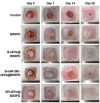
Changes in the wound closure areas in the rabbit ear at 0, 1, 2, and 4 weeks after surgery. BSSPD—bilayered thiolated alginate/PEG diacrylate hydrogel; B-sEVs—stem cell-secreted small extracellular vesicles; SRsEVs—small extracellular vesicles for sequential release. Adapted with permission from Shen et al., ACS Nano 15(4), 6352–6368 (2021). Copyright 2021 American Chemical Society.
FIG. 5.
FIG. 5.
GelMA-hypoCM accelerated wound healing in aged skin in vivo. Statistical analysis of the wound healing rate (a) and the average epidermis thickness (b). The results are presented as mean ± SD (***p < 0.001). (b) GelMA-hypoCM accelerates angiogenesis in aged skin in vivo. The blood perfusion at the wound areas of both groups was evaluated by a laser Doppler perfusion imaging system (c). Adapted with permission from Li et al., Biomater. Res. 27(1), 11 (2023). Copyright 2023 BMC, part of Springer Nature, licensed under a Creative Commons Attribution 4.0 International License.
FIG. 6.
FIG. 6.
Effects of ASC-derived exosomes in combination with HA transplantation on wound contraction rate. (a) The photographs represent the progressive healing of the wounds over time to compare the four experimental groups. (b) The percentage of wound contraction rate during the study period concerning the initial wound area. *p < 0.05, **p < 0.01, ***p < 0.001, ****p < 0.0001. Adapted with permission from Huldani et al., Tissue Cell 85, 102252 (2023). Copyright 2023 Elsevier.
FIG. 7.
FIG. 7.
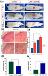
In vivo wound healing study of CMC/alginate-simvastatin in a rat model: (a) Images of wounds treated with CMC/alginate-simvastatin hydrogel at 0, 7, and 14 days, in comparison to the control (untreated wound). (b) Histopathological assessment of wound sections under various conditions using H&E staining after 14 days. Scale bar: 2 cm (a), scale bar: 10 and 100 μm (b). (c) Quantitative evaluation of the percentage of wound healing during 0, 7, and 14 days. (d) The mean thickness of the epidermis after 14 days, and (e) the mean diameter of the blood vessels at 14 days after treatment. The results are reported as the means (n = 3) ± standard deviation. (*p < 0.05). Adapted with permission from Hosseini et al., Eur. J. Pharmacol. 976, 176671 (2024). Copyright 2024 Elsevier.
FIG. 8.
FIG. 8.
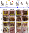
In vivo wound healing test of full-thickness wound in mice. (a) Experimental design for wounding and dressing. (b) Images representing MC_30K treated and control wounds at days 0, 6, 12, and 18. (c) Rate of wound closure in MC_30K treated and untreated wounds at different time points. Y error bar represents standard deviations, *signifies p < 0.05, **signifies p < 0.01. MC_30K represents hydrogel with a 30 mg/ml keratin concentration. Adapted with permission from Dixit et al., Int. J. Biol. Macromol. 263, 130073 (2024). Copyright 2024 Elsevier.
FIG. 9.
FIG. 9.
Antibacterial and proangiogenic composite PLGA dressings. (a) and (b) Representative images and quantitative analysis of MRSA colonies from a colony survival assay. (c) Wound images in four individual groups (blank control, gauze, PQHF-0, and PQHF-10) over 15 days. PQHF-0 and PQHF-10 correspond to dressings with 0 and 10 QC layers, respectively; (d) traces of wound closure. Scale bar: 5 mm; (e) statistical results of the wound healing rate. **p < 0.01, ***p < 0.001; scale bar: 200 μm. Adapted with permission from Wang et al., Chem. Eng. J. 404, 126525 (2021). Copyright 2021 Elsevier.
FIG. 10.
FIG. 10.
In vivo results on the healing of full-thickness burn wound in rabbits. (a) Full-thickness burn wound creation on the dorsal side of the rabbit model. (b) Macroscopic images of wounds treated with different formulations and observed after 0, 3, 7, and 14 days. The yellow square represents the wound size (2 cm2); the group treated with DSIS loaded with zinc oxide nanoparticles and vitamin C showed more rapid healing than all other group formulations. Adapted with permission from Singh et al., Int. J. Biol. Macromol. 255, 127810 (2024). Copyright 2024 Elsevier.
FIG. 11.
FIG. 11.
State-of-the-art of SOC technologies. Schematic diagram illustrating the derivation of a human skin equivalent and the subsequent construction of a skin-on-a-chip model. Adapted with permission from Cho et al., J. Invest. Dermatol. 144(8), 1707–1715 (2024). Copyright 2024 Elsevier.

Similar articles

References

    1. Sen C. K., Gordillo G. M., Roy S., Kirsner R., Lambert L., Hunt T. K., Gottrup F., Gurtner G. C., and Longaker M. T., “Human skin wounds: A major and snowballing threat to public health and the economy,” Wound Repair Regener. 17(6), 763–771 (2009).10.1111/j.1524-475X.2009.00543.x - DOI - PMC - PubMed
    1. Posnett and P. J. Franks J., “The burden of chronic wounds in the UK,” Nurs. Times 104(3), 44–45 (2008). - PubMed
    1. Tatar M., Bartke A., and Antebi A., “The endocrine regulation of aging by insulin-like signals,” Science 299(5611), 1346–1351 (2003).10.1126/science.1081447 - DOI - PubMed
    1. Lamberts S. W. J., Van Den Beld A. W., and Van Der Lely A.-J., “The endocrinology of aging,” Science 278(5337), 419–424 (1997).10.1126/science.278.5337.419 - DOI - PubMed
    1. Fulop T., Larbi A., Pawelec G., Khalil A., Cohen A. A., Hirokawa K., Witkowski J. M., and Franceschi C., “Immunology of aging: The birth of inflammaging,” Clin. Rev. Allergy Immunol. 64(2), 109–122 (2021).10.1007/s12016-021-08899-6 - DOI - PMC - PubMed