Review
. 2025 Jun 6;9(2):021502.
doi: 10.1063/5.0263236. eCollection 2025 Jun.

Mechanotransduction for therapeutic approaches: Cellular aging and rejuvenation

Affiliations
Review

Mechanotransduction for therapeutic approaches: Cellular aging and rejuvenation

Hye-Min Han et al. APL Bioeng. .

Abstract

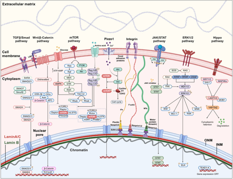
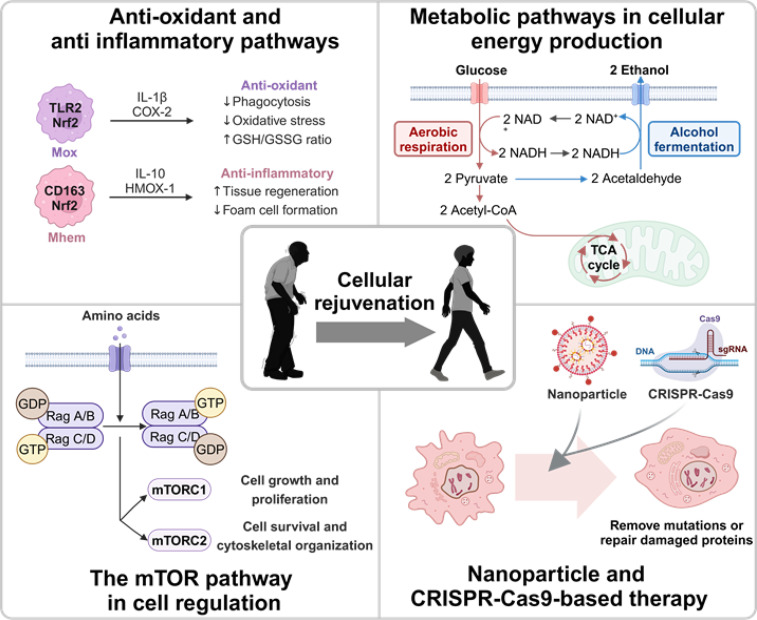
Mechanotransduction regulates cytoskeletal remodeling, nuclear mechanics, and metabolic adaptation, which are central to cellular aging and rejuvenation. These responses restore mechanical balance in aged cells, reprogram longevity-related gene expression, and alleviate age-related disorders, including neurodegeneration, musculoskeletal decline, and cardiovascular dysfunction. These insights indicate that mechanotransduction is pivotal in cellular and systemic processes underlying aging. The key signaling pathways, including the Hippo/Yes-associated protein (YAP), mechanistic target of rapamycin (mTOR), and transforming growth factor-beta (TGF-β)/Smad, have been explored in mediating age-related physiological decline, showing potential as therapeutic targets. Aging-dependent stiffening of the extracellular matrix (ECM) is associated with accelerated senescence. Interventions targeting ECM remodeling, such as mechanochemical therapies and nanoparticle delivery systems, provide promising strategies for counteracting cellular deterioration. Research progress has elucidated the critical role of mechanotransduction in organ-specific aging, enabling targeted interventions that align mechanical and biochemical therapeutic strategies. This review highlights the integration of mechanical modulation into therapeutic approaches, emphasizing its potential to restore cellular functionality, improve health, and extend lifespan. Advances in mechanomedicine have opened innovative frontiers in combating aging and age-associated diseases by addressing the interplay between mechanical forces and cellular processes. Cellular rejuvenation-the restoration of aged cells to a functionally younger state through the regulation of mechanotransduction pathways-involves the reversal of senescence-associated phenotypes, including nuclear deformation, mitochondrial alterations, and ECM stiffness. Furthermore, mechanotransduction plays a critical role in cellular rejuvenation by modulating YAP/TAZ activity, promoting autophagy, and maintaining cytoskeletal integrity.


Conflict of interest statement

The authors have no conflicts to disclose.

Figures

FIG. 1.
FIG. 1.
Pathways regulating cellular rejuvenation, including mechanotransduction and biochemical signaling. Key pathways, including TGF-β/Smad, Wnt/β-Catenin, JAK/STAT, ERK1/2, Hippo/YAP, and mTOR, coordinate biochemical and mechanical signals to restore cellular health and counter aging. The TGFβ/Smad and Wnt/β-Catenin pathways regulate transcription linked to cell fate and growth. JAK/STAT and ERK1/2 modulate immune responses and proliferation, whereas the Hippo pathway links cytoskeletal tension to YAP/TAZ activity. The mTOR pathway integrates nutrient sensing and mechanical cues to control growth and metabolism. Integrins and focal adhesions connect extracellular matrix (ECM) stiffness to intracellular signaling, thus affecting nuclear morphology and chromatin structure. These pathways highlight therapeutic targets for cellular rejuvenation. Created in BioRender. H. Han, see https://BioRender.com/m51s609 (2025).
FIG. 2.
FIG. 2.
The effect of shear stress and substrate stiffness on TAZ/YAP signaling and cellular morphology. (a) Representative immunofluorescence images showing DAPI (blue), TAZ (green), and F-actin (red) in control and shear stress conditions. (b) Quantification of TAZ subcellular localization in control and shear stress conditions. (c) Quantitative reverse transcription polymerase chain reaction (qRT-PCR) analysis showing relative fold induction of CTGF and Cyr61 in control and shear stress conditions. (d) Representative images of mineralized nodules in control and shear stress conditions, stained with Alizarin Red S. (e) qRT-PCR analysis of osteogenic marker genes (TAZ, Runx2, DLX5, Msx2) under control and shear stress conditions. Reproduced with permission from Kim et al., PLoS One 9, e92427 (2014). Copyright 2014 Authors, licensed under a Creative Commons Attribution (CC BY) license. (f) Immunofluorescence staining of Phalloidin (yellow) and YAP (white) in cells cultured on 5 and 29 kPa substrates under control and Talin 2 shRNA conditions. (g) Schematic representation of cell morphology changes in control and Talin 2 shRNA conditions. Reproduced with permission from Elosegui-Artola et al., Cell 171, 1397 (2017). Copyright 2017 Elsevier.
FIG. 3.
FIG. 3.
Effects of Nestin (NES) knockdown on cellular senescence, inflammatory cytokine expression, and nuclear architecture. (a) NES knockdown in A549 and H1299 cells induces an increase in senescence markers, as shown by SA-β-gal staining. (b) and (c) Increased mRNA expression levels of IL-6 and IL-8 in NES-knockdown A549 and H1299 cells. (d) Altered distribution of lamin B and H3K9me3 in NES-knockdown A549 and H1299 cells. Reproduced with permission from Zhang et al., Nat. Commun. 9, 3613 (2018). Copyright 2018 Authors, licensed under a Creative Commons Attribution (CC BY) license. (e) Quantification of SA β-galactosidase-positive cells in control and NES knockdown conditions. (f) Relative senescence-associated secretory phenotype (SASP) expression in control and NES knockdown conditions. (g) Representative fluorescence images of nuclear envelope integrity under different conditions (V: Vehicle, D: Drug, Q: Quiescence, D + Q: Drug + Quiescence). (h) Quantification of fluorescence intensity under different conditions. Reproduced with permission from Zhu et al., Aging Cell 14, 644–658 (2015). Copyright 2015 Authors, licensed under a Creative Commons Attribution (CC BY) license.
FIG. 4.
FIG. 4.
The effect of ECM stiffness and HDAC3 expression on osteoarthritis (OA) progression. (a) Aging markers (DAPI, p16, p21) observed in cartilage tissue under increased ECM stiffness. Reproduced with permission from Fu et al., Bone Res. 12, 32 (2024). Copyright 2024 Authors, licensed under a Creative Commons Attribution (CC BY) license. (b) Immunofluorescence staining shows alterations in F-actin (red) and vinculin (green) localization, along with nuclear DAPI staining (blue), demonstrating cytoskeletal reorganization under different conditions. (c) Quantification of F-actin fluorescence intensity reveals increased cytoskeletal tension in both young and aged cells following TGFβ1 treatment. (d) Vinculin fluorescence intensity indicates focal adhesion remodeling, highlighting differences between young and aged cells. Reproduced with permission from Zhu et al., Sci. Rep. 8, 2668 (2018). Copyright 2018 Authors, licensed under a Creative Commons Attribution (CC BY) license.
FIG. 5.
FIG. 5.
OA progression linked to ECM stiffness and HDAC3-related protein alterations. (a) HDAC3 staining in cartilage sections from control and OA models. (b) and (c) The percentage of HDAC3-positive cells was significantly higher in OA samples compared to controls, correlating with the severity of cartilage degeneration. OARSI scoring further highlights the progression of OA, showing significant increases in degeneration from control to moderate and severe conditions. Reproduced with permission from Fu et al., Bone Res. 12, 32 (2024). Copyright 2024 Authors, licensed under a Creative Commons Attribution (CC BY) license. (d) PDK1 inhibition reverses cellular senescence in human dermal fibroblasts by modulating the mTOR and NF-κB pathways, restoring the morphological and functional characteristics of youthful cells. Reproduced with permission from Proc. Natl. Acad. Sci. 117, 31535 (2020). Copyright 2020 National Academy of Sciences.
FIG. 6.
FIG. 6.
Therapeutic strategies targeting aging mechanisms and cellular rejuvenation. (a) Time-lapse imaging of cellular responses under different conditions, including normal and TGF-β stimulation at various time points (days 5, 7, 10, 13, 16). Heatmaps represent changes in fluorescence intensity over time. Reproduced with permission from Kim et al., Nat. Mater. 23, 290–300 (2023). Copyright 2023 Authors, licensed under a Creative Commons Attribution (CC BY) license. (b) Representative fluorescence images of cells treated with saline, VPQDs, CaNPs, and VPCaNPs. Nuclei are stained with DAPI (blue), membrane structures with DiD (red), and calcium deposits with Calcein (green). (c) X-ray diffraction (XRD) analysis of VPQDs, CaNPs, and VPCaNPs, displaying their crystalline structures. (d) Histological analysis of tissue samples treated with saline, VPQDs, CaNPs, and VPCaNPs. Reproduced with permission from Zhang et al., Nat. Commun. 15, 6783 (2024). Copyright 2024 Authors, licensed under a Creative Commons Attribution (CC BY) license.
FIG. 7.
FIG. 7.
Cellular rejuvenation strategy. Molecular mechanisms—including antioxidant and anti-inflammatory pathways, metabolic energy production, mTOR signaling, and nanoparticle-CRISPR-Cas9-based therapies—are depicted. The TLR2-Nrf2 and CD163-Nrf2 pathways promote tissue regeneration by reducing oxidative stress and modulating anti-inflammatory responses. Glucose metabolism through glycolysis, the TCA cycle, and alcohol fermentation optimize energy production and NAD+ synthesis. mTORC1 and mTORC2 regulate cell growth and survival through amino acid signaling, whereas nanoparticles enhance targeted delivery, and CRISPR-Cas9 restores cellular functions at the genetic level.

Similar articles

References

    1. Discher D. E., Janmey P., and Wang Y. L., “Tissue cells feel and respond to the stiffness of their substrate,” Science 310, 1139–1143 (2005).10.1126/science.1116995 - DOI - PubMed
    1. Lopez-Otin C., Blasco M. A., Partridge L., Serrano M., and Kroemer G., “The hallmarks of aging,” Cell 153, 1194–1217 (2013).10.1016/j.cell.2013.05.039 - DOI - PMC - PubMed
    1. Humphrey J. D., Dufresne E. R., and Schwartz M. A., “Mechanotransduction and extracellular matrix homeostasis,” Nat. Rev. Mol. Cell Biol. 15, 802–812 (2014).10.1038/nrm3896 - DOI - PMC - PubMed
    1. Ingber D. E., “Mechanical control of tissue morphogenesis during embryological development,” Int. J. Dev. Biol. 50, 255–266 (2006).10.1387/ijdb.052044di - DOI - PubMed
    1. Lammerding J. et al., “Lamin A/C deficiency causes defective nuclear mechanics and mechanotransduction,” J. Clin. Invest. 113, 370–378 (2004).10.1172/JCI200419670 - DOI - PMC - PubMed

LinkOut - more resources