Novel Approach in Treatment of Coarctation of the Aorta with Bifurcation Stenosis of the Left Subclavian Artery - The Road Less Traveled
- PMID: 40488154
- PMCID: PMC12139642
- DOI: 10.4103/heartviews.heartviews_64_24
Novel Approach in Treatment of Coarctation of the Aorta with Bifurcation Stenosis of the Left Subclavian Artery - The Road Less Traveled
Abstract
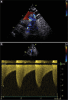
Endovascular stenting has emerged as the preferred treatment modality for coarctation of the aorta (CoA). However, CoA can sometimes extend beyond the aortic arch, involving adjacent vessels such as the left subclavian artery (LSA), which complicates conventional interventions. We present a case of CoA associated with proximal LSA stenosis which was successfully treated using a double-wire stent technique. The technique does not compromise the LSA flow and offers a promising alternative in complex CoA cases. A 19-year-old female presented with palpitations with dyspnea (New York Heart Association grade III) for 15 days. She also gave a history of intermittent claudication in the left upper limb for 3 years. Clinical examination revealed pallor and weak pulses in the left upper extremity and both lower extremities, with radio-radial and radio-femoral delays. Blood pressure measurements indicated significant gradients between the limbs, with readings of 244/112 mmHg in the right upper limb, 162/104 mmHg in the left upper limb, and 114/74 mmHg and 116/78 mmHg in the right and left lower limbs, respectively. Auscultation revealed normal S1 and S2 and a systolic murmur in the right interscapular area. Electrocardiogram revealed sinus arrhythmia with T-wave inversions in leads II, aVF, and V1-V6. Echocardiogram revealed severe postductal coarctation with a gradient of 84 mmHg. Computed tomography aortography confirmed a severe coarctation-preductal diameter of 12 mm and postductal diameter of 14 mm, with a concomitant LSA stenosis of 7 mm. The critical challenge in this case was stenting the coarctation without compromising the already symptomatic LSA stenosis. A novel endovascular approach was employed, utilizing two preplaced wires in both the aorta and LSA, followed by deployment of an uncovered stent and final kissing balloon angioplasty. This is the first instance in literature of such an approach being taken. Patients with CoA with associated bifurcation stenosis of the LSA are extremely rare and pose significant challenges for endovascular management. This case highlights a novel and effective interventional strategy, offering a tailored approach to preserve LSA patency while addressing the complex CoA anatomy.
Keywords: Angioplasty; case report; coarctation of the aorta; endovascular interventions; left subclavian artery; stenting.
Copyright: © 2025 Heart Views.
Conflict of interest statement
There are no conflicts of interest.
Figures


Similar articles
-
Stenting for peripheral artery disease of the lower extremities: an evidence-based analysis.Ont Health Technol Assess Ser. 2010;10(18):1-88. Epub 2010 Sep 1. Ont Health Technol Assess Ser. 2010. PMID: 23074395 Free PMC article.
-
Externalized Guidewires to Facilitate Fenestrated Endograft Deployment in the Aortic Arch.J Endovasc Ther. 2016 Feb;23(1):160-71. doi: 10.1177/1526602815614557. Epub 2015 Oct 28. J Endovasc Ther. 2016. PMID: 26511895 Free PMC article.
-
Percutaneous treatment for aneurysmal coarctation of the aorta with covered stenting: a case report.J Tehran Heart Cent. 2014;9(2):82-4. J Tehran Heart Cent. 2014. PMID: 25861324 Free PMC article.
-
Systematic review and meta-analysis of type B aortic dissection involving the left subclavian artery with a Castor stent graft.Front Cardiovasc Med. 2022 Nov 29;9:1052094. doi: 10.3389/fcvm.2022.1052094. eCollection 2022. Front Cardiovasc Med. 2022. PMID: 36523362 Free PMC article.
-
Coronary-subclavian steal syndrome: A case series and review of the literature.Vascular. 2024 Dec 13:17085381241307751. doi: 10.1177/17085381241307751. Online ahead of print. Vascular. 2024. PMID: 39673086 Review.
References
-
- Hartman EM, Groenendijk IM, Heuvelman HM, Roos-Hesselink JW, Takkenberg JJ, Witsenburg M. The effectiveness of stenting of coarctation of the aorta: A systematic review. EuroIntervention. 2015;11:660–8. - PubMed
-
- Baumgartner H, De Backer J, Babu-Narayan SV, Budts W, Chessa M, Diller GP, et al. 2020 ESC guidelines for the management of adult congenital heart disease. Eur Heart J. 2021;42:563–645. - PubMed
-
- Lampropoulos K, Budts W, Gewillig M. Dual wire technique for aortic coarctation stent placement. Catheter Cardiovasc Interv. 2011;78:425–7. - PubMed
-
- Singer MI, Rowen M, Dorsey TJ. Transluminal aortic balloon angioplasty for coarctation of the aorta in the newborn. Am Heart J. 1982;103:131–2. - PubMed
Publication types
LinkOut - more resources
Full Text Sources
