

Critical size defect in a rat calvaria model using trephination: An animal model for investigating potential bone regenerative scaffolds
- PMID: 40488170
- PMCID: PMC12141067
- DOI: 10.1016/j.mex.2025.103355
Critical size defect in a rat calvaria model using trephination: An animal model for investigating potential bone regenerative scaffolds
Abstract
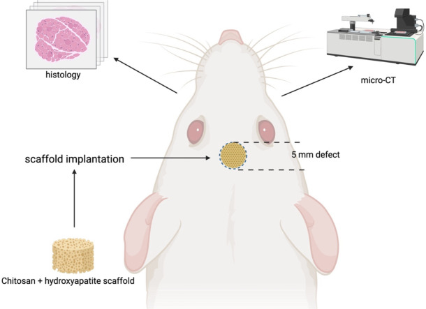
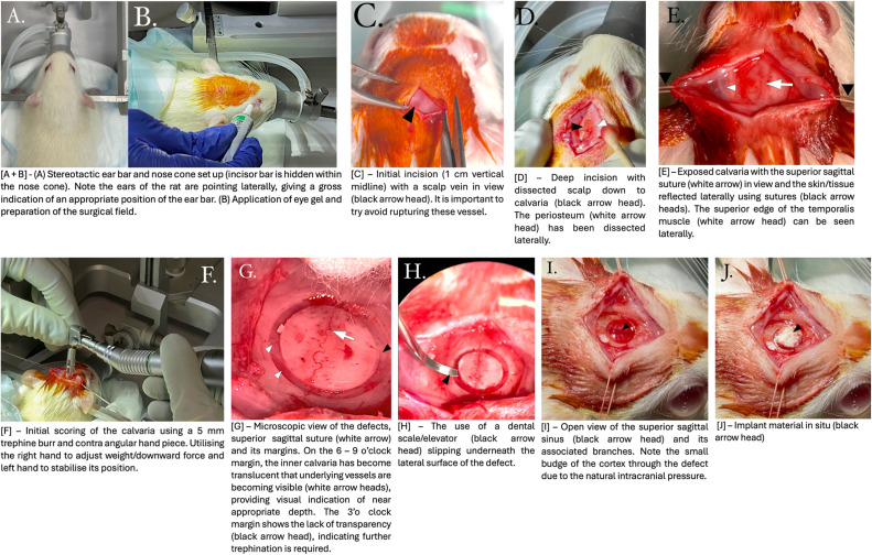
Animal studies have been a key in vivo investigation in translating bioregenerative material into human clinical trials. The critical size defect (CSD) in a rat's calvaria is a popular methodology for demonstrating and investigating the regenerative properties of a biomaterial. Various surgical approaches have been discussed in the literature on creating CSD and can vary based on species, age and anatomical location. This paper aims to provide a reliable and reproducible step-by-step guide for creating a critical-size defect on the calvaria of a laboratory rat using trephination. This paper will:•Cover the surgical approaches and management in the pre-, intra- and post-operative phases.•Provide figures to help visually guide readers in carrying out the operation.•Cover basic general anaesthetic issues and resolutions.
Keywords: Biomaterials; Bone regeneration; Bony defect; Calvaria; Critical size defect; Critical size defect trephination in rat calvaria using a chitosan-hydroxyapatite scaffold; Rat study; Small animal model; in vivo study.
© 2025 The Authors. Published by Elsevier B.V.
Conflict of interest statement
The authors declare that they have no known competing financial interests or personal relationships that could have appeared to influence the work reported in this paper.
References
-
- Vajgel A., et al. A systematic review on the critical size defect model. Clin. Oral. Implants Res. 2014;25(8):879–893. - PubMed
-
- Hollinger J.O., Kleinschmidt J.C. The critical size defect as an experimental model to test bone repair materials. J. Craniofacial Surg. 1990;1(1) - PubMed
-
- Baldwin P., et al. Autograft, allograft, and bone graft substitutes: clinical evidence and indications for use in the setting of orthopaedic trauma surgery. J. Orthop. Trauma. 2019;33(4) - PubMed
-
- Betz R.R. Limitations of autograft and allograft: new synthetic solutions. Orthopedics. 2002;25(5):S561–S570. - PubMed
-
- Buma P., Schreurs W., Verdonschot N. Skeletal tissue engineering—From in vitro studies to large animal models. Biomaterials. 2004;25(9):1487–1495. - PubMed
LinkOut - more resources
Full Text Sources
