. 2025 Jun 9:13:RP101338.
doi: 10.7554/eLife.101338.

Oviductin sets the species-specificity of the mammalian zona pellucida

Affiliations

Oviductin sets the species-specificity of the mammalian zona pellucida

Daniel de la Fuente et al. Elife. .

Abstract

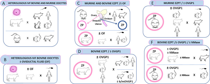
The zona pellucida (ZP) is vital for species-specific fertilization as this barrier mediates sperm-oocyte binding. Here, we determined whether sperm from distant mammalian orders (Carnivora, Primates, and Rodentia) could penetrate bovine oocytes by examining the role of bovine oviductal fluid and species-specific oviductal glycoprotein (OVGP1 or oviductin) from bovine, murine, or human sources in modulating the species-specificity of bovine and murine oocytes. Sperm from all the species were found to penetrate intact bovine ovarian oocytes to form hybrid embryos. However, contact with oviductal fluid or bovine, murine, or human OVGP1, conferred the ZP species-specificity, allowing only the penetration of the corresponding sperm regardless of the ZP's origin. Glycolytic and microstructural analyses revealed that OVGP1 covers the pores present in the ZP and that OVGP1 glycosylation determines sperm specificity. This suggests specific fertilization capacity is acquired in the oviduct through the ZP's incorporation of specific oviductin.

Keywords: OVGP1; bovine; developmental biology; fertilization; human; mouse; zona pellucida.


Conflict of interest statement

Dd, MM, YC, KC, RF, AM, JS, RB, PC, MA, DR, AG No competing interests declared

Figures

Figure 1.
Figure 1.. Schematic representation of global experimental design of homologous and heterologous IVF from diverse mammalian species.
(A) Represents design of experiment shown in Appendix 1—table 1, Appendix 1—table 2, Appendix 1—table 3, Figure 2A and B, Figure 2—figure supplement 1, Figure 2—figure supplement 2 and Figure 2—figure supplement 3. (B) Represents design of experiment shown in Appendix 1—table 4, Figure 2C. (C) Represents design of experiment shown in Appendix 1—table 5, Appendix 1—table 6, Figure 2D and E and Figure 3. (D) Represents design of experiment shown in Appendix 1—table 8, Appendix 1—table 9, Appendix 1—table 10, Figure 5 and Figure 7A. (E) Represents design of experiment shown in Appendix 1—table 11, and Figure 7B. (F) Represents design of experiment shown in Appendix 1—table 12, Appendix 1—table 13, Appendix 1—table 14 and Figures 8 and 9.
Figure 2.
Figure 2.. Heterologous IVF of bovine oocytes, mouse oocytes, or empty mouse ZPs, using human, mouse, or cat sperm, before and after contact with oviductal fluid.
(A) Embryo cleavage rates resulting from the IVF of bovine oocytes with human, murine, or feline sperm including bovine sperm as homologous IVF control, and parthenogenesis as a negative control of the IVF. (B) Sperm penetration rate after the IVF of IVM mouse oocytes with murine or bovine sperm, for homologous and heterologous IVF, respectively. (C) Embryo cleavage rates resulting from the IVF of bovine IVM oocytes, preincubated 30 min with bovine oviductal fluid, with human sperm IVF medium a=G-IVF PLUS medium (HeA); IVF medium b=Fert (HeB). (D) Penetration rates and average numbers of sperm bound to ZP after the empty zona penetration test (EZPT) using mouse ovarian IVM oocytes and murine, bovine, or human sperm. (E) Penetration rates and average numbers of sperm bound to ZP after the EZPT using mouse oviductal oocytes and murine, bovine, or human sperm. (F) Picture of an empty zona pellucida obtained from a mouse ovarian IVM oocyte after the EZPT using bovine sperm. Note the sperm has penetrated the zona. (G) Picture of an empty zona pellucida from a mouse oviductal oocyte after the EZPT using bovine sperm. Scale bar for ZP pictures = 50 µm. A non―fertilized parthenogenesis group is used as cleavage control in (A) and (C). Different letters above error bars (mean ± SD) indicate significant differences (p<0.05) among groups (ANOVA and Tukey’s post hoc test). Numbers of oocytes or ZPs used are indicated in Appendix 1—table 1, Appendix 1—table 2, Appendix 1—table 3, Appendix 1—table 4 and Appendix 1—table 5.
Figure 2—figure supplement 1.
Figure 2—figure supplement 1.. Heterologous IVF between bovine oocytes and human sperm.
Sperm-oocyte binding, pronuclear formation, and cleavage after homologous (Ho) (bovine sperm) and heterologous (He) IVF: human sperm capacitated with G-IVF PLUS medium (HeA) or Tyrode’s medium (HeB). Gametes were stained with Hoechst 33342. Ho: Bound bovine sperm after 2.5 hr of co-incubation with zona-intact bovine oocytes (A); pronuclear formation at 6 hpi (B); and embryo cleavage at 48 hpi (C). HeA and HeB: Bound human sperm after 2.5 hr of co-incubation of the gametes (D, G); pronuclear formation at 18 hpi (E, H); and hybrid-embryo cleavage at 48 hpi (F, I), respectively. Arrow points to sperm head chromatin. Images were captured with a 63 X objective. Scale bar 20 µm.
Figure 2—figure supplement 2.
Figure 2—figure supplement 2.. Heterologous IVF between bovine oocytes and murine sperm.
Sperm-oocyte binding, pronuclear formation, and cleavage after homologous (Ho, bovine sperm) and heterologous (He, mouse/rodent sperm) IVF. Gametes were stained with Hoechst 33342. Ho: Bound bovine sperm after 2.5 hr of co-incubation with zona-intact bovine oocytes (A); pronuclear formation at 6 hpi (B); and embryo cleavage at 24 hpi (C). He: Bound mouse sperm after 2.5 hr of co-incubation of the gametes (D); pronuclear formation at 18 hpi (E); and hybrid-embryo cleavage at 24 hpi (F). Arrow points to sperm head chromatin. Images were captured with a 63 X objective. Scale bar 20 µm.
Figure 2—figure supplement 3.
Figure 2—figure supplement 3.. Heterologous IVF between bovine oocytes and cat sperm.
Sperm-oocyte binding, pronuclear formation, and cleavage after homologous (Ho) (bovine sperm) and heterologous (He) (cat sperm capacitated in Tyrode’s medium (HeG)) IVF. Gametes were stained with Hoechst 33342. Ho: Bound bovine sperm after 2.5 hr of co-incubation with zona-intact bovine oocytes (A); pronuclear formation at 6 hpi (B); and embryo cleavage at 48 hpi (C). HeG: Bound cat sperm after 2.5 hr of co-incubation of the gametes (D); pronuclear formation at 18 hpi (E); and hybrid-embryo cleavage at 48 hpi (F). Arrow points to sperm head chromatin. Images were captured with a 63 X objective. Scale bar 20 µm.
Figure 2—figure supplement 4.
Figure 2—figure supplement 4.. Steps (A–E) of the method used to empty the bovine zone pellucida by removing the cytoplasmic contents of the oocyte containing all the organelles, nucleus and membranes, and removing also the polar body.
Details of this procedure can be found in the Materials and methods.
Figure 3.
Figure 3.. Incubation of empty ZPs obtained from bovine ovarian oocytes with oviductal fluid determines the specificity of spermatozoa capable of penetrating the zona.
The empty zona penetration test (EZPT) in ZPs obtained from IVM bovine oocytes was performed after homologous (bovine sperm) or heterologous (human or murine sperm) fertilization. Similar outcomes were observed when ZPs were not treated with oviductal fluid (A), but after incubation with oviductal fluid for 30 min, human and murine sperm were unable to penetrate the bovine ZPs (B) (six replicates per medium per semen sample). Different letters above error bars (mean ± SD) indicate significant differences (P<0.05) among groups (ANOVA and Tukey’s post hoc test). Numbers of ZPs used are indicated in Appendix 1—table 6. (C, D, I) Representative pictures of empty bovine ZPs penetrated by bovine sperm illustrating that the penetrating sperm (J) lack an acrosome, while those unable to penetrate the zona maintain the acrosome (K). (E, F, L) Empty bovine ZP penetrated by human sperm revealing that the penetrating sperm have lost the acrosome (M), whereas those not penetrating the zona maintain the acrosome (N). (G, H, O) Representative pictures of empty bovine ZPs penetrated by murine sperm illustrating that the penetrating sperm (P) lack an acrosome, while those unable to penetrate the zona maintain the acrosome (Q). In the absence of oviductal fluid, the empty bovine ZP can be penetrated by bovine (C, I), human (E, L), or murine (G, O) sperm; however, when the ZP has been in contact with oviductal fluid, it can only be penetrated by bovine sperm (D), and not by human (F) or mouse (H) sperm. Scale bar for ZP pictures = 50 µm. Scale bar for sperm pictures = 5 µm.
Figure 4.
Figure 4.. Structure of oviductin, western blots of OVGP1 recombinants and localization of these recombinants at the bovine or murine ZPs.
(A) Diagram showing the five regions (A, B, C, D, and E) present in some of the oviductin proteins of human (hOVGP1), murine (mOVGP1), and bovine (bOVGP1) mammalian species (adapted from Figure 1 from Avilés et al., 2010). (B) Western blots of the three OVGP1 recombinants proteins used in this study (human, murine, and bovine). Recombinant bovine protein was expressed in BHK-21 cells and purified, whereas recombinant murine and human proteins were purchased by Origene and had been produced in HEK293T. Then, proteins were separated by SDS-PAGE and analyzed by immunoblotting using rabbit polyclonal antibody to the human OVGP1. The following lanes of the gel contain the protein extracts from oviductal fluid of female mice in oestrus and anoestrus and from oviductal fluid of ovulated cows or anoestrus cows, indicating with an arrow the presence in estrous of the OVGP1 band for both species. ZPs from IVM murine (C) and bovine (D) oocytes were incubated for 30 min at RT with recombinants bOVGP1, mOVGP1, and hOVGP1. ZPs were fixed and imaged by confocal fluorescence and DIC microscopy using rabbit polyclonal antibody to the human OVGP1 for bOVGP1, and a monoclonal antibody against Flag―Tag for hOVGP1 and mOVGP1. Scale bars = 20 μm.
Figure 4—figure supplement 1.
Figure 4—figure supplement 1.. Sequence alignments of OVGP1 from Homo sapiens (NCBI AAI36407.1), Bos taurus (NP_001073685.1), and Mus musculus (AAI37996.1).
Sequences were aligned with the tool MUSCLE (Multiple Sequence Comparison by Log-Expectation) using Snapgene software. Consensus conserved amino acids are represented in the colored blocks: red = score over the 50% threshold and blue = less conserved. Amino acids corresponding to the consensus are indicated in light pink. N-glycosylation sites were predicted using the NetNGlyc 1.0 Server of DTU Health Tech Bioinformatic Services. The dark green boxes indicate possible asparagine (N) glycosylation. Residues adjacent to N in the sequons Asn-Xaa-Ser/Thr are circled in dark pink. Chitin-binding sites predicted by the UniProt server appear in turquoise boxes. Orange boxes locate the essential glutamic acid of CH18 family members.
Figure 4—figure supplement 2.
Figure 4—figure supplement 2.. Recombinant OVGP1 from human, murine and bovine recognized with anti-OVGP1 (A) and anti-His/anti-Flag (B).
Panel A is taken directly from Figure 4 in order to compare antibody recognition. In B, is represented the expression of recombinant proteins detected with an anti-His and anti-Flag co-incubated antibodies and developed simultaneously. Flag is detecting hOVGP1 and mOVGP1, while His is detecting bOVGP1.
Figure 4—figure supplement 3.
Figure 4—figure supplement 3.. Representation of immunofluorescence of control ZPs of IVM murine and bovine oocytes, ZPs incubated with murine or bovine oviductal fluid, and ZPs from murine superovulated (SOV).
IVM murine (A) and bovine (B) ZPs were fixed and imaged by conventional fluorescence and DIC microscopy using rabbit polyclonal antibody to the human OVGP1 (I, II) or a mouse monoclonal antibody against Flag-tag (III, IV). IVM ZPs from murine and bovine oocytes were incubated for 30 min with murine or bovine oviductal fluid (respectively) and imaged using anti-Flag tag (V, 1452 54 VI). ZPs obtained from oocytes of the oviducts of superovulated mice females (SOV) incubated with anti-OVGP1 (VII). Scale bars = 20 μm.
Figure 5.
Figure 5.. Incubation of empty ZPs obtained from bovine ovarian oocytes with OVGP1 determines the specificity of spermatozoa capable of penetrating the zona.
The empty zona penetration test (EZPT) was performed after homologous (bovine sperm) or heterologous (human or murine sperm) fertilization. Similar penetration rates were observed when ZPs were not treated with bOVGP1 protein (A) but after incubation with bOVGP1 for 30 min, human or murine sperm were unable to penetrate the bovine ZPs (B) A drastic reduction was also observed for fertilization by heterologous sperm, both in the average number of sperm penetrating the ZPs, and in the average of number of sperm binding to the ZPs, when fertilization without pretreatment of the ZP with bOVGP1 (A) was compared to fertilization after the zona had been in contact with bOVGP1 (B). (6 replicates per medium per semen sample). Different letters above error bars (mean ± SD) indicate significant differences (p<0.05) among groups (ANOVA and Tukey’s post hoc test). Numbers of ZPs used are indicated in Appendix 1—table 8.
Figure 6.
Figure 6.. Scanning electron micrographs (SEM) of the outer surface of the bovine ZP treated or not with OVGP1.
The ZP of an in vitro matured (IVM) bovine oocyte: Magnification x2000 (A) and x4000 (B); and bovine oocytes IVM in the presence of bovine OVGP1 (bOVGP1): Magnification x2000 (C) and x4000 (D). High magnification reveals the ultrastructural characteristics of the ZP’s pores on the IVM oocytes without bOVGP1 (B) and with bOVGP1 (D). Scale bars = 10 μm.
Figure 6—figure supplement 1.
Figure 6—figure supplement 1.. Scanning electron micrographs (SEM) of the outer surface of the bovine ZP.
SEM of the outer surface of the bovine ZP treated (B) or not (A) with bovine OF, mOVGP1 (C), or hOVGP1 (D). (E) Image showing bovine sperm on the surface of the bovine ZP after having been in contact with OVGP1, showing the comparison between the size of the sperm and the size of the pores of the ZP.
Figure 7.
Figure 7.. Species-specific OVGP1 confers complete species-specificity to the zona pellucida.
Experiment using the EZPT to analyze several combinations whereby ZPs from bovine and murine ovarian IVM oocytes were co-incubated with bovine, murine, or human oviductin, and exposed to sperm of all three species. The variables ZP penetration rate, average number of sperm penetrating the ZPs, and average number of sperm bound to ZPs were examined using bovine ZPs (A) or murine ZPs (B) and combinations of one of the three oviductins with sperm of one of the three species. Only by combining ZP, OVGP1 and sperm of the same species, was species-specific fertilization possible. However, when the same species was matched only with the oviductin and sperm, or only with the ZP and sperm, penetration and binding to the ZP of the spermatozoa also occurred, although in much smaller measure. No penetration was observed when only the ZP and OVGP1 species matched. Different letters above error bars (mean ± SD) indicate significant differences (p<0.05) among groups (ANOVA and Tukey’s post hoc test). Numbers of ZPs used are indicated in Appendix 1—table 10, Appendix 1—table 11.
Figure 8.
Figure 8.. Effect of neuraminidase (NMase) treatment of bovine or murine ZPs before and after contact with OVGP1 on sperm penetration.
Penetration and sperm binding rates were measured after homologous EZPT with bovine sperm using ZPs not subjected (A) or subjected (B) to 30 min of incubation with bOVGP1. Penetration and sperm binding rates were measured after homologous EZPT with murine sperm using ZPs not subjected (C) or subjected (D) to 30 min of incubation with mOVGP1. ZPs of both species were co-incubated with acetate buffer (PH 4.5) at 38 °C for 18 hr in the presence or absence of neuraminidase diluted at 5 UI/mL. Different letters above error bars (mean ± SD) indicate significant differences (p<0.05) among groups (ANOVA and Tukey’s post hoc test). Numbers of ZPs used are indicated in Appendix 1—table 12, Appendix 1—table 13.
Figure 9.
Figure 9.. Effect of neuraminidase (NMase) treatment of OVGP1 on EZPT using bovine ZPs with homologous or heterologous sperm.
(A) Penetration rate, average number of sperm within penetrated ZPs, and average of number of sperm bound to ZPs were measured after homologous EZPT with bovine sperm, using ZPs untreated with NMase and bOVGP1 either treated or untreated with NMase. (B) Penetration rate, average number of sperm within penetrated ZPs, and average of number of sperm bound to ZPs were measured after homologous (bovine sperm) or heterologous (mouse sperm) EZPT, using ZPs untreated with NMase, either without bOVGP1 or with bOVGP1 treated or untreated with NMase. Numbers of ZPs used are indicated in Appendix 1—table 14.
Figure 10.
Figure 10.. Model.
Schematic representation of the heterologous fertilization model via EZPT under multiple conditions.The figure illustrates how sperm penetration into the zona pellucida (ZP) occurs non-specifically among various mammalian species when the ZP has not been exposed to homologous oviductal fluid or OVGP1 (left diagram). However, this interaction becomes species-specific when the ZP is incubated with oviductal fluid or OVGP1 from the same species (central diagram) This specificity is lost when OVGP1 is heterologous to the ZP (right diagram).

Update of

  • doi: 10.1101/2024.07.01.601502
  • doi: 10.7554/eLife.101338.1
  • doi: 10.7554/eLife.101338.2
  • doi: 10.7554/eLife.101338.3

Similar articles

References

    1. Algarra B, Han L, Soriano-Úbeda C, Avilés M, Coy P, Jovine L, Jiménez-Movilla M. The C-terminal region of OVGP1 remodels the zona pellucida and modifies fertility parameters. Scientific Reports. 2016;6:32556. doi: 10.1038/srep32556. - DOI - PMC - PubMed
    1. Algarra B, Maillo V, Avilés M, Gutiérrez-Adán A, Rizos D, Jiménez-Movilla M. Effects of recombinant OVGP1 protein on in vitro bovine embryo development. The Journal of Reproduction and Development. 2018;64:433–443. doi: 10.1262/jrd.2018-058. - DOI - PMC - PubMed
    1. Araki Y, Kurata S, Oikawa T, Yamashita T, Hiroi M, Naiki M, Sendo F. A monoclonal antibody reacting with the zona pellucida of the oviductal egg but not with that of the ovarian egg of the golden hamster. Journal of Reproductive Immunology. 1987;11:193–208. doi: 10.1016/0165-0378(87)90057-x. - DOI - PubMed
    1. Avella MA, Baibakov B, Dean J. A single domain of the ZP2 zona pellucida protein mediates gamete recognition in mice and humans. The Journal of Cell Biology. 2014;205:801–809. doi: 10.1083/jcb.201404025. - DOI - PMC - PubMed
    1. Avilés M, Martínez-Menárguez JA, Castells MT, Madrid JF, Ballesta J. Cytochemical characterization of oligosaccharide side chains of the glycoproteins of rat zona pellucida: an ultrastructural study. The Anatomical Record. 1994;239:137–149. doi: 10.1002/ar.1092390204. - DOI - PubMed

LinkOut - more resources