

Computed tomography-based quantitative scoring system for rheumatoid arthritis-associated interstitial lung disease: a retrospective diagnostic accuracy study for progressive fibrosis detection
- PMID: 40488936
- PMCID: PMC12234623
- DOI: 10.1007/s10067-025-07511-y
Computed tomography-based quantitative scoring system for rheumatoid arthritis-associated interstitial lung disease: a retrospective diagnostic accuracy study for progressive fibrosis detection
Abstract
Objectives: To investigate the ability of quantitative parameters to assess the severity of rheumatoid arthritis (RA)-associated interstitial lung disease (ILD).
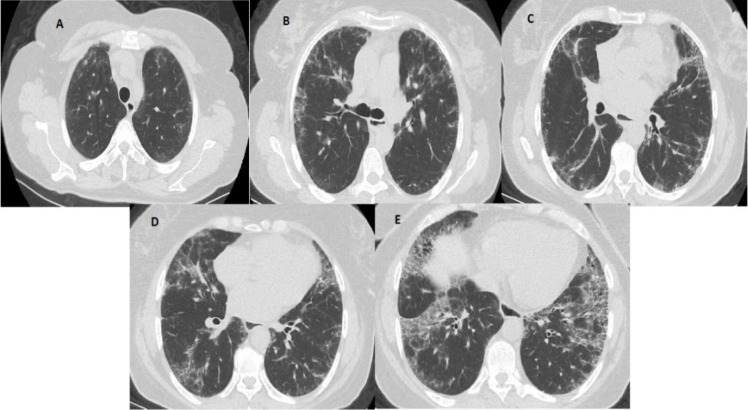
Methods: Thorax CT images and pulmonary function tests of RA patients followed up in a tertiary reference center were retrospectively examined. Radiologically, patients were divided into two groups as limited and extensive ILD, using two semiquantitative scoring systems. Two different methods (Method 1, Method 2) were used for quantitative image analysis, in which different Hounsfield unit values were selected as thresholds. Spearman's correlation test was used to evaluate the relationship between variables. The diagnostic performance of quantitative methods for the ability to distinguish limited ILD from extensive ILD was calculated using ROC analysis.
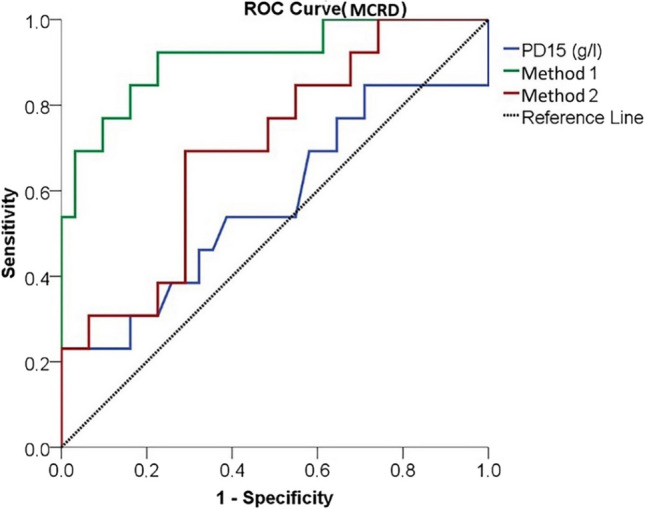
Results: Forty-four patients, 29 female and 15 male, were included in the study. A significant correlation was found between diffusion capacity of the lungs carbonmonoxide (DLCO) and total lung capacity (TLC), which are clinical markers, and both quantitative methods (p < 0.001). In terms of the performance of diagnostic tests, the power of Method 1 and Method 2 to distinguish limited and extensive disease classified based on semiquantitative scores was found to be strong (Method 1 AUC = 0.945, Method 2 AUC = 0.785 based on "ILD score"; Method 1 AUC = 0.911, Method 2 AUC = 0.700 based on "modified coarseness of reticular disease (MCRD) score").
Conclusion: This study demonstrates that quantitative methods (Method 1 [- 700 to - 200 HU] and Method 2 [- 800 to - 500 HU]) play a significant role in assessing the severity of RA-associated ILD (RA-ILD). Both methods showed significant correlations with pulmonary function tests (DLCO [r1 = - 0.338, p1 = 0.025; r2 = - 0.452, p2 = 0.002] and TLC [r1 = - 0.567, p1 < 0.001; r2 = - 0.576, p2 < 0.001), with Method 1 (AUCILD = 0.945 and AUCMCRD = 0.911) demonstrating superior performance in distinguishing between limited and extensive ILD. Our findings suggest that Hounsfield unit threshold-based quantitative CT analysis may serve as a more objective and reproducible alternative to semiquantitative systems (e.g., Goh score) in the standard evaluation of RA-ILD. Specifically, Method 1 may enable early detection of progression by mechanism, e.g., "tracking subtle density changes." However, further validation in multicenter prospective cohorts is needed to address limitations such as bias. Key Points • Interobserver and intraobserver variability poses a significant challenge in the objective assessment of rheumatoid arthritis-associated interstitial lung disease. • User independent quantitative methods can be used instead of user-dependent semiquantitative methods in assessing rheumatoid arthritis-associated interstitial lung disease. • Quantitative computed tomography analysis enables precise stratification of disease severity in rheumatoid arthritis-associated interstitial lung disease, distinguishing limited interstitial lung disease from extensive interstitial lung disease, which has a poor prognosis.
Keywords: Computer-assisted image analysis; Interstitial lung diseases; Multidetector computed tomography; Pulmonary function tests; Rheumatoid arthritis.
© 2025. The Author(s).
Conflict of interest statement
Declarations. Ethical approval and consent to participate.: Ethics committee approval was approved by the local ethics committee of Sütçü İmam University with the meeting, 01.02.2022, Decision No: 3. Consent for publication: Written informed consent was obtained from the all patient for publication of this study. Disclosures: None. Originality statement: The revised manuscript contains no plagiarized text passages, duplicated graphics, or previously published material without proper attribution.
Figures


References
MeSH terms
LinkOut - more resources
Full Text Sources
Medical
