. 2025 Jun 9;36(1):49.
doi: 10.1007/s10856-024-06845-4.

Interlamellar keratoplasty for implantation of decellularized porcine corneal lenticule in a rabbit for corneal thickening

Affiliations

Interlamellar keratoplasty for implantation of decellularized porcine corneal lenticule in a rabbit for corneal thickening

Lays Fernanda Nunes Dourado et al. J Mater Sci Mater Med. .

Abstract

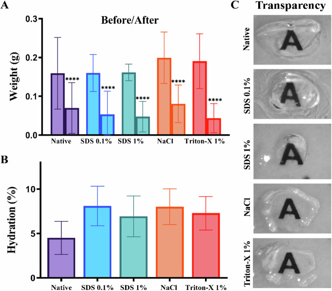
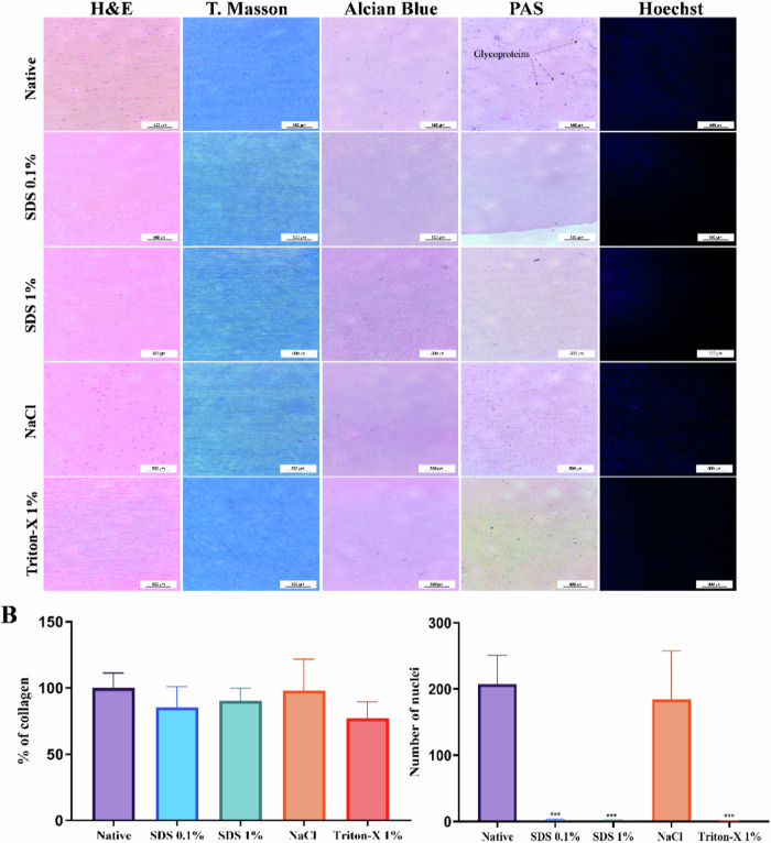
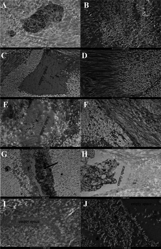
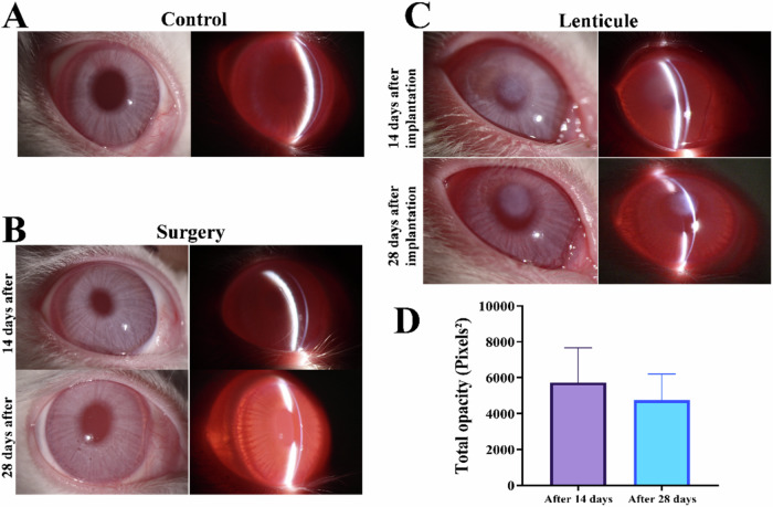
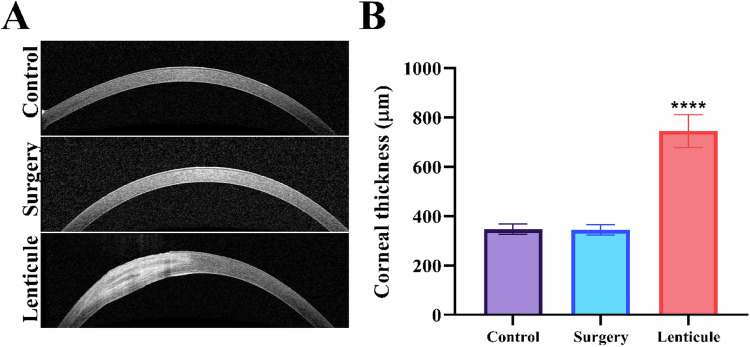
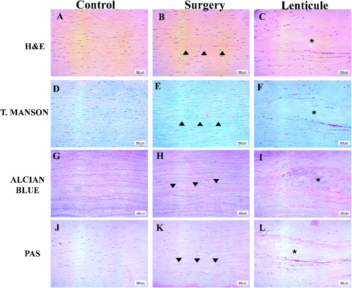
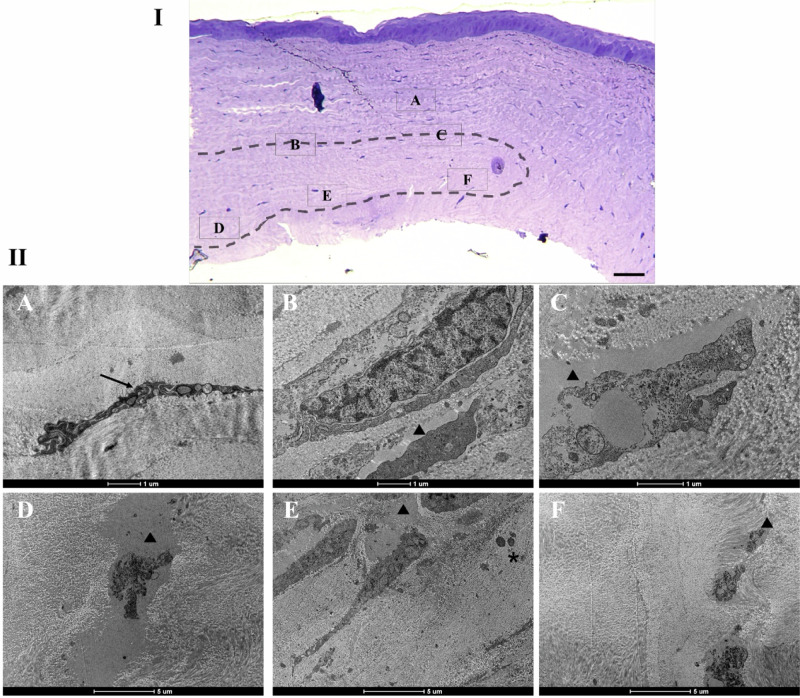
Changes in collagen orientation and distribution on the corneas lead to the development of diseases characterized by progressive thinning, such as keratoconus. Part of people diagnosed with keratoconus require a corneal graft, which has availability as a major limiting factor. In this scenario, new approaches have been tested to obtain substitute tissues. Porcine cornea has been receiving increasing attention due to its ease of obtaining, biomechanical properties similar to those of human tissue and lower antigenicity. Based on this, the objective of this study was to evaluate the biocompatibility of porcine stroma decellularized by sodium dodecyl sulfate (SDS) through interlamellar implantation in rabbit corneas. The obtained results showed that the lenticule intrastromal implantation was successfully performed and did not elicit rejection. Furthermore, the implanted stroma was able to promote an increase in the thickness of the host cornea. Microscopic analyses revealed that the tissue was well-adhered and the collagen fibrils were more aligned on its periphery. Therefore, it is concluded that the implantation of decellularized porcine stroma occurred satisfactorily and represents a promising alternative to replace human tissue.


Conflict of interest statement

Compliance with ethical standards. Conflict of interest: The authors declare no competing interests.

Figures

Fig. 1
Fig. 1
Measurement of weight variation and hydration of stromas after the decellularization process. The graph shows the initial weight and final weight in grams (A) or Hydration percentage (B) in the native, SDS 0.1%, SDS 1%, NaCl, and Triton-X 1% groups. C Representative images from each group after dehydration with glycerol. Values are expressed as mean ± standard deviation (n = 8)
Fig. 2
Fig. 2
Microscopic assessments of decellularized treatments in porcine stromas. A Representative images of histological section with hematoxylin and eosin, Masson’s Trichrome (T. Masson), Alcian Blue, and Periodic Acid Schiff (PAS) stainings show the effects of decellularization processes. B All the processes changed the collagen fiber, mainly in stroma treated by Triton-x. C In Hoechst 33258 marked images, it is demonstrated that stroma prepared by SDS 0.1%, SDS 1%, and Triton-X 1% have reduced number of nuclei, while the NaCl treatment presented intact nuclei visible, similar to the native group. Scale bar: 50 µm. Values are expressed as mean ± standard deviation (n = 6)
Fig. 3
Fig. 3
Transmission electron microscopy (TEM) micrographs of decellularized corneal stroma. TEM images of the stroma of native cornea (A, B). Corneas treated with NaCl (G, H) show the presence of cellular components (indicated by black arrow), while in the images of SDS 0.1%, SDS 1%, and Triton-X 1% (CF, I, J, respectively), no cellular debris were observed. The collagen fibrils showed a lower density in Triton-X 1% (indicated by asterisks in (J)). Scale bar: 500 nm
Fig. 4
Fig. 4
Slit lamp and opacity quantification after surgery procedure. Representative slit-lamp photographs of (A) native cornea (control group), (B) corneas that underwent interlamellar keratoplasty procedure without (surgery group), and (C) with insertion of decellularized porcine corneal lenticule (lenticule group) after 14 and 28 days of procedure. D Quantification of central cornea haze after 14 and 28 days of interlamellar keratoplasty. Values are expressed as mean ± standard deviation (n = 5)
Fig. 5
Fig. 5
OCT images and Thickness of the cornea. A OCT images of native and postoperative cornea show the absence of changes in the cornea after the surgical process and the stroma in the corneas that received the decellularized porcine lenticule after 28 days. B Quantification of corneal thickness for all groups. Statistical significance was obtained by comparing control to surgery or lenticule groups. Values are expressed as mean ± standard deviation (n = 5). ****p < 0.005
Fig. 6
Fig. 6
Representative histological sections of cornea native and 28 days after interlamellar keratoplasty. Sections were stained with H&E (AC), T. Manson (DF), Alcian Blue (GI), and PAS (JL). Scale bar: 500 μm (AL). The black arrowheads show the surgical place on the corneal stroma in the surgery group (B, E, H, K) and the asterisks highlight the decellularized porcine lenticule (C, F, I, L)
Fig. 7
Fig. 7
Ultrastructure of the corneal stroma at 28 days after interlamellar keratoplasty. I—Optical micrograph of the cornea stained with toluidine blue to demonstrate in the MET the areas around the lenticule. The dotted area indicates the insertion site of the decellularized lenticule. II—TEM images of the indicated areas in I are shown in (AF). A Black arrow indicates keratocyte between the collagen fibrils. Empty cell space (arrowhead) shows cellular structures upper (B, C) and under (DF) the boundaries of lenticule. D In the decellularized lenticule a fibril disorganization was observed. E Granulocytes (asterisks) were observed in transplanted lenticule. F The structure of the collagen fibrils was more organized in lenticule periphery

Similar articles

References

    1. Lorenzo-Martín E, Gallego-Muñoz P, Mar S, Fernández I, Cidad P, Martínez-García MC. Dynamic changes of the extracellular matrix during corneal wound healing. Exp Eye Res 2019;186:107704. 10.1016/j.exer.2019.107704 - PubMed
    1. Ma J, Wang Y, Wei P, Jhanji V. Biomechanics and structure of the cornea: implications and association with corneal disorders. Surv Ophthalmol 2018;63:851–61. 10.1016/j.survophthal.2018.05.004 - PubMed
    1. Ahearne M. Corneal extracellular matrix decellularization. 2020:81–95. 10.1016/bs.mcb.2019.10.013 - PubMed
    1. Ehlers N, Hjortdal J. Corneal thickness: measurement and implications. Exp Eye Res 2004;78:543–8. 10.1016/j.exer.2003.09.017 - PubMed
    1. Michelacci YM. Collagens and proteoglycans of the corneal extracellular matrix. Braz J Med Biol Res 2003;36:1037–46. 10.1590/S0100-879X2003000800009 - PubMed

LinkOut - more resources