. 2026 Jan 1;21(1):156-172.
doi: 10.4103/NRR.NRR-D-24-00550. Epub 2024 Oct 22.

Cerebellar microglia: On the edge between neuroinflammation and neuroregulation

Affiliations

Cerebellar microglia: On the edge between neuroinflammation and neuroregulation

Marina S Dukhinova et al. Neural Regen Res. .

Abstract

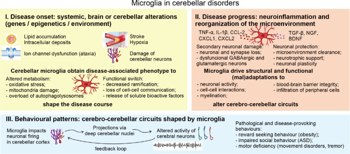
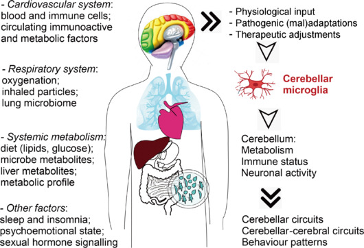
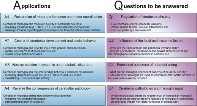
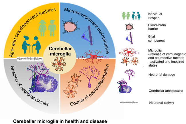
The cerebellum is receiving increasing attention for its cognitive, emotional, and social functions, as well as its unique metabolic profiles. Cerebellar microglia exhibit specialized and highly immunogenic phenotypes under both physiological and pathological conditions. These immune cells communicate with intrinsic and systemic factors and contribute to the structural and functional compartmentalization of the cerebellum. In this review, we discuss the roles of microglia in the cerebellar microenvironment, neuroinflammation, cerebellar adaptation, and neuronal activity, the associated molecular and cellular mechanisms, and potential therapeutic strategies targeting cerebellar microglia in the context of neuroinflammation. Future directions and unresolved questions in this field are further highlighted, particularly regarding therapeutic interventions targeting cerebellar microglia, functional mechanisms and activities of microglia in the cerebellar circuitry, neuronal connectivity, and neurofunctional outcomes of their activity. Cerebellar morphology and neuronal performance are influenced by both intrinsic and systemic factors that are actively monitored by microglia in both healthy and diseased states. Under pathological conditions, local subsets of microglia exhibit diverse responses to the altered microenvironment that contribute to the structural and functional compartmentalization of the cerebellum. Microglia in the cerebellum undergo early maturation during the embryonic stage and display specialized, highly immunogenic phenotypes. In summary, cerebellar microglia have the capacity to serve as regulatory tools that influence outcomes across a wide range of neurological and systemic conditions, including neurodevelopmental, neurodegenerative, metabolic, and stress-related disorders.

Keywords: Purkinje cells; brain regeneration; cerebellar diseases; cerebellum; innate immunity; macrophages; metabolism; microglia; neuroinflammation; neuropathology.


Conflict of interest statement

Conflicts of interest: The authors declare no conflicts of interest.

Figures

Figure 1
Figure 1
Timeline of research on cerebellar microglia. Since the 1960s, extensive research has been conducted on the phenotypes of cerebellar microglia in both healthy and diseased states. Concurrently, ongoing exploration into therapeutic interventions designed to modulate microglial activity with the aim of mitigating cerebellar inflammation and dysfunction is ongoing, with particular focus on future prospects. ASD: Autism specturm disorder.
Figure 2
Figure 2
Anatomy of the cerebellum and cerebellar microglia. (A) Microglia are located in all compartments of the cerebellum, including gray matter, white matter, and perivascular areas. (B) Under baseline conditions, cerebellar microglia exhibit a pronounced immunogenic phenotype and play a key role in regulating the microenvironment and functions of the cerebellum. BBB: Blood–brain barrier; CD40: cluster of differentiation 40; Clec: C-type lectin-like domain; CX3CL1: C-X3-C motif chemokine ligand 1; CX3CR1: C-X3-C motif chemokine receptor 1; IL: interleukin; PC: Purkinje cells; Stat: signal transducer and activator of transcription; TNF-α: tumor necrosis factor α; TREM: triggering receptors expressed on myeloid cells.
Figure 3
Figure 3
General mechanisms of microglial activity in cerebellar pathology. Microglia (orange) transition from a ramified state to an activated and/or impaired state in response to inflammation and changes in the microenvironment. Under disease progression, microglia drive immunogenic responses and exhibit altered modes of communication with other cerebellar cells, including neurons (violet), oligodendrocytes (blue), and components of the blood–brain barrier (brown). IL: Interleukin; NF-κB: nuclear factor kappa B; STING: stimulator of the interferon genes; TNFα: tumor necrosis factor α.
Figure 4
Figure 4
Cerebellar circuits are wired by microglia. (A, B) The cerebral cortex sends signals to the pontine nucleus, which relays them to the cerebellar cortex via mossy fibers (dark blue). These mossy fibers form synapses with GCs, which convey signals to PCs through parallel fibers (blue). PCs, in turn, project to the deep cerebellar nuclei, which also receive excitatory inputs from both mossy fibers and climbing fibers (violet). (B, C) Microglia (red) employ various strategies to regulate cerebellar functions, including direct communication with neurons, release of cytokines with neuroactive properties (red box), and shaping of neuronal circuits. (C) Detailed mechanisms of microglia-mediated modulation of the cerebellum are as follows: 1) Microglia interact with PCs (violet) by releasing neuroactive cytokines. 2) Microglial cells regulate the morphology and viability of GCs (blue). 3) Interactions of microglia with the nodes of Ranvier influence neuronal membrane polarization. Furthermore, microglia maintain subsets of oligodendrocytes (dark blue) and support axon myelination. (D) Cerebellar microglia are integrated into both cerebellar and cerebral–cerebellar circuits, thereby regulating essential neuronal functions. CCL2: Chemokine ligand 2; CCR2: chemokine receptor 2; CXCL: chemokine (C–X–C motif) ligand; CXCR: chemokine (C–X–C motif) receptor; EGF: epidermal growth factor; GCs: granular cells; IL: interleukin; Mbp: myelin basic protein; Mog: myelin oligodendrocyte glycoprotein; PCs: Purkinje cells; Sk2: small-conductance, Ca2+-activated K+ channel subtype 2; TNF-α: tumor necrosis factor α.
Figure 5
Figure 5
Involvement of microglia in cerebellar disorders. Cerebellar microglia are involved in disease pathogenesis from the early stages (I. Disease onset, yellow color). Fluctuations in the brain microenvironment caused by various intrinsic or environmental factors trigger changes in the metabolic and immune profiles of microglia. With disease progression (II. Disease progression, pink color), microglia continue to alter the cerebellar microenvironment, which impacts the activity of cerebellar neurons, particularly Purkinje cells. Proinflammatory polarized microglia lose direct cell–cell interactions and are unable to sustain neuronal viability. Additionally, excessive release of neuroactive cytokines can selectively affect the activity of GABAergic and glutamatergic cerebellar neurons, regulating neuronal firing in the cerebellar cortex. This activity projects via the deep cerebellar nuclei and ultimately alters cerebral neuron function. Cerebral neurons, in turn, provide feedback to the cerebellum, creating a feedback loop that rewires both cerebellar and cerebral-cerebellar circuits, ultimately leading to changes in disease-associated behavior patterns (III. Behavioral patterns, blue). ASD: Autism spectrum disorder; BDNF: brain-derived neurotrophic factor; CCL-2: chemokine ligand 2; CXCL: chemokine (C–X–C motif) ligand; IL: interleukin; NGF: nerve growth factor; TGF-β: tumor growth factor β; TNF-α: tumor necrosis factor-α.
Figure 6
Figure 6
Potential routes for systemic regulation of cerebellar microglia. Systemic factors can provoke pathogenic activation of microglia (Table 2) or serve as potential targets for therapy of cerebellar disorders. Possible connections between cerebellar microglia and microglia in other organs, including lung (blue), heart (dark pink), liver (dark red), digestive system, and intestinal microbiota, are shown.
Figure 7
Figure 7
Key prospective directions in research on cerebellar microglia. Applications (A) of cerebellar microglia as a potential target for addressing cerebellar dysfunction and neuronal loss are shown in blue. Key questions (Q) regarding the mechanisms by which cerebellar microglia respond to various pathogenic factors and modulate neuronal activity that remain unclear (indicated in violet).
Figure 8
Figure 8
Cerebellar microglia shape structure and functions of the cerebellum in health and disease. Cerebellar microglia are involved in maintenance and modulation of cerebellar microenvironment in development, adulthood and aging. Microglia contribute to age- and sex-specific aspects of cerebellar neuronal performance. Microglia regulate blood–brain barrier and glial cells of the cerebellum and are integrated into the neuronal circuits. In pathogenic conditions cerebellar microglia become activated or can undergo cell damage. Dysfunction of cerebellar microglia leads to neurodegeneration, loss of tissue integrity, and impairment of neuronal circuitry and cerebellar and cerebral functions.

Similar articles

References

    1. Abel JM, Witt DM, Rissman EF. Sex differences in the cerebellum and frontal cortex: roles of estrogen receptor alpha and sex chromosome genes. Neuroendocrinology. 2011;93:230–240. - PMC - PubMed
    1. Agyemang AA, Sveinsdóttir K, Vallius S, Sveinsdóttir S, Bruschettini M, Romantsik O, Hellström A, Smith LEH, Ohlsson L, Holmqvist B, Gram M, Ley D. Cerebellar exposure to cell-free hemoglobin following preterm intraventricular hemorrhage: causal in cerebellar damage? Transl Stroke Res. 2017;8:461–473. - PMC - PubMed
    1. Akang E, Dosumu O, Afolayan S, Agumah R, Akanmu AS. Modeling cerebellar limb dysmetria and impaired spatial memory in rats using lamivudine: a preliminary study. J Chem Neuroanat. 2020;109:101838. - PMC - PubMed
    1. Alekseeva OS, Gilerovich EG, Kirik OV, Korzhevskii DE. Structure and spatial organization of microgliocytes in the molecular layer of the cerebellar cortex in rabbits. Neurosci Behav Physiol. 2017;47:637–640.
    1. Ament SA, Cortes-Gutierrez M, Herb BR, Mocci E, Colantuoni C, McCarthy MM. A single-cell genomic atlas for maturation of the human cerebellum during early childhood. Sci Transl Med. 2023;15:1283. - PubMed

LinkOut - more resources